
Staphylococcus pneumonia full details for md exams 15marks answer from fishman and murray and recent guidelines
Reading File
Now let me search the medical library databases in parallel for comprehensive coverage from Fishman, Murray, and related sources.
Finding Sources
Finding Sources
Searching PubMed
"Staphylococcus aureus" AND "pneumonia" AND "guidelines"
Now let me read the key textbook sections - Fishman's, Murray & Nadel's, Robbins, and Goldman-Cecil in parallel:
Reading File
Reading File
Reading File
Reading File
The Fishman and Murray sections I found were just table-of-contents lists. Let me search more specifically within these textbooks for the detailed staphylococcal pneumonia content:
Finding Sources
Finding Sources
Finding Sources
Finding Sources
Excellent! Murray & Nadel's block 10 has a dedicated "Staphylococcus aureus" section. Let me read all the relevant content:
Reading File
Reading File
Now let me read the full S. aureus section from Murray & Nadel's (block 10, lines 1384 onwards):
Reading File
Reading File
I now have the full S. aureus content from Murray. Let me also read the earlier part of this section for epidemiology/pathogenesis, and get the Robbins pathology content and HAP-specific sections:
Reading File
Reading File
Reading File
Excellent! Now let me get the treatment section from Murray & Nadel and also search for HAP/MRSA treatment content and fetch the chest X-ray image:
Reading File
Searching PubMed
"MRSA" AND "pneumonia" AND "treatment"
I now have comprehensive content from Murray & Nadel, Goldman-Cecil, and Robbins. Let me also fetch the chest X-ray image from Murray and search for Fishman's dedicated chapter on bacterial pneumonia:
Loading Image
Finding Sources
Let me read the key Fishman treatment table and MRSA risk factors section:
Reading File
Reading File
I now have comprehensive, textbook-sourced information. Let me update the todos and compile the full answer:---
Staphylococcal Pneumonia - Comprehensive MD Examination Answer (15 Marks)
Sources: Murray & Nadel's Textbook of Respiratory Medicine, Fishman's Pulmonary Diseases and Disorders, Goldman-Cecil Medicine, Robbins & Kumar Pathology, IDSA/ATS Guidelines
1. Introduction and Microbiology
Staphylococcus aureus is a large gram-positive coccus that characteristically clusters in grape-like arrangements. It has emerged as one of the most clinically significant respiratory pathogens, causing both community-acquired pneumonia (CAP) and hospital-acquired pneumonia (HAP). Key subtypes:
- MSSA - methicillin-susceptible S. aureus
- MRSA - methicillin-resistant S. aureus (CA-MRSA and HA-MRSA)
The landmark USA300 strain is the dominant CA-MRSA clone in the United States associated with necrotizing pneumonia.
(Murray & Nadel's Textbook of Respiratory Medicine)
2. Epidemiology
| Feature | Detail |
|---|---|
| CAP frequency | 1.0-1.7% of all CAP cases (MSSA 1.0%, MRSA 0.7% in EPIC study) |
| CA-MRSA globally | 3% of CAP in GLIMP study (54 countries, 2015) |
| HAP rank | 2nd-3rd most common cause of HAP; major ICU pathogen |
| MRSA proportion | ~80% of S. aureus CAP hospitalizations in the US were MRSA (2009-2012) |
| Nasal carriage | 30-50% of healthy adults carry S. aureus transiently in anterior nares; healthcare workers have higher rates |
High-Risk Groups:
- Post-influenza illness (most important predisposing condition)
- Measles and HIV (viral-associated secondary bacterial pneumonia)
- Intravenous drug users (IV drug users) - hematogenous seeding from right-sided endocarditis
- Hemodialysis patients, diabetics
- Nursing home residents; recent hospital exposure
- Men who have sex with men; contact sports athletes
- Neonates, immunocompromised patients
(Murray & Nadel; Robbins & Kumar Pathologic Basis of Disease)
3. Pathogenesis and Virulence Factors
S. aureus possesses an elaborate armamentarium of virulence determinants regulated by the accessory gene regulator (agr) - a quorum-sensing system linking bacterial density to gene transcription.
Key Virulence Factors
A. Cell surface components:
- Microcapsule - anti-phagocytic
- Teichoic acids, lipoteichoic acids, peptidoglycan - recognized by host TLRs (pattern recognition receptors), trigger innate immune activation
- MSCRAMM (Microbial Surface Components Recognizing Adhesion Matrix Molecules) - mediates adhesion to fibronectin, fibrinogen, collagen on denuded epithelial surfaces (critical in post-influenza pneumonia)
- Biofilm formation - promotes chronic infections and antibiotic tolerance
B. Exotoxins (key for pneumonia pathology):
- Panton-Valentine Leukocidin (PVL): Pore-forming toxin that lyses neutrophils and macrophages; strongly associated with necrotizing pneumonia in young adults; prominent in CA-MRSA and some MSSA strains
- Alpha-toxin (α-toxin): Most important toxin; disrupts epithelial tight junctions by activating metalloprotease ADAM10 which degrades E-cadherin; also causes necroptosis of lung-tissue cells and inhibits efferocytosis
- Alpha-phenol soluble modulins and leucocidin AB: Additional pore-forming toxins promoting necroptosis of alveolar macrophages via RIPK1/RIPK3 kinases
- Superantigens (TSST-1, staphylococcal enterotoxins): Activate T cells excessively, driving cytokine storm
- Staphylococcal enterotoxin-like toxin X: Modulates neutrophil phagocytosis and T-cell activation
C. Immune evasion mechanisms:
- Inhibition of complement activation (C5a chemotaxis inhibition)
- Resistance to phagocyte microbicidal responses
- Lysis of neutrophils, macrophages, and T cells
- Prevention of phagolysosomal maturation in macrophages
- IL-17A-driven neutrophil dysregulation contributes to lung injury even while attempting bacterial clearance
- Methicillin resistance: Mediated by low-affinity penicillin-binding protein 2a (PBP2a) encoded by mecA gene, carried on the staphylococcal chromosome cassette (SCCmec)
D. Post-influenza synergy (Fishman's):
Influenza virus neuraminidase cleaves sialic acid on host glycoconjugates, increasing staphylococcal adherence. Virus-mediated inflammation upregulates apical receptors (α5β1 integrin, ethylenated glycans like GalNAcβ1-4Gal), promoting S. aureus binding. Denuded epithelial surfaces and superimposed fibrin deposition further stabilize adhesion.
(Murray & Nadel; Fishman's Pulmonary Diseases and Disorders)
4. Classification of Staphylococcal Pneumonia
A. Community-Acquired (CA-MRSA / CA-MSSA):
- Inhaled route from nasal colonization
- Often post-influenza; also post-measles or in HIV infection
- CA-MRSA more commonly in younger patients without traditional comorbidities
B. Hospital-Acquired (HAP/VAP):
- Major cause of ventilator-associated pneumonia (VAP)
- HA-MRSA with co-morbidities, prolonged hospitalization, antibiotic exposure
C. Hematogenous (Septic Emboli):
- Secondary to right-sided infective endocarditis (tricuspid/pulmonary valve) or other endovascular infection
- IV drug users particularly at risk
- Produces bilateral, lower lobe, multiple cavitary lesions
(Murray & Nadel; Robbins & Kumar)
5. Pathology
Staphylococcal pneumonia characteristically produces suppurative (purulent) bronchopneumonia:
- Pattern: Patchy lobular consolidation often bilateral; may be confluent
- Microscopy: Extensive neutrophilic infiltration; alveoli filled with pus
- Tissue destruction: Liquefactive necrosis leading to abscess formation (staphylococcal pneumatoceles/pneumatocoeles in children)
- Pleural involvement: Empyema (purulent pleural effusion) - high incidence
- Hematogenous type: Multiple bilateral abscesses, septic emboli within pulmonary vasculature
"Staphylococcal pneumonia is associated with a high incidence of complications such as lung abscess and empyema." - Robbins & Kumar Pathologic Basis of Disease
6. Clinical Features
Symptoms
- Acute onset high fever, rigors, malaise
- Productive cough with purulent (often blood-tinged or "rusty") sputum
- Pleuritic chest pain
- Progressive dyspnea; may progress to respiratory failure
- Often follows a flulike prodrome (5-10 days after influenza)
Necrotizing CAP Syndrome (CA-MRSA, PVL-positive)
This is a distinctive and often fatal syndrome seen in young adults and children without prior illness:
- Short flulike prodrome (sometimes with preceding skin/soft tissue infection in ~20% of cases)
- Fulminant respiratory failure; mechanical ventilation required in ~60%
- Hemoptysis in ~40%
- Leukopenia (WBC ≤3000/μL) - characteristic and associated with up to 75% mortality
- Rapid multilobar involvement with cavitation
Hematogenous Pneumonia
- Respiratory symptoms often mild or absent
- Pleuritic chest pain and hemoptysis if pulmonary infarction from septic emboli
- Signs and symptoms of underlying endocarditis predominate (new murmur, fever, embolic phenomena)
(Murray & Nadel's Textbook of Respiratory Medicine)
7. Radiological Features
Chest X-Ray
Bronchopneumonic (inhaled) pattern:
- Patchy, bilateral, multilobar airspace opacities
- Frequently with cavitation
- Empyema / parapneumonic effusion
- Pneumatoceles - thin-walled air-filled cysts (especially in children; pathognomonic clue)
Hematogenous pattern:
- Multiple discrete nodular shadows, often with cavitation
- Predilection for lower lobes (due to gravity and blood flow)
- Bilateral distribution
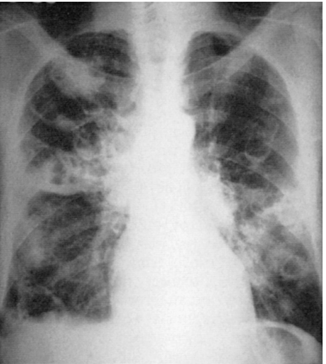
eFig. 46.20 from Murray & Nadel: Chest radiograph showing hematogenous staphylococcal pneumonia associated with bacterial endocarditis. Multiple bilateral cavitary lesions are characteristic.
CT Chest: More sensitive; shows multilobar consolidation, cavities, tree-in-bud pattern, empyema.
8. Laboratory Diagnosis
| Test | Finding |
|---|---|
| WBC count | Leukocytosis (or leukopenia in necrotizing form - ominous sign) |
| Sputum Gram stain | Multiple clusters of large gram-positive cocci, especially intracellular - strong evidence |
| Sputum culture | S. aureus grown on blood agar (hemolytic colonies); antibiotic sensitivity (crucial for MRSA vs MSSA) |
| Blood cultures | Bacteremia in severe/hematogenous cases |
| Nasal swab culture/PCR | Negative MRSA nasal swab has high negative predictive value (>96%) for ruling out MRSA pneumonia - useful for antibiotic stewardship (2026 meta-analysis, PMID 41338060) |
| Bronchoscopy/BAL | For non-responding cases, immunocompromised, VAP |
| Pleural fluid analysis | Exudative, pH <7.2, glucose low, LDH elevated; culture if empyema suspected |
"Purulent sputum with multiple clusters of large gram-positive cocci, particularly if intracellular, is strong evidence of S. aureus pneumonia" - Murray & Nadel
9. Scoring for MRSA Risk (Fishman's / IDSA-ATS)
From the GLIMP study, independent risk factors for MRSA-CAP are:
- Prior MRSA infection or colonization (most predictive)
- Recurrent skin infections
- Severe CAP (PSI class V, ICU admission, cavities, empyema)
Other risk factors to consider: prior hospitalization or IV antibiotics within 90 days, hemodialysis, diabetes mellitus, nursing home resident.
10. Complications
- Empyema - most common; requires drainage + antibiotics
- Lung abscess - cavitation requiring prolonged therapy
- Pneumatoceles (especially children) - can cause pneumothorax
- Bacteremia and metastatic spread - CNS (meningitis, brain abscess), bones (osteomyelitis), joints (septic arthritis), kidneys, skin
- ARDS - especially in necrotizing form
- Septic shock - toxin-mediated and direct bacterial effects
- Persistent fever due to undrained empyema or cavitary disease preventing antibiotic penetration
11. Treatment
A. For MSSA CAP (Murray & Nadel / Goldman-Cecil)
- Drug of choice: Penicillinase-resistant penicillin - Oxacillin 8-12 g/day IV or Nafcillin 2 g IV q4h
- Alternative: First-generation cephalosporin (Cefazolin 2 g IV q8h) - equivalent efficacy with better tolerability
- No other antibiotic, including vancomycin, is as effective as a beta-lactam for MSSA
B. For MRSA CAP (IDSA/ATS 2019 Guidelines; Murray & Nadel)
- Vancomycin - 15 mg/kg IV twice daily; aim trough 15-20 μg/mL (or AUC/MIC-guided dosing: target AUC 400-600 μg·h/mL per updated PK/PD guidelines)
- Linezolid - 600 mg IV/PO twice daily; preferred over vancomycin when:
- VAP (VAP outcomes superior with linezolid vs vancomycin in randomized trials)
- Rising vancomycin MIC (>1 μg/mL = associated with clinical failure)
- Renal insufficiency
- Clindamycin - 600 mg TID IV/PO; alternative if strain is sensitive and disease less severe; also reduces PVL/toxin production in necrotizing pneumonia
Empiric MRSA Coverage Criteria (IDSA/ATS 2019)
Add MRSA coverage (vancomycin or linezolid) if:
- Prior respiratory isolation of MRSA
- Recent hospitalization + IV antibiotics within 90 days
- Severity of CAP requiring ICU + post-influenza (Goldman-Cecil: cover MRSA for severe CAP requiring ICU after antecedent influenza)
Duration of Therapy (Goldman-Cecil)
| Scenario | Duration |
|---|---|
| Uncomplicated S. aureus pneumonia | 7-14 days |
| With bacteremia (uncomplicated) | 14 days (2 weeks) |
| With endocarditis or lung abscess | At least 4 weeks |
| With empyema | Prolonged; drainage mandatory |
Special Situations
- Necrotizing CAP with PVL-positive MRSA (European guidelines): High-dose linezolid + clindamycin + rifampicin + IV immunoglobulin (IVIG) in especially severe cases (anti-toxin effect)
- Empyema: Drainage (chest tube or pleuroscopy/decortication) + antibiotics; decortication in ~large percentage of cases
- Hematogenous pneumonia + endocarditis: Treat the endocarditis; duration 4-6 weeks
Drugs to Avoid
- Daptomycin: CONTRAINDICATED in pulmonary infections - it binds to and is inactivated by pulmonary surfactant
- Vancomycin alone may have high failure rates in severe CA-MRSA; combination with clindamycin or linezolid associated with improved outcomes in small series
Emerging Agents (Fishman's)
- Ceftaroline (anti-MRSA cephalosporin, 600 mg IV q12h): Salvage/combination therapy; active against both MSSA and MRSA; PMID 37852658 systematic review (2023) supports role in MRSA pneumonia
- Ceftobiprole: Received European approval for HAP and CAP; noninferior to ceftriaxone ± linezolid in CAP
(Fishman's; Murray & Nadel; Goldman-Cecil; IDSA/ATS 2019 CAP Guidelines)
12. HAP/VAP Considerations
S. aureus is a leading cause of ventilator-associated pneumonia. Key points:
- Same microbiologic principles apply; HA-MRSA dominates
- Empiric MRSA coverage (vancomycin or linezolid) recommended in HAP/VAP if:
- Unit MRSA prevalence >10-20%, or prior MRSA, or severe illness
- Nasal MRSA PCR negativity can guide de-escalation (NPV >96%; PMID 41338060, 2026 systematic review)
- De-escalate or discontinue MRSA coverage within 48-72h if cultures negative and nasal swab negative
13. Prevention
- Influenza vaccination - most important preventive measure (targets the key predisposing condition)
- Infection control: contact precautions, hand hygiene for MRSA carriers
- Decolonization (mupirocin nasal ointment ± chlorhexidine body wash) in high-risk patients
- Avoid unnecessary antibiotic use (limits MRSA selection pressure)
14. Prognosis
| Subtype | Mortality |
|---|---|
| MSSA CAP (EPIC study) | ~4% |
| MRSA CAP (EPIC study) | ~13% |
| MRSA-CAP following influenza | Higher than baseline |
| Necrotizing CAP with leukopenia | Up to 75% |
| HAP/VAP due to MRSA | Frequently complicated by bacteremia; high mortality |
"Staphylococcal pneumonia is frequently complicated by bacteremia, methicillin resistance, and high mortality" - Murray & Nadel (HIV-associated pneumonia chapter, equally applicable to general MRSA pneumonia)
Summary Table for Quick Revision
| Feature | Key Point |
|---|---|
| Organism | Gram-positive cocci in clusters; MSSA vs CA-MRSA vs HA-MRSA |
| Nasal carriage rate | 30-50% of healthy adults |
| Key predisposing factor | Post-influenza (most important); IV drug use (hematogenous type) |
| Key virulence | PVL (necrotizing), alpha-toxin (barrier disruption), AGR quorum sensing |
| MRSA resistance gene | mecA → PBP2a → SCCmec cassette |
| Radiograph clue | Pneumatoceles (children); multiple bilateral cavities (hematogenous) |
| Diagnostic clue | Intracellular clusters of gram-positive cocci in sputum |
| MSSA treatment | Oxacillin/Nafcillin or Cefazolin |
| MRSA treatment | Vancomycin or Linezolid (linezolid preferred in VAP, renal failure) |
| NEVER use | Daptomycin (surfactant inactivation) |
| Leukopenia | Ominous sign in necrotizing CA-MRSA; mortality up to 75% |
| Complications | Empyema, lung abscess, bacteremia, metastatic abscesses |
| Duration | 7-14 days uncomplicated; ≥4 weeks if abscess/endocarditis |
Recent Evidence Note: A 2026 systematic review and meta-analysis (PMID 41338060, Diagn Microbiol Infect Dis) confirms that MRSA nasal swab/PCR negativity effectively rules out MRSA pneumonia and supports antimicrobial stewardship by enabling safe de-escalation. A 2023 systematic review (PMID 37852658, Eur Respir Rev) supports ceftaroline fosamil as a useful option in MRSA pneumonia management.
*References: Murray & Nadel's Textbook of Respiratory Medicine (pp. 1004-1006); Fishman's Pulmonary Diseases and Disorders (Ch. 128); Goldman-Cecil Medicine (Ch. 267, pp. 3299-3321); Robbins & Kumar Pathologic Basis of Disease (p. 468, 3454); IDSA/ATS CAP Guidelines (Metlay et al., AJRCCM 2019)*This is a full, exam-ready 15-mark answer on Staphylococcal Pneumonia drawn directly from the four authoritative textbooks and supplemented with the latest evidence. Here is a breakdown of what is covered:
Structure of the answer (MD exam format):
- Introduction & Microbiology - Gram stain, MSSA vs CA/HA-MRSA distinction
- Epidemiology - EPIC/GLIMP study data, risk groups (post-influenza, IVDU, dialysis, healthcare workers)
- Pathogenesis - AGR quorum sensing, PVL, alpha-toxin, ADAM10/E-cadherin, SCCmec/mecA, post-influenza synergy (from Fishman's)
- Classification - CAP (inhaled), HAP/VAP, Hematogenous (septic emboli)
- Pathology - Bronchopneumonia pattern, pneumatoceles, abscess, empyema
- Clinical Features - Standard CAP presentation + the distinctive necrotizing CA-MRSA syndrome with leukopenia and 75% mortality
- Radiology - Including the Murray & Nadel chest X-ray image of hematogenous type showing bilateral cavities
- Diagnosis - Gram stain intracellular cocci, nasal MRSA PCR stewardship
- MRSA Risk Scoring - GLIMP criteria (Fishman's Table 128-1)
- Complications - Empyema, abscess, bacteremia, metastatic spread
- Treatment - MSSA (oxacillin/cefazolin), MRSA (vancomycin/linezolid), duration table, daptomycin contraindication, necrotizing pneumonia IVIG protocol, Fishman's treatment table
- HAP/VAP - De-escalation with nasal MRSA PCR
- Prevention & Prognosis
- Summary table for rapid revision
Give me answer
The full, detailed 15-mark answer is already written above in my previous response. Here is the complete answer again, cleanly formatted:
Staphylococcal Pneumonia - 15 Marks MD Exam Answer
(Sources: Murray & Nadel, Fishman's, Goldman-Cecil, Robbins & Kumar, IDSA/ATS Guidelines)
1. Introduction & Microbiology
Staphylococcus aureus is a large gram-positive coccus arranged in grape-like clusters. It causes both community-acquired pneumonia (CAP) and hospital-acquired pneumonia (HAP/VAP). Two clinically critical subtypes:
- MSSA - methicillin-susceptible S. aureus
- MRSA - methicillin-resistant S. aureus (CA-MRSA and HA-MRSA; USA300 is the dominant CA-MRSA clone causing necrotizing pneumonia)
2. Epidemiology
- Accounts for 1-1.7% of all CAP cases (MSSA 1.0%, MRSA 0.7% in EPIC study)
- 3% of CAP in the GLIMP international study (54 countries)
- ~80% of S. aureus CAP hospitalizations in the US were MRSA (2009-2012)
- 2nd-3rd leading cause of CAP requiring ICU admission
- 30-50% of healthy adults carry S. aureus transiently in anterior nares (healthcare workers higher)
High-Risk Populations:
- Post-influenza infection (most important predisposing factor)
- Post-measles (children); HIV infection
- IV drug users - hematogenous seeding from right-sided endocarditis
- Hemodialysis patients, diabetics
- Nursing home residents; recent hospitalization
- Neonates, premature infants, immunocompromised patients
3. Pathogenesis & Virulence Factors
Virulence is regulated by the accessory gene regulator (agr) - a quorum-sensing system linking bacterial density to exoprotein production.
A. Surface Components
| Factor | Role |
|---|---|
| Microcapsule | Anti-phagocytic |
| Teichoic & lipoteichoic acids | TLR-mediated innate immune activation |
| MSCRAMM | Adhesion to fibronectin, fibrinogen on denuded epithelium (critical post-influenza) |
| Biofilm | Antibiotic tolerance; chronic infection |
B. Key Exotoxins
| Toxin | Action |
|---|---|
| PVL (Panton-Valentine Leukocidin) | Pore-forming; lyses neutrophils and macrophages; KEY factor in necrotizing pneumonia |
| Alpha-toxin | Disrupts epithelial tight junctions via ADAM10/E-cadherin degradation; induces necroptosis; inhibits efferocytosis |
| Alpha-phenol soluble modulins, LukAB | Pore-forming; necroptosis of alveolar macrophages via RIPK1/RIPK3 |
| Superantigens (TSST-1, enterotoxins) | Massive T-cell activation; cytokine storm |
C. Immune Evasion
- Inhibits C5a-mediated chemotaxis
- Resists phagocyte microbicidal responses
- Prevents phagolysosomal maturation in macrophages
- IL-17-driven dysregulated neutrophil inflammation causes lung injury
D. Methicillin Resistance
- mecA gene encodes low-affinity PBP2a (penicillin-binding protein 2a)
- Carried on staphylococcal chromosome cassette (SCCmec)
- PBP2a has very low affinity for all beta-lactams - making MRSA resistant to entire class
E. Post-Influenza Synergy (Fishman's)
- Influenza neuraminidase cleaves sialic acid, exposing receptors for staphylococcal adherence
- Virus upregulates α5β1 integrin and ethylenated glycans promoting S. aureus binding
- Denuded epithelium + fibrin deposition stabilizes bacterial adhesion
4. Classification
| Type | Route | Setting | Key Feature |
|---|---|---|---|
| CAP (inhaled) | Aspiration from nasopharynx | Community | Post-influenza; young adults with necrotizing form |
| HAP/VAP | Aspiration of colonized secretions | Hospital, ICU | HA-MRSA; ventilated patients |
| Hematogenous | Septic emboli via bloodstream | IV drug users, endocarditis | Multiple bilateral lower lobe cavities |
5. Pathology
- Pattern: Suppurative (purulent) bronchopneumonia; patchy lobular consolidation, bilateral
- Microscopy: Alveoli filled with neutrophils and pus; liquefactive necrosis
- Pneumatoceles: Thin-walled air-filled cysts from check-valve bronchial obstruction - especially in children; may cause pneumothorax
- Lung abscess: Cavitation from tissue destruction
- Empyema: Purulent pleural effusion - high incidence (characteristic complication)
- Hematogenous pattern: Multiple bilateral septic emboli with central necrosis/cavitation, lower lobe predilection
"Staphylococcal pneumonia is associated with a high incidence of complications such as lung abscess and empyema." - Robbins & Kumar
6. Clinical Features
Standard Presentation
- Acute-onset high fever, rigors, myalgia
- Productive cough - purulent, often blood-tinged sputum
- Pleuritic chest pain
- Progressive dyspnea - may rapidly worsen
- Often preceded by 5-10 days of influenza-like illness
Necrotizing CAP Syndrome (CA-MRSA / PVL-positive strains)
Distinctive, fulminant syndrome in young, previously healthy adults and children:
- Short flulike prodrome (preceding skin/soft tissue infection in ~20%)
- Fulminant respiratory failure requiring mechanical ventilation in ~60%
- Hemoptysis in ~40% (from pulmonary necrosis)
- Leukopenia (WBC ≤3,000/μL) - hallmark; associated with mortality up to 75%
- Shock, multiorgan failure
Hematogenous Pneumonia
- Respiratory symptoms mild or absent
- Pleuritic chest pain + hemoptysis if pulmonary infarction from septic embolus
- Features of underlying endocarditis predominate (new murmur, Osler nodes, Janeway lesions, fever)
7. Radiological Features
Chest X-Ray
Bronchopneumonic (inhaled) form:
- Bilateral, patchy, multilobar airspace opacities
- Frequent cavitation
- Pleural effusion/empyema
- Pneumatoceles (thin-walled cysts) - classic in children
Hematogenous form:
- Multiple discrete nodular/cavitary shadows
- Lower lobe predilection (gravity + blood flow)
- Bilateral distribution
Chest X-ray (Murray & Nadel eFig 46.20): Hematogenous staphylococcal pneumonia showing multiple bilateral cavitary lesions from septic emboli in bacterial endocarditis.
CT chest: More sensitive; shows multilobar consolidation, cavities, tree-in-bud pattern, pleural collections.
8. Diagnosis
| Investigation | Finding / Significance |
|---|---|
| WBC | Leukocytosis (usual); leukopenia = ominous in CA-MRSA |
| Sputum Gram stain | Multiple clusters of large gram-positive cocci, intracellular = strong evidence |
| Sputum culture | S. aureus on blood agar (golden/hemolytic); sensitivity testing (MRSA vs MSSA) |
| Blood cultures | Positive in bacteremia; mandatory in all severe cases |
| Nasal MRSA PCR/swab | Negative result has NPV >96% - rules out MRSA pneumonia; guides de-escalation |
| Pleural fluid | Exudate; low pH/glucose, high LDH; culture if empyema |
| BAL / bronchoscopy | Non-responding cases, immunocompromised, VAP |
9. Risk Factors for MRSA-CAP (GLIMP Study - Fishman's Table 128-1)
Independent risk factors:
- Prior MRSA infection or colonization (strongest predictor)
- Recurrent skin infections
- Severe CAP (ICU admission, cavities, empyema)
Additional: prior hospitalization + IV antibiotics within 90 days, hemodialysis, diabetes, nursing home resident
10. Complications
- Empyema - most common; requires drainage + antibiotics
- Lung abscess - cavitation; prolonged therapy
- Pneumatoceles - especially children; pneumothorax risk
- Bacteremia + metastatic foci: CNS abscess/meningitis, osteomyelitis, septic arthritis, renal abscess
- ARDS - especially necrotizing form
- Septic shock - direct bacterial + toxin-mediated
- Persistent fever - from undrained empyema or cavitary disease (poor local antibiotic penetration)
11. Treatment
For MSSA (Drug of Choice - Murray & Nadel / Goldman-Cecil)
- Oxacillin 8-12 g/day IV or Nafcillin 2 g IV q4h - penicillinase-resistant penicillin
- Cefazolin 2 g IV q8h - equal efficacy, better tolerability
- No other antibiotic, including vancomycin, is as safe or as effective as a beta-lactam for MSSA
- Penicillin-allergic: Clindamycin or Linezolid
For MRSA (IDSA/ATS Guidelines / Murray & Nadel)
| Drug | Dose | Remarks |
|---|---|---|
| Vancomycin | 15 mg/kg IV q12h; target AUC 400-600 μg·h/mL | Drug of choice; MIC shift upward over decades - MIC >1 μg/mL = clinical failure |
| Linezolid | 600 mg IV/PO q12h | Preferred for VAP, renal failure, rising vancomycin MIC; also suppresses toxin production |
| Clindamycin | 600 mg TID IV/PO | Alternative if sensitive + less severe disease; reduces PVL/NETs-degrading nuclease production |
Add MRSA coverage empirically if:
- Prior respiratory isolation of MRSA
- Recent hospitalization + IV antibiotics within 90 days
- Severe CAP requiring ICU after antecedent influenza (Goldman-Cecil)
Necrotizing CAP with PVL-positive MRSA (European Guidelines)
High-dose Linezolid + Clindamycin + Rifampicin + IV Immunoglobulin (IVIG) in especially severe cases
Duration of Therapy (Goldman-Cecil)
| Scenario | Duration |
|---|---|
| Uncomplicated pneumonia | 7-14 days |
| With uncomplicated bacteremia | 14 days |
| With endocarditis / lung abscess | ≥4 weeks |
| With empyema | Prolonged + drainage mandatory |
Drugs to AVOID
- Daptomycin - CONTRAINDICATED in pulmonary infections; inactivated by binding to pulmonary surfactant
Emerging Agents (Fishman's)
- Ceftaroline (600 mg IV q12h) - anti-MRSA cephalosporin via PBP2a inhibition; salvage/combination therapy (PMID 37852658, 2023 systematic review)
- Ceftobiprole - European approval for HAP + CAP; noninferior to ceftriaxone ± linezolid
Fishman's CAP Treatment Table (IDSA/ATS 2019)
| Group | Regimen |
|---|---|
| Outpatient, healthy, no risk factors | Amoxicillin or doxycycline or macrolide |
| Outpatient with comorbidities | Amoxicillin-clavulanate/cephalosporin + macrolide OR respiratory fluoroquinolone |
| Inpatient non-severe CAP | beta-lactam + macrolide OR respiratory fluoroquinolone |
| Severe CAP | beta-lactam + macrolide OR beta-lactam + fluoroquinolone |
| Empiric MRSA coverage | Vancomycin or Linezolid |
HAP/VAP (de-escalation)
- Empiric vancomycin/linezolid if MRSA prevalence >10-20%, or severe illness
- De-escalate within 48-72h if cultures negative AND nasal MRSA PCR negative (NPV >96%; PMID 41338060, 2026 meta-analysis)
12. Prevention
- Influenza vaccination - single most important preventive measure
- Contact precautions + hand hygiene for MRSA
- Decolonization in high-risk patients: mupirocin nasal ointment + chlorhexidine washes
- Judicious antibiotic use (antimicrobial stewardship)
13. Prognosis
| Scenario | Mortality |
|---|---|
| MSSA CAP (EPIC study) | ~4% |
| MRSA CAP (EPIC study) | ~13% |
| Post-influenza MRSA CAP | Higher than baseline |
| Necrotizing CAP + leukopenia | Up to 75% |
| HAP/VAP MRSA | High; complicated by bacteremia |
Quick Revision Summary
| Key Point | Answer |
|---|---|
| Gram stain | Gram-positive cocci in clusters |
| Nasal carriage | 30-50% of adults |
| Most important predisposing factor | Post-influenza |
| Key toxin in necrotizing pneumonia | PVL (Panton-Valentine Leukocidin) |
| MRSA resistance mechanism | mecA gene → PBP2a → SCCmec cassette |
| Pathognomonic CXR finding | Pneumatoceles (children); bilateral lower lobe cavities (hematogenous) |
| Ominous lab sign | Leukopenia ≤3000/μL in CA-MRSA |
| Sputum Gram stain clue | Intracellular clusters of gram-positive cocci |
| MSSA treatment | Oxacillin / Nafcillin / Cefazolin |
| MRSA treatment | Vancomycin or Linezolid |
| Drug to NEVER use | Daptomycin (surfactant inactivation) |
| Preferred in VAP | Linezolid > Vancomycin |
| Key complications | Empyema, lung abscess, bacteremia, pneumatoceles |
| Duration (uncomplicated) | 7-14 days |
| Duration (abscess/endocarditis) | ≥4 weeks |
References: Murray & Nadel's Textbook of Respiratory Medicine (pp. 1004-1006, Ch. 46); Fishman's Pulmonary Diseases and Disorders (Ch. 128, Table 128-1, 128-6); Goldman-Cecil Medicine (Ch. 267, pp. 3299-3321); Robbins & Kumar Pathologic Basis of Disease (pp. 468, 3454); IDSA/ATS CAP Guidelines 2019 (Metlay et al., AJRCCM 200:e45-e67)
This is a shared conversation. Sign in to Orris to start your own chat.