
All urology surgical instruments with labelled diagrams and explanation and mechanics... Teach me for my exam point of view all urology surgical instruments along with novel instrument s
Reading File
Now let me search the medical library and web simultaneously for comprehensive urology surgical instruments content.
Finding Sources
Finding Sources
Searching the Web
urology surgical instruments names diagrams explained exam
Finding Sources
Finding Sources
Reading Web Page
https://regalinstruments.com/blogs/articles/urology-surgical…
Finding Sources
Searching the Web
urology instruments cystoscope resectoscope TURP ureteroscope nephroscope diagram labeled
Reading File
Reading File
Loading Image
Loading Image
Loading Image
Finding Sources
Finding Sources
Reading File
Loading Image
I now have excellent material from Hinman's Atlas of Urologic Surgery and Campbell-Walsh Wein Urology, plus great diagrams. Let me now compile the comprehensive exam-oriented answer.
Complete Guide to Urology Surgical Instruments - Exam Ready
PART 1: ENDOSCOPIC INSTRUMENTS (Most Exam-Important)
1. CYSTOSCOPE

Definition: An endoscope inserted transurethrally to directly visualize the urethra, bladder neck, trigone, ureteral orifices, and bladder mucosa.
Parts (numbered on diagram):
| Number | Part | Function |
|---|---|---|
| 1 | Ocular funnel | Eye attachment point for surgeon's eye or camera |
| 2 | Ocular window | Where image is viewed |
| 3 | Light pillar | Connects fiber-optic light source |
| 4 | Number denoting sheath size | Usually in French gauge (Fr) |
| 5 | Irrigation inlet/outlet | Inflow/outflow of irrigant fluid |
| 6 | Number denoting size of ureteric catheter that can be passed | |
| 7 | Stopcock of working channel | Controls access to working channel |
| 8 | Working channel | Allows passage of instruments (biopsy forceps, catheters) |
| 9 | Color code disk | Identifies telescope angle |
| 10 | Beak | Tapered end inserted into urethra |
| 11 | Markings on sheath (1 cm apart) | Estimate depth of insertion |
| 12 | Objective window | Distal lens |
Mechanics:
- Telescope has a rod-lens (Hopkins lens) system that transmits magnified image
- Light transmitted through fiber-optic bundles running alongside the telescope
- Irrigation channels allow continuous inflow/outflow to distend bladder and clear blood/debris
- Available in different telescope angles: 0° (forward), 12°, 30°, 70° (most common for bladder), 120° (retrograde view)
- French size (Fr) = outer diameter in mm x 3; standard diagnostic cystoscope is 17-21 Fr
Types:
- Rigid cystoscope - metal sheath, better optics, used in OR
- Flexible cystoscope - fiber optic or digital chip-tip, office-based, better for anterior wall visualization, more patient comfort
- Continuous flow cystoscope - two-channel: irrigant in one, out the other simultaneously
Clinical use:
- Hematuria evaluation, bladder tumor diagnosis/surveillance, ureteral catheterization, foreign body removal, urodynamics
2. RESECTOSCOPE
Definition: A modified cystoscope with an added cutting loop electrode used for transurethral resection procedures.
Components:
| Part | Description |
|---|---|
| Outer sheath | 24-26-28 Fr; contains irrigation channels |
| Inner sheath | Continuous flow inner sheath for irrigation |
| Telescope | Usually 30° or 12° Hopkins rod lens |
| Working element | The frame that holds and drives the loop |
| Loop electrode (cutting loop) | Wire loop that carries electrical current to cut tissue |
| Rollerball/button electrode | Used for coagulation/vaporization |
| Obturator | Blind or visual; used to introduce sheath into bladder atraumatically |
Mechanics:
- The working element has a spring-return mechanism - the loop is pushed forward into tissue by the surgeon's thumb, then snaps back automatically
- Monopolar resectoscope: Uses non-ionic irrigant (glycine 1.5%, sorbitol/mannitol) because saline disperses monopolar current. Risk of TUR syndrome (dilutional hyponatraemia, water intoxication from glycine absorption)
- Bipolar resectoscope: Active and return electrodes are both on the same loop - current flows between them only through the tissue at the tip. Can use normal saline as irrigant. Eliminates TUR syndrome. This is the modern preferred approach.
Key Exam Points:
- Monopolar TURP: glycine irrigant, risk of TUR syndrome
- Bipolar TURP: saline irrigant, safer, especially in large prostates
- Loop is advanced UNDER vision, cutting on withdrawal (not on forward movement)
- Verumontanum is the anatomical landmark to avoid distal sphincter injury
Used in:
- TURP (Transurethral Resection of Prostate)
- TURBT (Transurethral Resection of Bladder Tumour)
- TUIP (Transurethral Incision of Prostate)
- Transurethral resection of ureterocele
- Urethral stricture (internal urethrotomy variant)
3. URETEROSCOPE
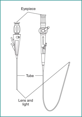
Definition: A long, slender endoscope passed transurethrally through the bladder and up the ureter (and into the renal pelvis).
Types:
| Type | Fr Size | Deflection | Use |
|---|---|---|---|
| Semi-rigid ureteroscope | 6-9 Fr | None (slight flex) | Distal/mid ureter stones |
| Flexible ureteroscope (FURS) | 7.5-9 Fr | Active up to 180-270° deflection | Upper ureter, renal pelvis, calyces |
| Digital ureteroscope | Same | Same | Chip-on-tip camera, no fiber bundle degradation |
Parts (flexible):
- Eyepiece/camera port at proximal end
- Handle with deflection lever (active deflection of tip)
- Irrigation port (inflow)
- Working channel (2.4-3.6 Fr) - for laser fibers, baskets, biopsy forceps
- Long insertion tube (40-45 cm)
- Distal tip with lens and light
Mechanics:
- Active deflection via a pull-wire mechanism (Bowden cable): pulling the lever bends the distal tip
- Fiber-optic bundles transmit light and image (or digital chip transmits image directly)
- Access sheath (ureteral access sheath, UAS) used to facilitate repeated insertion/removal, reduces intrarenal pressure
- Holmium:YAG laser fiber passed through working channel for lithotripsy
Used in:
- Ureterorenoscopy (URS) for ureteral/renal stones - laser lithotripsy
- Upper tract urothelial carcinoma - biopsy, fulguration
- Ureteral stricture assessment/management
4. NEPHROSCOPE
Definition: A short, wide-bore endoscope used for percutaneous nephroscopy (PCNL - Percutaneous Nephrolithotomy).
Types:
| Type | Size | Use |
|---|---|---|
| Standard rigid nephroscope | 24-28 Fr | Standard PCNL |
| Mini-PCNL nephroscope | 14-20 Fr | Smaller tracts |
| Ultra-mini/Micro-PCNL | 4.8-12 Fr | Paediatric/mini PCNL |
| Offset rigid nephroscope | Varies | HoLEP (holmium laser prostate enucleation) |
Parts:
- Short rigid shaft (15-20 cm)
- Wide working channel (3-4 mm) for large stone forceps, EHL/laser probes, ultrasonic probes
- Offset eyepiece (to allow instruments to be passed in a straight line while viewing obliquely)
- Irrigation in/out ports
Mechanics:
- Introduced through a percutaneous tract dilated in the flank/back
- Alligator stone forceps, three-prong graspers passed through working channel
- Ultrasonic lithotripter: hollow probe that transmits ultrasound vibrations to fragment stone, suction through center removes fragments
- Electrohydraulic lithotripsy (EHL): spark-gap probe creates shock waves
- Pneumatic lithotripter: compressed air fires a probe to fragment stone mechanically
Used in:
- PCNL for large (>2 cm) or complex renal stones
- Calyceal diverticulum access
- Endopyelotomy
PART 2: DILATORS
5. URETHRAL DILATORS
Types and Mechanics:
A. Van Buren Sounds (Metal Sounds)
- Solid, curved, metal instruments in graduated sizes (8-34 Fr)
- S-shaped curve follows the male urethra (penile + perineal + prostatic)
- Used for: urethral stricture dilation, assessment of urethral calibre
- Passed by "sounding" - following the urethral curve with gentle forward pressure
B. Clutton Sounds
- Longer, more gently curved than Van Buren
- Used in females (shorter, straighter urethra - no need for S-curve)
C. Filiform Bougies + Followers
- Filiforms: Very fine (1-5 Fr), flexible, with a screw-tip; used when urethra is nearly completely obstructed and a sound cannot pass
- Followers: Hollow dilators that screw onto the filiform to gradually dilate over the guidewire
- Mechanics: Pass filiform past stricture, screw on progressively larger followers
D. Hegar Dilators
- Straight, solid dilators
- Used primarily for cervical/urethral dilation in females, also used in paediatric urethral dilation
E. Balloon Dilators
- Angioplasty-type catheter with an inflatable balloon at the tip
- Used for ureteral strictures, urethral strictures, ureteropelvic junction (UPJ) obstruction
- Mechanics: Placed over a guidewire, inflated to a preset pressure (ATM) to dilate stricture radially without shear force
PART 3: CATHETERS
6. FOLEY CATHETER
- Self-retaining urethral catheter with an inflatable balloon (5-30 mL)
- Drainage eye at tip, balloon inflation port, drainage port
- Standard sizes: 12-24 Fr
- Three-way Foley: Has a third channel for continuous irrigation (used post-TURP)
7. COUNCIL CATHETER
- Has a through-and-through lumen from tip to balloon port
- Passed over a guidewire (wire exits through the tip)
- Used when the urethra is difficult/traumatised
8. COUDE CATHETER
- Has a slightly bent (elbow) tip
- Helps navigate the curved male urethra / enlarged prostate / bladder neck obstruction
- Named from French "coude" = elbow
9. SUPRAPUBIC CATHETER (SPC)
- Introduced percutaneously through the abdominal wall into the bladder
- Used when urethral catheterization is impossible/contraindicated
- Placed using a trocar/cannula system under ultrasound guidance
PART 4: STONE INSTRUMENTS (Intracorporeal Lithotripsy)
10. STONE BASKETS / DORMIA BASKET
- Wire basket (3-4 wires) opened around a stone and then withdrawn to trap/extract it
- Available in: flat wire (rigid), nitinol (flexible, shape-memory for flexible ureteroscopy), tip-deflecting
- Mechanics: Passed beyond stone in collapsed state, opened to surround stone, withdrawn with stone trapped
11. ELLIK EVACUATOR
- A glass or plastic bulb device with two channels
- Mechanics: Creates a Venturi effect - squeezing the bulb forces irrigant into bladder, releasing it creates suction that draws chips/clots into the bulb's lower reservoir
- Used after TURP, TURBT: evacuates prostatic chips and blood clots from bladder
- Quoted directly from Hinman's Atlas: "the Ellik evacuator produces a Venturi effect, allowing chips of prostate tissue to settle to the bottom of the bulb"
12. RANDALL STONE FORCEPS
- Four curved forceps of progressively increasing angulation (one straight + three curved)
- Key distinguishing feature: no locking ratchet mechanism (unlike hemostatic clamps)
- Used to retrieve stone fragments from renal calyces through a nephroscope
- The progressive angles allow access to differently oriented calyces
PART 5: OPEN/LAPAROSCOPIC INSTRUMENTS
13. VASCULAR CLAMPS (Urological)
| Clamp | Feature | Use |
|---|---|---|
| DeBakey clamp | Longitudinal interlocking ridges, atraumatic jaw | Renal artery/vein clamping in partial nephrectomy |
| Satinsky clamp | Side-biting, partial occlusion | IVC/renal vein partial clamping |
| Bulldog clamp | Small, spring-loaded | Temporary renal artery occlusion (warm ischemia) |
| Wylie "J" clamp | J-shaped jaw | Deep vessel access |
| Glover clamp | Atraumatic curved jaw | General vascular |
Mechanics: Very fine interlocking longitudinal ridges grip vessel wall without crushing - prevents slipping while maintaining vessel integrity for anastomosis.
14. KIDNEY PEDICLE CLAMPS
- Extra-long (30+ cm), right-angled or curved
- Designed to control the renal hilum en masse during nephrectomy
- Examples: Herrick kidney pedicle clamp
15. RETRACTORS
| Retractor | Type | Use |
|---|---|---|
| Young (prostatic) retractor | Self-retaining, open ring | Perineal prostatectomy |
| Lowsley tractor | Solid curved handle with curved blades | Guides prostate during perineal prostatectomy |
| Balfour retractor | Self-retaining, abdominal | Open nephrectomy/cystectomy |
| Finochietto retractor | Rib spreader | Flank/thoracoabdominal approach |
| Smith-Buie retractor | Anal retractor | Perineal procedures |
| Bookwalter | Modular self-retaining | Radical cystectomy, open nephrectomy |
16. CIRCUMCISION CLAMPS
| Clamp | Mechanism |
|---|---|
| Gomco clamp | Bell placed over glans; base plate locks over foreskin stretched over bell; foreskin excised along base |
| Plastibell | Plastic ring left under foreskin; ligature tied over it; foreskin tied off and falls off |
| Mogen clamp | Flat, slit clamp; foreskin pulled through slit and clamped; excised flush with clamp |
PART 6: SPECIAL/NOVEL INSTRUMENTS
17. HOLMIUM:YAG LASER SYSTEM (HoLEP / RIRS)
- Wavelength 2140 nm (absorbed by water)
- Equipment (from Hinman's Atlas): Holmium laser (minimum 100 W), 550 μm end-firing quartz fiber, fiber-stabilizing catheter
- Used in HoLEP (Holmium Laser Enucleation of Prostate): the laser precisely enucleates the prostate adenoma in anatomical planes
- Also used for lithotripsy in flexible ureteroscopy (RIRS - Retrograde Intrarenal Surgery)
- Thulium fiber laser (TFL): Newer, wavelength 1940 nm, superior absorption by water, finer dust fragmentation (dusting mode), being rapidly adopted
18. TISSUE MORCELLATOR
- Used after HoLEP to fragment the enucleated prostate lobe inside the bladder
- Introduced through an offset nephroscope working channel
- Reciprocating blades macerate tissue, fragments aspirated through suction
- Allows retrieval of large enucleated prostatic lobes that cannot exit through the urethra
- Quoted from Hinman's Atlas: "A standard tissue morcellator with reciprocating blades is introduced through the working channel of an offset nephroscope"
19. SINGLE-USE DIGITAL FLEXIBLE URETEROSCOPE
- Examples: LithoVue (Boston Scientific), Uscope PU3022 (Pusen)
- Chip-on-tip digital sensor eliminates optical fiber degradation
- Single-use eliminates repair costs, sterilization issues, cross-infection
- Enhanced deflection (up to 270°) and irrigation compared to reusable scopes
20. ROBOTIC SURGICAL SYSTEM (da Vinci)
- EndoWrist instruments: 7 degrees of freedom (more than human wrist)
- Tremor filtration, motion scaling
- Used for: Robot-assisted radical prostatectomy (RARP), robot-assisted radical cystectomy (RARC), robot-assisted partial nephrectomy (RAPN), robotic pyeloplasty
- Currently considered standard of care for radical prostatectomy in high-volume centers
21. URETERAL ACCESS SHEATH (UAS)
- Two-component (inner dilator + outer sheath) hollow tube placed in the ureter (10-16 Fr outer diameter)
- Allows repeated passage of flexible ureteroscope without repeated ureteral dilation
- Reduces intrarenal pressure during flexible ureteroscopy (prevents pyelovenous backflow/sepsis)
- Examples: Cook Navigator, Bard Optitip UAS
22. DOUBLE-J (DJ) STENT / URETERAL STENT
- Polyurethane or silicone tube with coiled (pigtail) ends - one in renal pelvis, one in bladder
- The coils prevent migration
- Used for: ureteric obstruction relief, post-ureteroscopy drainage, post-pyeloplasty splinting
- Retrieval string or flexible cystoscopy + grasping forceps used for removal
23. NEPHROSTOMY TUBE
- Placed percutaneously into renal pelvis under ultrasound/fluoroscopic guidance
- Drains obstructed kidney directly externally
- Also serves as access for PCNL (later upsized)
24. OPTICAL URETHROTOME (Sachse)
- Cold knife (or laser) attached to cystoscope working channel
- The cold knife is a guarded, forward-cutting blade
- Used for internal urethrotomy (Otis/Sachse urethrotomy) of urethral strictures
- Mechanics: Blade deployed under direct vision, incision made at 12 o'clock position through the stricture
PART 7: QUICK EXAM REVISION TABLE
| Instrument | Key Exam Points |
|---|---|
| Rigid cystoscope | Hopkins rod-lens; 17-21 Fr; 30°/70° lens angles |
| Flexible cystoscope | Better for anterior wall; office-based; can do without general anaesthesia |
| Monopolar resectoscope | Glycine irrigant; TUR syndrome risk; loop cuts on withdrawal |
| Bipolar resectoscope | Saline irrigant; no TUR syndrome; active+return electrode on loop |
| Semi-rigid ureteroscope | 6-9 Fr; distal/mid ureter; no tip deflection |
| Flexible ureteroscope | Active deflection via pull-wire; laser lithotripsy; 40-45 cm long |
| Standard nephroscope | 24-28 Fr; PCNL; offset eyepiece; large working channel |
| Van Buren sounds | S-shaped; graduated 8-34 Fr; male urethral stricture dilation |
| Filiform + follower | Densely obstructed urethra; screw-thread connection |
| Ellik evacuator | Venturi effect; post-TURP chip evacuation |
| Randall stone forceps | No ratchet; 4 angles; calyceal stone retrieval through nephroscope |
| Dormia basket | Wire basket; stone entrapment; nitinol for flexible scopes |
| Gomco clamp | Bell + base plate; circumcision |
| HoLEP setup | 100 W Holmium laser; 550 μm fiber; tissue morcellator; offset nephroscope |
| DJ stent | Coiled both ends (J-J); anti-migration; ureteral drainage |
| Ureteral access sheath | Reduces intrarenal pressure; allows repeated ureteroscope passage |
| da Vinci robotic system | 7 DOF; tremor filtration; RARP standard of care |
PART 8: MNEMONICS FOR THE EXAM
Resectoscope components - "STOLE":
- Sheath (outer)
- Telescope (30° lens)
- Obturator (blind/visual)
- Loop electrode (cutting/coagulation)
- Element (working element with spring return)
Cystoscope angles you must know:
- 0° = forward view (urethra, bladder neck)
- 30° = slightly forward (most diagnostic use)
- 70° = lateral/upward (bladder dome, lateral walls, ureteral orifices)
- 120° = retrograde (bladder neck from inside)
TUR syndrome - "TURP done WRONG":
- Too much glycine absorption → dilutional hyponatraemia
- Treat: IV hypertonic saline (3%) if symptomatic, furosemide
Sources: Hinman's Atlas of Urologic Surgery (Elsevier, 4th Ed.); Campbell-Walsh-Wein Urology (3-Volume Set, 12th Ed.); Purdue University CIS Module on Urology Instruments (2024)
This is a shared conversation. Sign in to Orris to start your own chat.