
Chest x ray image's in different respiratory illness conditions
Chest radiographs showing common respiratory illnesses: lobar pneumonia, pulmonary edema, pneumothorax, pleural effusion, tuberculosis, COPD and ARDS
chest radiography AND pneumonia
Radiopaedia chest x-ray pneumonia pneumothorax pleural effusion pulmonary edema COPD radiographic examples
Chest radiograph lobar pneumonia consolidation air bronchograms
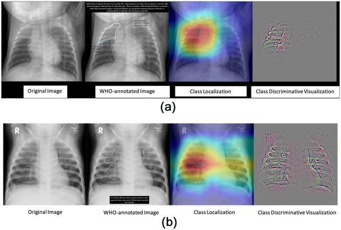
This diagnostic image comparison displays pediatric frontal chest radiographs across two cases (a and b), categorized by World Health Organization (WHO) standards for pneumonia. Each row includes the original radiograph, the WHO-annotated image, a deep learning class localization heat map, and a class discriminative visualization. Row (a) depicts Primary Endpoint Pneumonia (PEP), characterized by a dense, focal consolidation in the right upper lobe with visible air bronchograms. Despite moderate patient rotation to the right, the localized opacity remains clear. Row (b) illustrates 'other-infiltrates,' showing bilateral, diffuse, and patchy opacities that lack the well-defined density of endpoint pneumonia. These infiltrates are more dispersed throughout the lung fields rather than lobar. The visual provides a comparison between high-density lobar consolidation (PEP) and less dense, non-focal interstitial or alveolar infiltrates. This educational material is designed to train clinical recognition of diagnostic patterns in pediatric respiratory infections and demonstrates the application of artificial intelligence in localizing pulmonary pathology.
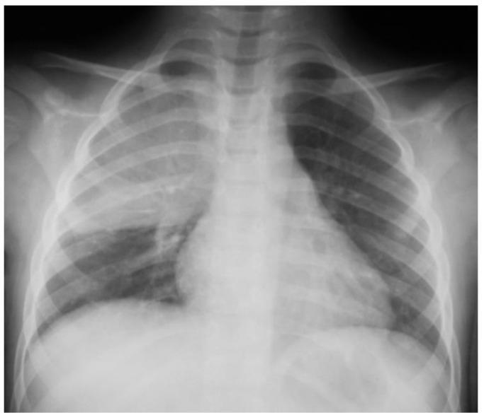
Anteroposterior (AP) pediatric chest radiograph demonstrating a prominent, dense opacity in the right upper and middle lung zones. The finding is consistent with lobar consolidation, showing an ill-defined superior border and a relatively sharp inferior margin at the horizontal fissure. The presence of air bronchograms within the opacity indicates alveolar replacement by fluid, inflammatory exudate, or cells, characteristic of bacterial pneumonia. The left lung field remains clear with normal vascular markings. The mediastinal silhouette and cardiac shadow appear within normal limits for a pediatric patient, although the right heart border is partially obscured by the adjacent consolidation (positive silhouette sign). The skeletal structures, including the ribs and clavicles, are intact. This diagnostic image serves as a classic example of community-acquired pneumonia in a child, illustrating the radiographic hallmarks of pulmonary consolidation and its anatomical localization.
Chest x ray pleural effusion meniscus sign
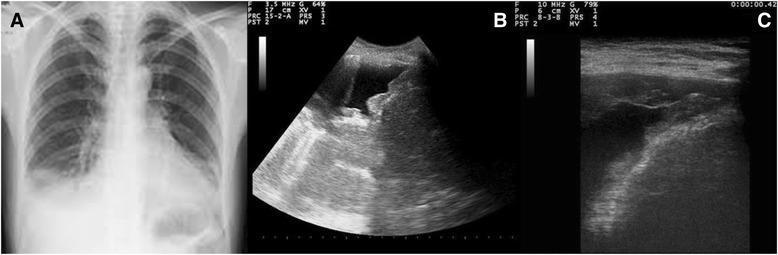
This composite educational graphic compares radiographic and ultrasonographic findings of a pleural effusion. Panel A is a posteroanterior chest X-ray showing a right-sided pleural effusion, characterized by a homogeneous opacity in the lower lung zone with blunting of the costophrenic angle and a visible meniscus sign. Panels B and C are bedside chest ultrasound images (sonograms) of the same patient. These point-of-care ultrasound (POCUS) views provide a more detailed assessment of the pleural space, showing an anechoic (fluid-filled) space consistent with effusion. Notably, the ultrasound images reveal irregular, hyperechoic nodular variations and thickening along the parietal pleura, diaphragm, and costophrenic sinus. These morphological irregularities, which are not visible on the conventional X-ray, are highly suggestive of neoplastic pleural involvement or malignant pleural disease. This comparison illustrates the increased diagnostic sensitivity of ultrasound for evaluating pleural surface morphology in the context of persistent effusion.
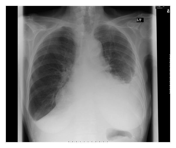
This diagnostic image is a posterior-anterior (PA) chest X-ray of an adult patient. The primary finding is a large, homogenous opacity in the left lower and middle hemithorax, which completely obscures the left costophrenic angle and left hemidiaphragm silhouette. This appearance is characteristic of a large left-sided pleural effusion, showing a classic meniscus sign where the fluid level curves upwards along the lateral chest wall. The opacity also partially silhouettes the left heart border. In contrast, the right lung field appears clear with sharp costophrenic and cardiophrenic angles. The mediastinal structures are centrally located, though the cardiac silhouette is obscured on the left. This radiograph serves as a clinical example of significant pleural fluid accumulation, useful for teaching radiographic signs such as silhouette sign and meniscus formation. The clinical context suggests an exudative effusion potentially associated with carbimazole therapy, a rare adverse effect.
Chest radiograph pulmonary edema bat wing pattern cardiomegaly
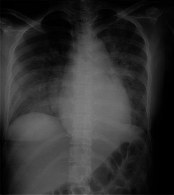
Anteroposterior (AP) chest radiograph demonstrating classic radiological signs of acute cardiogenic pulmonary edema in a pregnant patient. The cardiac silhouette is significantly enlarged, consistent with cardiomegaly. There is marked prominence and enhancement of the pulmonary vasculature, indicating venous congestion. The lung parenchyma shows diffuse, bilateral fluffy and patchy opacities, primarily distributed in a perihilar ('bat-wing') pattern, suggesting alveolar edema. The diaphragm is visible, and the skeletal structures, including the ribs and spine, are unremarkable. Clinically, this imaging correlates with acute heart failure, secondary to severe mitral regurgitation from infective endocarditis. The radiograph serves as a diagnostic tool for identifying pulmonary fluid overload and evaluating the severity of congestive heart failure in a critical obstetric and cardiovascular context.
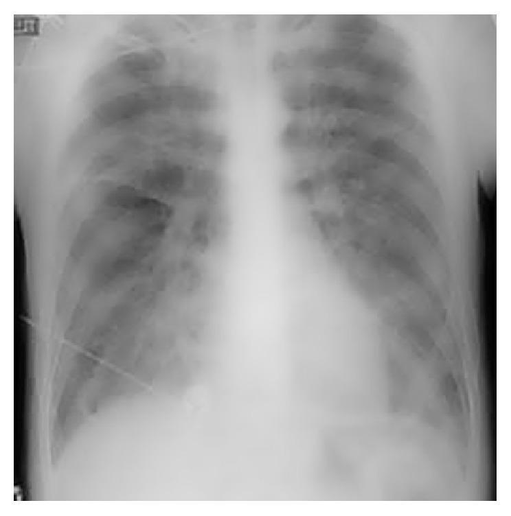
This diagnostic image is a posterior-anterior (PA) or anterior-posterior (AP) chest radiograph. The primary finding is bilateral, diffuse, 'fluffy' opacities throughout both lung fields, consistent with acute pulmonary edema. These opacities are more pronounced in the perihilar regions and mid-to-lower zones, obscuring normal vascular markings and demonstrating a 'bat-wing' or 'angel-wing' distribution pattern. Despite the extensive pulmonary alveolar and interstitial edema, the cardiac silhouette maintains a normal size and shape, without evidence of cardiomegaly or contour abnormalities. This discordance between severe pulmonary edema and a normal cardiac size is often characteristic of non-cardiogenic pulmonary edema or acute cardiogenic failure before ventricular dilation occurs. The mediastinal structures appear central, and the costophrenic angles are partially obscured by the overlying opacities. This image serves as a clinical example of pulmonary congestion and respiratory distress findings in an acute care or emergency medicine context.
Chest x ray COPD emphysema hyperinflation flattened diaphragms
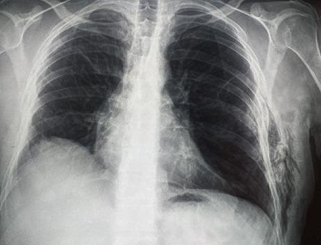
This diagnostic image is an anteroposterior (AP) chest X-ray demonstrating parietal subcutaneous gas and signs of underlying pulmonary pathology. The most prominent finding is the presence of subcutaneous emphysema along the left lower chest wall and axillary region, appearing as linear and mottled radiolucencies (dark streaks) dissecting through the soft tissue planes. Both lung fields exhibit hyperlucency and flattened diaphragms, characteristic of hyperinflation consistent with chronic obstructive pulmonary disease (COPD) or emphysema. The right lung appears more hyperlucent than the left. Bony structures, including the ribs, clavicles, and scapulae, are clearly visualized, with the ribs showing a more horizontal orientation typical of hyperinflation. The mediastinum remains centrally located. This radiograph is an educational example of subcutaneous emphysema resulting from thoracic wall air leakage, often seen in the context of pulmonary hernias or previous thoracic surgeries.
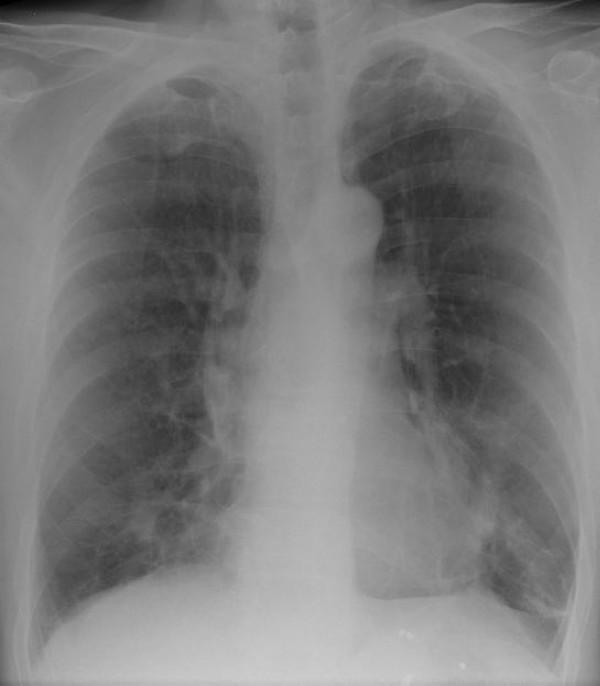
A posterior-anterior (PA) chest X-ray of a human adult. The primary finding is an ill-defined, mass-like opacity located in the right middle lung zone, positioned medially near the right heart border. This density lacks sharp margins and displays a non-uniform, heterogeneous texture. Throughout both lung fields, there is evidence of increased lucency and a subtle reticular pattern, particularly in the upper lobes, which is suggestive of emphysematous changes and hyperinflation. The diaphragms appear slightly flattened, consistent with chronic obstructive pulmonary disease (COPD) or emphysema. There are also scattered, faint, spiculated opacities visible bilaterally in the lung parenchyma. The mediastinal contour and cardiac silhouette are otherwise within normal limits for this view. This image demonstrates findings clinically associated with talc granulomatosis and secondary emphysema, presenting as a mass-like lesion that may mimic malignancy or infectious processes.
Chest x ray tuberculosis upper lobe cavitation
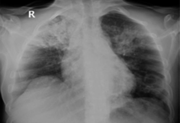
This diagnostic image is a frontal (posteroanterior) chest X-ray demonstrating significant bilateral pulmonary pathology. In the right lung, there is a dense, heterogeneous lobar consolidation primarily occupying the right upper lobe. Within this opacification, a radiolucent area is visible, highly suggestive of cavitation. The left lung displays sublobar, patchy infiltrates and consolidation, also predominantly in the upper zone, with subtle features indicating potential central cavitation. The cardiac silhouette appears within normal limits, though the mediastinal borders are partially obscured by the adjacent pulmonary opacities. These findings are classically associated with infectious processes such as pulmonary tuberculosis (TB) or necrotizing pneumonia. In the provided clinical context of a co-infection involving COVID-19 and tuberculosis, the imaging illustrates the characteristic upper-lobe predilection and cavitary nature of reactivated TB, while the more diffuse patchy elements may represent overlapping viral pneumonia patterns.
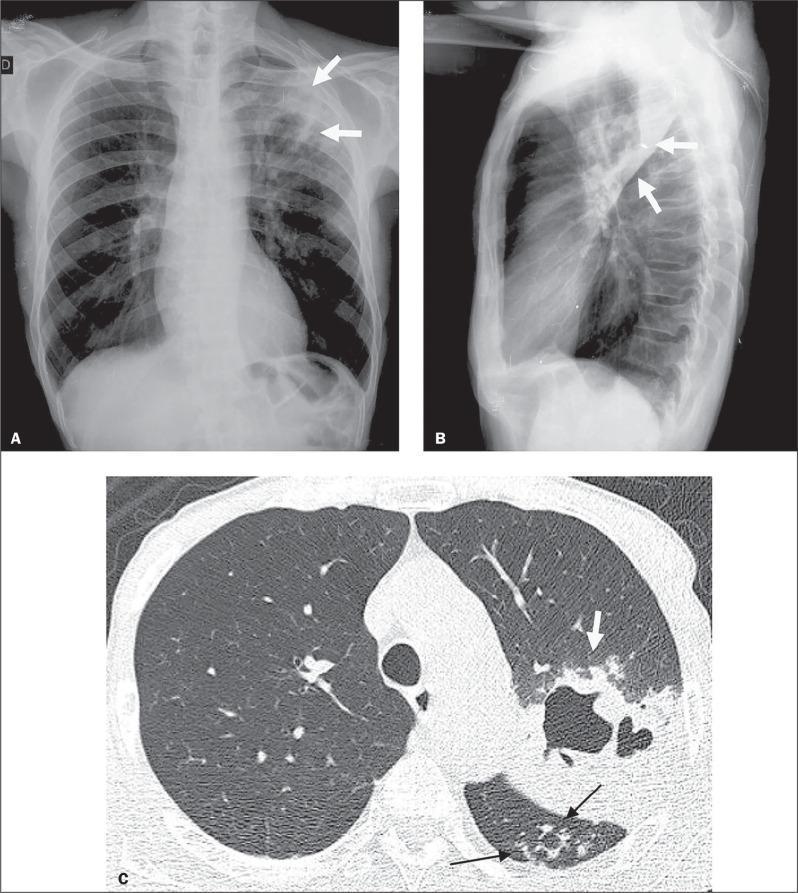
Diagnostic imaging composite of a 66-year-old male with active pulmonary tuberculosis (TB), comprising a posteroanterior chest X-ray (A), a lateral chest X-ray (B), and an axial High-Resolution Computed Tomography (HRCT) scan (C). Panels A and B demonstrate increased opacity in the left upper lung zone indicative of consolidation, with radiolucent areas representing cavitation (white arrows). Panel C, the HRCT at the level of the lower lobes, provides a detailed view of the pulmonary pathology. It reveals a focal area of dense consolidation containing a central air-filled cavity (white arrow) in the left lung. Additionally, characteristic 'tree-in-bud' patterns are visible (black arrows), consisting of small centrilobular nodules and branching linear opacities that indicate endobronchial spread of infection within the upper segment of the left lower lobe. These imaging findings are hallmark radiological features of secondary or active tuberculosis, illustrating parenchymal destruction and small airway involvement.
Common chest X-ray appearances in respiratory illnesses
| Condition | Typical chest X-ray findings | Example |
|---|---|---|
| Lobar pneumonia | Focal dense air-space opacity, often limited to a lobe or segment. Air bronchograms may be visible. Adjacent heart or diaphragm borders can disappear, called the silhouette sign. | |
| Pleural effusion | Homogeneous opacity in the lower hemithorax, blunting of the costophrenic angle, and a curved upper border called the meniscus sign. A large effusion can shift the mediastinum away. | |
| Pneumothorax | A sharp visceral pleural line with no lung markings beyond it toward the chest wall. In tension pneumothorax, look for mediastinal shift away from the affected side. This is an emergency. | 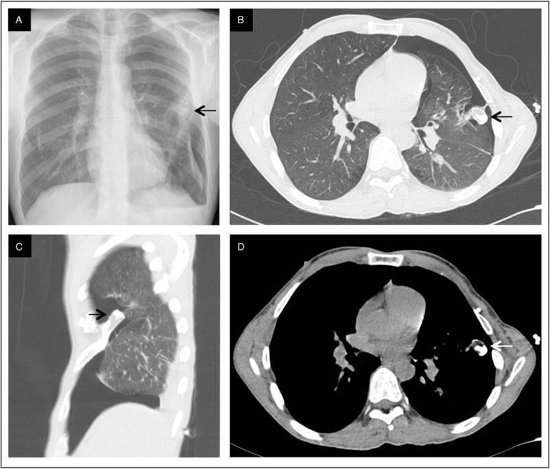 |
| Cardiogenic pulmonary edema | Enlarged cardiac silhouette, upper-lobe venous diversion, bilateral interstitial or fluffy alveolar opacities, often central and perihilar in a bat-wing pattern. Pleural effusions may coexist. See this radiology review of pulmonary edema. | |
| COPD / emphysema | Hyperinflated, hyperlucent lungs; low, flattened diaphragms; increased retrosternal air space on lateral film; a narrow, vertically oriented heart may be seen. A normal X-ray does not exclude COPD. | |
| Post-primary pulmonary tuberculosis | Upper-lobe or apical patchy opacities, fibrosis, volume loss, and sometimes cavitation. X-ray supports suspicion but microbiologic testing is needed to confirm TB. | |
| ARDS / non-cardiogenic edema | Diffuse bilateral patchy air-space opacities. Unlike typical cardiogenic edema, the heart may be normal in size and pleural effusions may be absent. | |
Quick pattern guide
- White localized opacity: consolidation, often pneumonia
- White lower-zone opacity with meniscus: pleural effusion
- Very dark peripheral area without markings: pneumothorax
- Bilateral central fluffy opacities: pulmonary edema
- Large dark lungs with flat diaphragms: emphysema/COPD
- Upper-lobe cavities or infiltrates: consider tuberculosis