
Detailed anatomy and physiology of thyroid gland and parathyroid gland in simplified manner for university 20 markers long answer according to standard ENT textbook
Reading File
Now let me search the medical library for thyroid and parathyroid anatomy and physiology in ENT textbooks.
Finding Sources
Finding Sources
Finding Sources
I found the key ENT textbooks (Cummings Otolaryngology, Scott-Brown's, K.J. Lee's). Let me now search for thyroid anatomy specifically in ENT books and read all the relevant sections.
Finding Sources
Finding Sources
Finding Sources
Now let me read the key sections - Cummings block2 (Thyroid Gland anatomy section) and block28 (Thyroid Surgery with parathyroid anatomy), plus Scott-Brown's thyroid hormones and surgery sections, and Guyton physiology.
Reading File
Reading File
Reading File
Let me read the main Cummings Thyroid Gland anatomy section and also the Scott-Brown's thyroid surgery section (which has detailed anatomy).
Reading File
Reading File
Let me now get the detailed thyroid anatomy and parathyroid anatomy sections from Cummings block28 and the Guyton physiology section.
Reading File
Reading File
Excellent! I now have very rich content. Let me read more of the Guyton physiology section and also look for parathyroid anatomy section in Cummings.
Reading File
Finding Sources
Now let me read the Cummings Parathyroid Anatomy section (the most important ENT source) and continue the Guyton physiology section on parathyroid.
Reading File
Reading File
Now let me get the parathyroid hormone physiology from Guyton and also search for images.
Reading File
Searching Images
thyroid gland anatomy diagram lobes isthmus
Let me also get the Guyton parathyroid physiology section and fetch the thyroid anatomy diagram from Guyton.
Finding Sources
Loading Image
Excellent! I now have the beautiful thyroid anatomy diagram from Guyton. Let me also read the Guyton parathyroid hormone physiology and the Cummings thyroid surgery section for the recurrent laryngeal nerve relationship.
Reading File
Reading File
Let me read the Guyton parathyroid hormone/calcium section and also get the thyroid surgery anatomy from Cummings block 28 (which will have RLN, blood supply etc.).
Reading File
Reading File
I now have enough material from all key sources to write a comprehensive 20-marker answer. Let me also quickly fetch the thyroid hormone synthesis diagram.
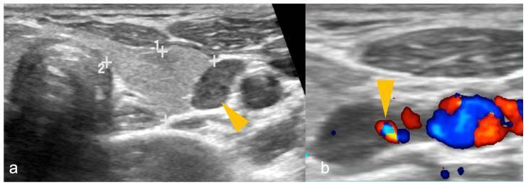
I now have comprehensive information from all the key textbooks (Cummings Otolaryngology, Guyton & Hall, Scott-Brown's). I have the anatomy diagrams too. Let me now write the comprehensive, exam-quality 20-marker answer.
Anatomy and Physiology of the Thyroid Gland and Parathyroid Gland
PART A: THYROID GLAND
1. Embryology
The thyroid gland develops from an endodermal downgrowth at the base of the tongue (foramen cecum) around the 4th week of embryonic life. It descends in the midline of the neck along the thyroglossal duct, passing anterior to the hyoid bone, reaching its final position anterior to the trachea by the 7th week. The thyroglossal duct normally obliterates; failure to do so results in a thyroglossal duct cyst, most commonly found just below the hyoid bone. Ectopic thyroid tissue may persist anywhere along this migratory path, including a lingual thyroid at the foramen cecum. C cells (parafollicular cells), which secrete calcitonin, are derived from the ultimobranchial bodies (4th pharyngeal pouch / neural crest).
2. Gross Anatomy
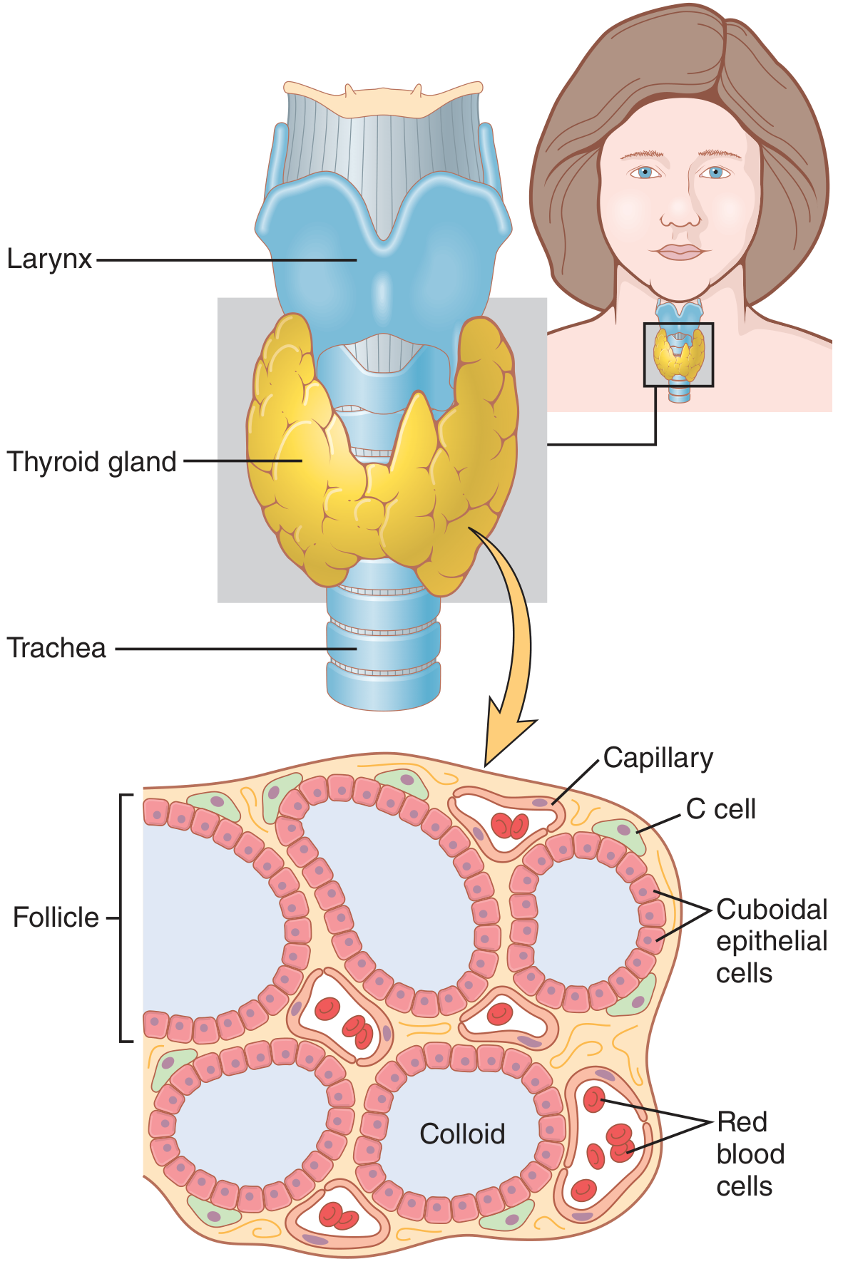
Figure 77.1 - Guyton & Hall Textbook of Medical Physiology: Anatomy and microscopic appearance of the thyroid gland showing follicles, colloid, cuboidal epithelial cells, and C cells.
Position and Shape:
The thyroid gland is the largest endocrine gland in the body, shaped like a "butterfly" or "H," consisting of:
- Two lateral lobes (right and left)
- An isthmus connecting them anteriorly across the 2nd and 3rd tracheal rings
- A pyramidal lobe extending superiorly from the isthmus in ~50% of individuals (a remnant of the thyroglossal duct)
It lies within the anterior leaves of the middle layer of deep cervical fascia (visceral space), anterior and lateral to the thyroid cartilage, cricoid cartilage, and upper tracheal rings. (Cummings Otolaryngology Head and Neck Surgery)
Dimensions:
- Each lobe: ~5 cm long, 3 cm wide, 2 cm thick
- Weight: approximately 20-30 g in adults
- Isthmus: ~1.25 cm wide, ~1.25 cm tall
Capsule:
The gland is enclosed in a true fibrous capsule, outside of which lies a false capsule formed by the pretracheal fascia. Between these two layers run the blood vessels, lymphatics, and parathyroid glands. Fibrous septa from the true capsule pass inward to divide the gland into irregular lobules.
3. Anatomical Relations
| Direction | Structure |
|---|---|
| Anterior | Strap muscles (sternohyoid, sternothyroid, omohyoid), sternocleidomastoid |
| Posterior/medial | Trachea, esophagus, recurrent laryngeal nerve (in tracheo-esophageal groove) |
| Posterolateral | Common carotid artery, internal jugular vein, vagus nerve (carotid sheath) |
| Superior | Thyroid cartilage, inferior pharyngeal constrictor |
The recurrent laryngeal nerve (RLN) runs in the tracheo-esophageal groove posteriorly and is the most surgically important structure related to the thyroid. On the right, the RLN loops around the right subclavian artery; on the left, it loops around the arch of the aorta. The nerve lies in close proximity to the inferior thyroid artery and the inferior pole of each lobe, making it vulnerable during thyroid surgery.
The external branch of the superior laryngeal nerve runs medial to the superior thyroid vessels and innervates the cricothyroid muscle, responsible for voice pitch. It is at risk during ligation of the superior thyroid artery.
4. Blood Supply
Arterial Supply:
- Superior thyroid artery: First branch of the external carotid artery; enters the superior pole
- Inferior thyroid artery: Branch of the thyrocervical trunk (from subclavian artery); enters the posterior aspect near the middle of each lobe; most important blood supply - also supplies the parathyroid glands
- Thyroidea ima artery (inconstant): Arises from brachiocephalic or aortic arch, enters at the isthmus (~1-3% of people)
- Blood flow is approximately 5 times the weight of the gland per minute, one of the highest blood flows per gram of any tissue in the body (Guyton & Hall)
Venous Drainage:
- Superior thyroid vein drains into the internal jugular vein
- Middle thyroid vein drains into the internal jugular vein
- Inferior thyroid vein(s) drain into the brachiocephalic veins
Lymphatic Drainage:
- Superior part drains to upper deep cervical nodes
- Inferior part and isthmus drain to pretracheal and paratracheal (Level VI) nodes
- Important in thyroid cancer staging
5. Nerve Supply
- Adrenergic fibers from the middle cervical ganglion (sympathetic) and parasympathetic from the vagus nerve control blood flow and modulate hormone secretion
- The gland is NOT innervated by motor fibers; secretion is primarily under hormonal control (TSH)
6. Histology / Microanatomy
The thyroid parenchyma consists of:
- Follicles: The functional unit. Each follicle is a closed sphere (100-300 µm diameter) lined by a single layer of cuboidal to columnar follicular epithelial cells. The lumen is filled with gelatinous colloid (mainly thyroglobulin).
- When the gland is active: follicular cells become tall (columnar), colloid is reduced
- When the gland is inactive: cells are flat (squamous), colloid is abundant
-
Follicular cells (thyrocytes): Synthesize thyroglobulin and produce T3/T4. They have a well-developed rough ER, Golgi apparatus, and abundant mitochondria.
-
Parafollicular cells (C cells): Located in the interfollicular stroma and between follicular cells, but do not border the follicular lumen. They secrete calcitonin, which lowers serum calcium. They are derived from neural crest cells.
7. Physiology of the Thyroid Gland
A. Thyroid Hormones
The thyroid produces two main hormones:
- Thyroxine (T4): 93% of secreted hormone; tetraiodothyronine; prohormone; half-life ~6-7 days
- Triiodothyronine (T3): ~7% secreted; 3-4x more potent than T4; most active form; half-life ~1 day
- Calcitonin: from C cells
B. Hormone Synthesis (Step-by-Step)
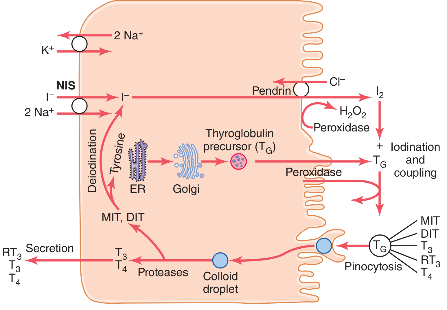
Figure 77.2 - Guyton & Hall: Thyroid cellular mechanisms for iodine transport, thyroxine and T3 formation, and secretion. NIS = sodium-iodide symporter; MIT = monoiodotyrosine; DIT = diiodotyrosine.
Step 1 - Iodide Trapping (Iodide Pump):
- Iodide (I⁻) from blood is actively transported into follicular cells by the Sodium-Iodide Symporter (NIS) on the basolateral membrane - co-transports 2 Na⁺ with 1 I⁻ against concentration gradient
- Energy comes from Na⁺-K⁺-ATPase
- Normal gland: intracellular I⁻ = 30 × plasma concentration; active gland: up to 250 ×
- Stimulated by TSH
Step 2 - Thyroglobulin Synthesis:
- Follicular cells synthesize thyroglobulin (MW ~335,000 Da; contains ~70 tyrosine residues) in the RER and Golgi
- Secreted by exocytosis into the follicular lumen
Step 3 - Oxidation of Iodide:
- I⁻ exits the apical membrane via pendrin (chloride-iodide counter-transporter) into the lumen
- Thyroid peroxidase (TPO) + H₂O₂ oxidizes I⁻ → I₂ (active iodine) at the apical membrane
Step 4 - Organification (Iodination of Tyrosine):
- Active iodine binds tyrosine residues on thyroglobulin:
- 1 iodine + tyrosine → Monoiodotyrosine (MIT)
- 2 iodines + tyrosine → Diiodotyrosine (DIT)
Step 5 - Coupling Reaction:
- TPO catalyzes coupling:
- MIT + DIT → T3 (triiodothyronine)
- DIT + DIT → T4 (thyroxine)
- The hormones remain stored within the thyroglobulin molecule as colloid (enough for ~2-3 months supply)
Step 6 - Secretion (Release):
- TSH stimulates follicular cells to engulf colloid by pinocytosis/endocytosis
- Lysosomes fuse with endosomes → proteases cleave thyroglobulin → releases MIT, DIT, T3, T4
- MIT and DIT are deiodinated intracellularly (iodine recycled); T3 and T4 enter the bloodstream
- In blood: T3 and T4 are bound to thyroxine-binding globulin (TBG, ~70%), albumin, and transthyretin
Peripheral Conversion:
- ~80% of circulating T3 is produced by peripheral deiodination of T4 by deiodinase enzymes (mainly in liver and kidney)
C. Actions of Thyroid Hormones
| System | Effect |
|---|---|
| Metabolic | Increases basal metabolic rate; increases O₂ consumption |
| Cardiovascular | Increases heart rate, stroke volume, cardiac output |
| CNS | Promotes brain development (fetal); increases alertness, reflex speed |
| Growth | Promotes normal growth; essential for skeletal maturation |
| Protein | Increases protein synthesis (anabolic at physiological levels) |
| Fat | Increases fat mobilization; decreases cholesterol |
| Carbohydrate | Increases glucose absorption and utilization |
| Temperature | Increases heat production (calorigenic effect) |
D. Regulation of Thyroid Secretion (HPT Axis)
The hypothalamus-pituitary-thyroid axis regulates hormone output via a classic negative feedback loop:
- TRH (thyrotropin-releasing hormone): Released from the paraventricular nucleus of the hypothalamus → travels via portal blood to anterior pituitary
- TSH (thyroid-stimulating hormone / thyrotropin): Released from thyrotrophs of the anterior pituitary (MW ~28,000; glycoprotein)
- Stimulates all steps of thyroid hormone synthesis and release
- Mechanism: TSH binds G-protein coupled receptor → adenylyl cyclase → cAMP → protein kinase A → multiple phosphorylations
- Effects: increased iodide trapping, thyroglobulin proteolysis, iodination, cell growth, columnar transformation
- Negative feedback: Rising T3/T4 inhibits both TRH release (hypothalamus) and TSH secretion (pituitary)
- Cold/stress can stimulate TRH release, increasing thyroid output
E. Calcitonin
- Secreted by C cells in response to hypercalcemia
- Lowers serum calcium by inhibiting osteoclast activity (decreasing bone resorption)
- Physiologically less important in adults; more relevant in children during bone formation
PART B: PARATHYROID GLANDS
1. Embryology
- Superior parathyroid glands (parathyroid IV): Derived from the 4th pharyngeal pouch, along with the thyroid C cells
- Inferior parathyroid glands (parathyroid III): Derived from the 3rd pharyngeal pouch along with the thymus; they descend with the thymus and may therefore be found anywhere from the angle of the jaw to the anterior mediastinum (ectopic locations common)
- This crossed origin explains why the inferior glands have more variable locations than the superior glands
2. Gross Anatomy
Number: Typically 4 glands (2 superior + 2 inferior)
- 84-87% of people have all 4 glands
- 3% to 6% have only 3
- Supernumerary (5th or more) glands occur in a small percentage; can be found in the mediastinum (Cummings Otolaryngology)
Size and Appearance:
- Weight: 30-50 mg each
- Shape: Oval or bean-shaped
- Color: Yellow to brown (darker in young, more yellow in elderly due to increasing fat content)
- Dimensions: approximately 5 mm × 3 mm × 1 mm
Location:
The glands are normally found posterior to the thyroid lobes, just deep to the true fibrous capsule of the thyroid. Location relative to the RLN is the key surgical landmark:
- Superior parathyroid (PT IV): Located above the level where the RLN crosses the inferior thyroid artery; typically at the level of the cricoid cartilage or upper thyroid lobe, posterior surface; more consistent in position
- Inferior parathyroid (PT III): Located below the RLN-inferior thyroid artery crossing; near the inferior pole of the thyroid or in the thyrothymic ligament; more variable in location and may be in the anterior mediastinum
3. Vascular Anatomy (Clinically Critical)
Arterial Supply:
- Both superior and inferior parathyroid glands receive their primary blood supply from the inferior thyroid artery (branch of thyrocervical trunk from subclavian artery)
- 10% of inferior parathyroid glands derive dominant supply from a branch of the superior thyroid artery
- Rich anastomoses exist with arteries of the larynx, pharynx, esophagus, and trachea, so ligation of the inferior thyroid artery does not always devascularize the parathyroid glands (Cummings Otolaryngology)
Clinical Note: Transient hypoparathyroidism from ischemia may occur in up to 20% of patients after total thyroidectomy and usually resolves within weeks.
Venous Drainage: Via the thyroid venous plexus
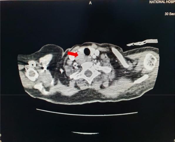
CT neck (CECT): Well-defined enhancing mass (red arrow) posterior to right thyroid lobe - classic location for a superior parathyroid adenoma.
4. Histology of the Parathyroid Gland
The parathyroid gland is enclosed in a thin fibrous capsule with septa that divide it into lobules. It contains three cell types:
- Chief cells (principal cells): Most abundant; small, polygonal cells with pale cytoplasm; synthesize and secrete PTH; arranged in cords and nests with a rich capillary network
- Oxyphil cells: Larger than chief cells; deeply eosinophilic (more mitochondria - take up Tc-99m in nuclear scans); fewer in number; function uncertain; increase in number with age
- Fat cells (stromal adipocytes): Increase with age (up to 50% of gland by middle age); serve as an indicator of gland activity (decreased fat = hyperactive gland)
5. Physiology of Parathyroid Glands
A. Parathyroid Hormone (PTH)
- An 84-amino acid polypeptide; the most important regulator of calcium homeostasis
- Synthesized by chief cells as pre-pro-PTH → pro-PTH → PTH (active 1-84 peptide)
- Half-life: ~4 minutes in blood
- Secretion triggered by: low plasma Ca²⁺ (hypocalcemia); also mildly stimulated by hypomagnesemia
- Secretion suppressed by: high plasma Ca²⁺ (hypercalcemia), detected via the Calcium-Sensing Receptor (CaSR) on chief cell surface
B. Actions of PTH (Three Target Organs)
1. Bone (primary reservoir of calcium):
- Rapid phase (minutes to hours): Activates existing osteocytes via the osteocytic membrane system → osteolysis, releasing calcium and phosphate from bone fluid into ECF
- Slow phase (days to weeks): Proliferation and activation of osteoclasts → bone resorption → release of Ca²⁺ and phosphate into blood
- PTH acts on osteoblasts (which have PTH receptors) → they release RANKL → stimulates osteoclast differentiation and activity
- Result: ↑ serum Ca²⁺ and ↑ serum phosphate from bone
2. Kidney (calcium retention, phosphate wasting):
- Increases calcium reabsorption in the distal convoluted tubule
- Decreases phosphate reabsorption in the proximal tubule → phosphaturia (phosphate excretion in urine; prevents precipitation of Ca²⁺-phosphate)
- Stimulates 1-alpha-hydroxylase enzyme → converts 25-hydroxycholecalciferol to 1,25-dihydroxycholecalciferol (active Vitamin D / calcitriol)
- Net renal effect: ↑ serum Ca²⁺, ↓ serum phosphate
3. Intestine (indirect, via Vitamin D):
- PTH stimulates renal activation of Vitamin D (calcitriol)
- Calcitriol acts on intestinal epithelial cells → increases synthesis of calbindin (calcium-binding protein) → promotes intestinal absorption of Ca²⁺ and phosphate
- This effect takes 1-2 days to develop
Summary of PTH effects on blood levels:
| Mineral | PTH Effect |
|---|---|
| Calcium | ↑ (raises serum Ca²⁺) |
| Phosphate | ↓ (lowers serum phosphate, despite releasing it from bone, because renal excretion > bone release) |
C. Regulation of PTH Secretion (Calcium Sensing)
The Calcium-Sensing Receptor (CaSR) on chief cells is the key regulator:
- Normal serum calcium: 8.5-10.5 mg/dL (2.1-2.6 mmol/L)
- Hypocalcemia → CaSR less activated → PTH secretion increases → Ca²⁺ rises back to normal
- Hypercalcemia (>9-10 mg/dL) → CaSR activated → PTH secretion suppressed
- Hypermagnesemia also suppresses PTH; severe hypomagnesemia paradoxically impairs PTH secretion (by depleting G-protein signaling)
- Vitamin D (calcitriol) also inhibits PTH gene transcription (long-loop feedback)
D. Clinical Correlations
| Condition | PTH | Calcium | Phosphate | Cause |
|---|---|---|---|---|
| Primary hyperparathyroidism | ↑ | ↑ | ↓ | Parathyroid adenoma (~80%) |
| Secondary hyperparathyroidism | ↑↑ | Low/Normal | ↑ | Chronic kidney disease |
| Hypoparathyroidism | ↓ | ↓ | ↑ | Post-thyroidectomy, autoimmune |
| Pseudohypoparathyroidism | ↑ | ↓ | ↑ | End-organ resistance to PTH |
Tetany results from hypocalcemia: Trousseau's sign (carpopedal spasm with BP cuff inflation), Chvostek's sign (facial twitch on tapping facial nerve). Severe hypocalcemia causes laryngospasm and life-threatening airway compromise.
Summary Table
| Feature | Thyroid Gland | Parathyroid Gland |
|---|---|---|
| Location | Anterior neck, C5-T1 level | Posterior surface of thyroid |
| Number | 1 (bilobed) | Usually 4 (2 superior, 2 inferior) |
| Weight | 20-30 g | 30-50 mg each |
| Blood supply | Superior + inferior thyroid arteries | Inferior thyroid artery (mainly) |
| Embryology | Thyroid diverticulum (floor of pharynx) | 3rd pouch (inferior), 4th pouch (superior) |
| Histology | Follicles + colloid + C cells | Chief cells + oxyphil cells |
| Key hormone | T3, T4, Calcitonin | PTH |
| Main function | Metabolism, growth, development | Calcium & phosphate homeostasis |
| Venous drainage | Superior, middle, inferior thyroid veins | Thyroid venous plexus |
Sources:
- Cummings Otolaryngology Head and Neck Surgery (Chapters 8, 123, 124) - primary ENT reference
- Scott-Brown's Otorhinolaryngology Head & Neck Surgery Vol. 1 (Chapters 5, 6)
- Guyton and Hall Textbook of Medical Physiology (Chapters 77, 80) - thyroid and parathyroid physiology
This is a shared conversation. Sign in to Orris to start your own chat.