
Hii iam a veterinary doctor
Hello! Welcome! It's great to have a veterinary professional here.
I'm Orris, your AI assistant. I can help you with a wide range of topics relevant to your practice, including:
- Clinical questions - diagnoses, treatment protocols, drug dosages for animals
- Pharmacology - veterinary drugs, interactions, dosing by species
- Pathology & anatomy - species-specific details
- Research & evidence - latest studies, guidelines, clinical trials
- Case discussions - working through differential diagnoses
- General information - anything else you need!
What can I help you with today?
I want inn detailed info about the scrotal ablaation in dogs with images of procedure
Reading File
Scrotal ablation (scrotectomy) in dogs is a surgical procedure to remove the scrotal sac, typically performed alongside castration in cases of scrotal trauma, neoplasia, severe dermatitis, or when the scrotum is pendulous and at risk. Let me search the library and recent literature simultaneously.
Searching sources...
Finding Sources
Finding Sources
Searching the Web
scrotal ablation scrotectomy dogs surgical procedure technique images veterinary
Searching PubMed
scrotal ablation scrotectomy dogs surgical technique
Good - found images and relevant sources. Let me fetch the procedure images and extract detailed content simultaneously.
Loading Image
Loading Image
Loading Image
Loading Image
Reading Web Page
https://rastetterdvm.com/scrotal-ablation-in-dogs/
Searching PubMed
castration scrotectomy canine complications
Excellent - I have great procedural images and detailed content. Let me also get the veteriankey image and fetch more detailed surgical technique info.
Loading Image
Searching the Web
scrotal ablation dogs step by step surgical technique suture closure anesthesia veteriankey
I now have comprehensive information and images. Let me compile the full detailed answer.
Scrotal Ablation (Scrotectomy) in Dogs - Complete Surgical Guide
Definition
Scrotal ablation (SA) refers to the complete surgical removal of the scrotal sac and its associated skin/dartos tissue. It is typically performed concurrently with or following castration (orchiectomy). While standard castration leaves the scrotal sac intact, scrotal ablation excises the entire scrotal tissue, eliminating it as a potential source of post-operative complications or disease.
Indications
Scrotal ablation is indicated in the following scenarios:
| Category | Specific Conditions |
|---|---|
| Neoplasia | Scrotal tumors (mast cell tumors - most common at 54.6%, round cell neoplasms 58.6% of all scrotal neoplasms, melanocytic, mesenchymal) |
| Trauma | Bite wounds, lacerations, degloving injuries to the scrotum |
| Infection / Dermatitis | Severe scrotal dermatitis, chronic infections, non-healing wounds, abscesses |
| Elective / Preventive | Older or large-breed dogs with pendulous, heavy scrotum at risk for seroma or hematoma after standard neuter |
| Aesthetic | Owner request to prevent the "empty scrotal sac" appearance post-castration |
| Swelling / Vascular issues | Persistent or non-resolving scrotal swelling, scrotal hematoma |
Per a 2014 study of 676 canine scrotal tumors, the most common scrotal neoplasms in dogs are round cell tumors (58.6%), mesenchymal tumors (13.6%), and melanocytic neoplasms (11.8%), with mast cell tumors comprising 54.6% of all round cell tumors.
Pre-Operative Preparation
Patient Evaluation
- Complete physical exam and assessment of scrotal tissue
- Blood work: CBC, biochemistry panel (especially in older dogs or those with neoplasia)
- Fine-needle aspirate (FNA) of any scrotal mass prior to surgery to guide margins
- Thoracic radiographs if malignant neoplasia is suspected (to rule out metastasis)
- Clipping: wide surgical clip of the entire perineal and inguinal region
- Positioning: dorsal recumbency, hindlimbs pulled cranially and secured
Anesthesia Protocol
- General anesthesia (intubated) with IV catheter placed
- Pre-medication: opioids (e.g., methadone, hydromorphone) + acepromazine or dexmedetomidine
- Induction: propofol or alfaxalone
- Maintenance: isoflurane/sevoflurane
- Loco-regional analgesia is highly recommended:
- Intratesticular/intrafunicular block: 2% lidocaine or 0.5% bupivacaine
- Splash block at closure: epinephrine (1 mg/mL) 1:9 diluted with 2% lidocaine (vasoconstriction + analgesia)
- Splash block reduces post-op drainage and provides hemostasis of scrotal vasculature
Pre-surgical Planning
- Ink/mark the intended elliptical incision margins on the scrotal skin before draping
- Assess skin laxity - always err on the side of removing less skin initially, as you can always excise more but cannot replace removed skin
- Ensure the planned closure is tension-free
Surgical Technique - Step by Step
Step 1 - Patient Positioning and Draping
The dog is placed in dorsal recumbency. The scrotum and testes are draped into the surgical field, creating a sterile environment.


Step 2 - Castration First
If the dog is intact, routine castration is performed first before scrotal ablation:
- Either prescrotal (standard) or scrotal approach to orchiectomy
- Both testicles are removed and spermatic cord stumps ligated (open or closed technique)
- Testicular vessels are ligated with transfixation ligatures using absorbable suture (e.g., PDS, Monocryl, or Vicryl)
- This step is completed before beginning the scrotal excision to keep tissue planes clean
Step 3 - Planning and Incising the Scrotal Ellipse
- An elliptical incision is planned at the base of the scrotum
- The ellipse should encompass ALL scrotal skin (both pouches), with the long axis oriented cranio-caudally
- Mark the margins with a surgical pen before incising
- The incision is made through skin and dartos muscle layer
- Key rule: Leave slightly more skin than you think you need - tension-free closure is paramount
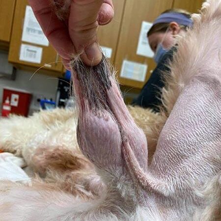
Step 4 - Tissue Dissection and Scrotal Removal
- Mosquito hemostatic forceps are placed into the right and left scrotal pouches to elevate the skin and dartos, protecting the underlying structures during dissection
- Careful blunt and sharp dissection is used to free the scrotal tissue
- The urethra runs immediately deep to the scrotum - protect it at all times
- Subcutaneous hemorrhage is controlled with:
- Electrocautery (preferred)
- Ligation
- Temporary mosquito forceps application
- Once the scrotal tissue is excised, the underlying urethra becomes readily visible
Step 5 - Urethral Identification and Protection
After scrotal removal, pause and confirm:
- Identify the urethra visually (it lies in the perineal midline)
- Confirm no urethral trauma (assess by gentle palpation)
- If urethral injury has occurred, a scrotal urethrostomy may need to be performed at this point
Step 6 - Closure
Closure proceeds in layers:
| Layer | Technique | Suture Material |
|---|---|---|
| Deep fascia (separate scrotal fascial incisions) | Simple interrupted or continuous | Absorbable monofilament (PDS 2-0 or 3-0, Monocryl) |
| Deep subcutaneous tissue (SQ) | Simple interrupted or simple continuous | Absorbable (Vicryl 3-0) |
| Intradermal / skin | Intradermal (preferred) OR external interrupted | Absorbable intradermal (Monocryl 3-0-4-0) or non-absorbable skin sutures |
- Reassess skin tension throughout closure and again with the patient undraped at the end
- The final closure should leave a small ridge of tissue (not a perfectly flat surface) - this is intentional to accommodate post-surgical swelling and improve comfort

Step 7 - Compression Bandage (Optional but Recommended)
For large-breed dogs or those with a pendulous scrotum:
- Place a small piece of sterile gauze directly over the surgical site
- Wrap with self-adhesive bandage material (e.g., VetRap/Coban)
- This scrotal wrap:
- Applies compression to reduce drainage
- Allows quantification of any post-op drainage
- Provides hemostasis of scrotal vasculature
- Prevents environmental contamination and self-trauma
Post-Operative Care
Immediate (0-24 hours)
- Monitor recovery from anesthesia closely
- Pain management is essential:
- NSAIDs (meloxicam, carprofen, grapiprant)
- Opioids as needed (buprenorphine patch or oral transmucosal)
- A small amount of serosanguinous drainage is expected for the first few hours
- Apply cold packs (wrapped in a towel) for 10-15 min every 4-6 hours to reduce swelling
Short-term (Days 1-14)
- Elizabethan collar (E-collar) mandatory - prevent licking/chewing at incision
- Strict exercise restriction for 10-14 days (leash walks only)
- Keep incision clean and dry - no bathing
- Check incision daily for: redness, swelling, discharge, dehiscence, odor
- Antibiotics: indicated if surgery was for infected tissue; not routinely needed for clean elective cases
- Recheck at 14 days for suture removal (if external sutures used) and wound assessment
Warning Signs Requiring Immediate Attention
- Excessive bleeding or rapidly expanding hematoma
- Purulent discharge or strong odor
- Complete wound dehiscence
- Signs of urethral injury: straining to urinate, dysuria, urine pooling
Complications
| Complication | Description | Management |
|---|---|---|
| Seroma | Fluid accumulation under closure | Compression wrap, aspiration if large |
| Hematoma | Blood accumulation; more common in large dogs | Small - conservative; Large - surgical drainage |
| Wound dehiscence | Closure breakdown, often from tension or self-trauma | Reclose if clean; open wound management if infected |
| Infection | Post-op site infection | Antibiotics, wound lavage, possible debridement |
| Urethral damage | Rare but serious - can occur during dissection | Scrotal urethrostomy; urinary catheter placement |
| Excessive swelling | Common in first 3-5 days | Cold compress, compression wrap, rest |
| Skin necrosis | Due to excessive tension on closure | Debride, allow secondary healing or reconstructive closure |
Comparison: Standard Castration vs. Scrotal Ablation
| Feature | Standard Prescrotal Castration | Scrotal Ablation + Castration |
|---|---|---|
| Scrotum removed? | No | Yes |
| Incision site | Prescrotal (cranial to scrotum) | Elliptical at scrotal base |
| Recovery time | 7-10 days | 10-14 days |
| Post-op swelling | Scrotum may fill with fluid | Eliminated risk of scrotal fluid accumulation |
| Indicated for | Routine neuters | Large/old dogs, neoplasia, trauma, dermatitis |
| Aesthetic result | Empty scrotal sac remains | No visible scrotal tissue |
| Complication risk | Higher in large/pendulous scrotum | Reduced |
Special Considerations
- Cryptorchid dogs: If retained testes are present, the approach changes (inguinal or abdominal approach for the retained testis), but scrotal ablation may still be performed for the descended testis and pendulous scrotal tissue
- Scrotal neoplasia: Wide surgical margins are needed; FNA or incisional biopsy should ideally precede surgery; histopathology of excised tissue is mandatory
- Scrotal urethrostomy: If severe urethral obstruction or damage exists, scrotal ablation may be combined with a permanent scrotal urethrostomy to create a new urethral opening at the perineum
- Older intact dogs: These are the most commonly indicated patients - large pendulous scrotum in older dogs almost always warrants ablation at the time of late-in-life neutering
Key Surgical Pearls
- Plan your incision margins before draping - use a surgical marker
- Always protect the urethra - it is immediately beneath the scrotal tissue
- Leave adequate skin for tension-free closure - err conservative
- Electrocautery is your best friend for scrotal hemostasis (very vascular tissue)
- Compression wraps significantly reduce post-op seroma/hematoma in large dogs
- Intradermal closure reduces self-trauma compared to external skin sutures
- Submit all excised tissue for histopathology - scrotal neoplasms are common and unexpected
- Reassess tension with patient undraped before final skin closure
The Veterian Key surgical reference and Vet Surgery Online are excellent free resources for reviewing the technique further with additional intraoperative figures.
This is a shared conversation. Sign in to Orris to start your own chat.