
Management and nursing management of patient with eclampsia
eclampsia management magnesium sulfate
eclampsia management magnesium sulfate seizure obstetrics
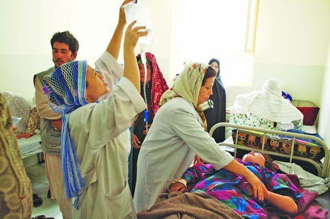
This clinical photograph depicts a healthcare setting in a resource-limited environment, specifically documenting the administration of intravenous (IV) therapy for an obstetric emergency. A female patient, identified by the context as having pre-eclampsia, is lying supine in a hospital bed. A healthcare professional in a white clinical coat is actively managing the IV setup, holding a clear plastic infusion bag (containing magnesium sulphate) elevated above the patient to facilitate gravity-fed flow. The IV tubing is visible, extending from the bag to an insertion point on the patient's arm. The image illustrates critical maternal health interventions and the clinical management of hypertensive disorders of pregnancy. The surrounding individuals and their attire, including headscarves and traditional garments, provide cultural context for the clinical setting in Afghanistan. This visual serves as an educational resource for global health, emergency obstetric care, and the practical application of magnesium sulphate protocols in preventing eclamptic seizures.
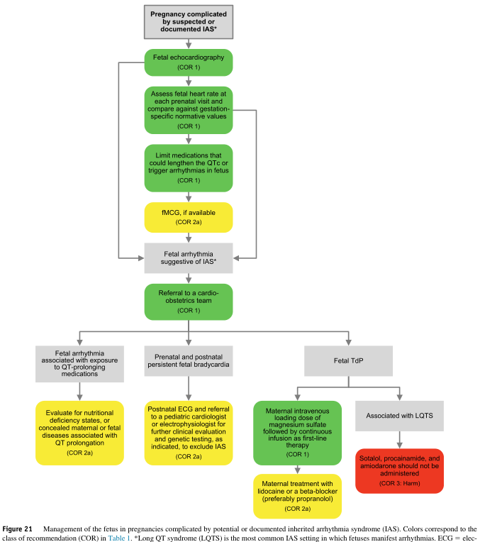
Summary : This flowchart outlines the management of fetuses in pregnancies complicated by suspected or documented inherited arrhythmia syndrome (IAS), with a focus on long QT syndrome (LQTS) and fetal arrhythmias. It details recommended steps, decision points, and treatments, using color codes to indicate the class of recommendation (COR). flowchart: # Nodes : • Start (rectangle): "Pregnancy complicated by suspected or documented IAS*" • Rectangle: "Fetal echocardiography (COR 1)" • Rectangle: "Assess fetal heart rate at each prenatal visit and compare against gestation-specific normative values (COR 1)" • Rectangle: "Limit medications that could lengthen the QTc or trigger arrhythmias in fetus (COR 1)" • Rectangle (yellow): "fMCG, if available (COR 2a)" • Diamond: "Fetal arrhythmia suggestive of IAS*" • Rectangle: "Referral to a cardio-obstetrics team (COR 1)" • Rectangle: "Fetal arrhythmia associated with exposure to QT-prolonging medications" • Rectangle (yellow): "Evaluate for nutritional deficiency states, or concealed maternal or fetal diseases associated with QT prolongation (COR 2a)" • Rectangle: "Prenatal and postnatal persistent fetal bradycardia" • Rectangle (yellow): "Postnatal ECG and referral to a pediatric cardiologist or electrophysiologist for further clinical evaluation and genetic testing, as indicated, to exclude IAS (COR 2a)" • Rectangle: "Fetal TdP" • Rectangle: "Maternal intravenous loading dose of magnesium sulfate followed by continuous infusion as first-line therapy (COR 1)" • Rectangle (yellow): "Maternal treatment with lidocaine or a beta-blocker (preferably propranolol) (COR 2a)" • Rectangle: "Associated with LQTS" • Rectangle (red): "Sotalol, procainamide, and amiodarone should not be administered (COR 3: Harm)" # Connectors : • Downward arrows connect each step in sequence. • After "Fetal echocardiography", arrows lead to "Assess fetal heart rate..." and then to "Limit medications...". • From "Limit medications...", arrow leads to "fMCG, if available". • From "fMCG, if available", arrow leads to "Fetal arrhythmia suggestive of IAS*". • From "Fetal arrhythmia suggestive of IAS*", arrow leads to "Referral to a cardio-obstetrics team". • From "Referral to a cardio-obstetrics team", three branches: – Left: "Fetal arrhythmia associated with exposure to QT-prolonging medications" → "Evaluate for nutritional deficiency states..." – Middle: "Prenatal and postnatal persistent fetal bradycardia" → "Postnatal ECG and referral..." – Right: "Fetal TdP" → "Maternal intravenous loading dose..." → "Maternal treatment with lidocaine or a beta-blocker..." → "Associated with LQTS" → "Sotalol, procainamide, and amiodarone should not be administered" • Some steps loop back to earlier steps (e.g., assessment at each prenatal visit). # Layout : • Vertical flow from top to bottom, with a major split into three branches after referral to cardio-obstetrics team. • Color coding: Green (COR 1: strong recommendation), Yellow (COR 2a: moderate recommendation), Red (COR 3: harm). • Decision diamond for arrhythmia presence. • Merges and branches for different arrhythmia scenarios. # Analysis : • The flowchart emphasizes early and repeated fetal heart assessment, medication management, and specialist referral. • It provides clear pathways for different arrhythmia findings: exposure to QT-prolonging drugs, persistent bradycardia, and fetal TdP. • Strong recommendations (green) are given for initial steps and magnesium sulfate therapy; moderate recommendations (yellow) for additional diagnostics and treatments; and explicit harm warnings (red) for certain drugs in LQTS. • The structure supports rapid identification and tailored management of fetal arrhythmias, with escalation to advanced therapies and genetic evaluation as needed.
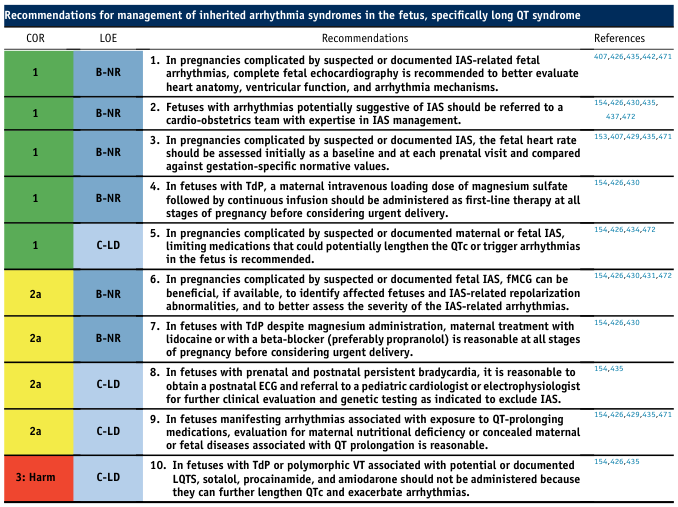
Recommendations for management of inherited arrhythmia syndromes in the fetus, specifically long QT syndrome <table><thead><tr><th>COR</th><th>LOE</th><th>Recommendations</th><th>References</th></tr></thead><tbody><tr><td>1</td><td>B-NR</td><td>1. In pregnancies complicated by suspected or documented IAS-related fetal arrhythmias, complete fetal echocardiography is recommended to better evaluate heart anatomy, ventricular function, and arrhythmia mechanisms.</td><td>407,426,435,442,471</td></tr><tr><td>1</td><td>B-NR</td><td>2. Fetuses with arrhythmias potentially suggestive of IAS should be referred to a cardio-obstetrics team with expertise in IAS management.</td><td>154,426,430,435,437,472</td></tr><tr><td>1</td><td>B-NR</td><td>3. In pregnancies complicated by suspected or documented IAS, the fetal heart rate should be assessed initially as a baseline and at each prenatal visit and compared against gestation-specific normative values.</td><td>153,407,429,435,471</td></tr><tr><td>1</td><td>B-NR</td><td>4. In fetuses with TdP, a maternal intravenous loading dose of magnesium sulfate followed by continuous infusion should be administered as first-line therapy at all stages of pregnancy before considering urgent delivery.</td><td>154,426,430</td></tr><tr><td>1</td><td>C-LD</td><td>5. In pregnancies complicated by suspected or documented maternal or fetal IAS, limiting medications that could potentially lengthen the QTc or trigger arrhythmias in the fetus is recommended.</td><td>154,426,434,472</td></tr><tr><td>2a</td><td>B-NR</td><td>6. In pregnancies complicated by suspected or documented fetal IAS, fMCG can be beneficial, if available, to identify affected fetuses and IAS-related repolarization abnormalities, and to better assess the severity of the IAS-related arrhythmias.</td><td>154,426,430,431,472</td></tr><tr><td>2a</td><td>B-NR</td><td>7. In fetuses with TdP despite magnesium administration, maternal treatment with lidocaine or with a beta-blocker (preferably propranolol) is reasonable at all stages of pregnancy before considering urgent delivery.</td><td>154,426,430</td></tr><tr><td>2a</td><td>C-LD</td><td>8. In fetuses with prenatal and postnatal persistent bradycardia, it is reasonable to obtain a postnatal ECG and referral to a pediatric cardiologist or electrophysiologist for further clinical evaluation and genetic testing as indicated to exclude IAS.</td><td>154,435</td></tr><tr><td>2a</td><td>C-LD</td><td>9. In fetuses manifesting arrhythmias associated with exposure to QT-prolonging medications, evaluation for maternal nutritional deficiency or concealed maternal or fetal diseases associated with QT prolongation is reasonable.</td><td>154,426,429,435,471</td></tr><tr><td>3: Harm</td><td>C-LD</td><td>10. In fetuses with TdP or polymorphic VT associated with potential or documented LQTS, sotalol, procainamide, and amiodarone should not be administered because they can further lengthen QTc and exacerbate arrhythmias.</td><td>154,426,435</td></tr></tbody></table>
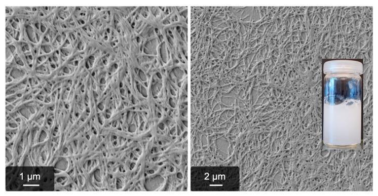
This composite image illustrates the microscopic and macroscopic characteristics of magnesium sulfate (MgSO4)-induced fibrinogen fibrillogenesis, a process relevant to understanding non-enzymatic protein aggregation and blood coagulation research. The image features two scanning electron microscopy (SEM) micrographs and an inset clinical photograph. The SEM images, shown at 1 μm and 2 μm scales, reveal a dense, interconnected network of long, fibrous protein strands. These fibers exhibit a random orientation and form a complex mesh-like architecture with visible interstitial pores. Morphologically, the fibers appear relatively thick and irregular, characteristic of 'pseudo-fibrin' formation where fibrinogen self-assembles without thrombin catalysis. The inset photograph shows the macroscopic result of this process: a homogeneous, turbid, milky-white gel within a transparent glass vial. The opacity of the solution confirms the light-scattering properties of the dense protein fiber network. This visual material is primarily used in hematology and biochemistry to study alternative pathways of fibrin formation and the influence of ionic environments on protein structural dynamics.
Management and Nursing Management of Eclampsia
Definition
- Tintinalli's Emergency Medicine, p. 674; Creasy & Resnik's Maternal-Fetal Medicine
Symptoms Preceding Eclampsia
| Symptom | Frequency (%) |
|---|---|
| Headache | 83 |
| Hyperreflexia | 80 |
| Proteinuria | 80 |
| Edema | 60 |
| Clonus | 46 |
| Visual signs | 45 |
| Epigastric pain | 20 |
- Creasy & Resnik's Maternal-Fetal Medicine, p. 1071
Medical Management
1. Immediate Priorities
- Control and prevent seizures - magnesium sulfate
- Control severe hypertension - antihypertensives
- Deliver the fetus - emergency obstetric consultation
2. Seizure Control: Magnesium Sulfate (First-Line)
- Loading dose: 4-6 g IV over 15-30 minutes
- Maintenance infusion: 1-2 g/hour continuous infusion for at least 24 hours
- Mechanically controlled infusion pump is mandatory
- Creasy & Resnik, p. 1071; Brenner & Rector's The Kidney, p. 2157; Tintinalli's Emergency Medicine, p. 674
Magnesium Toxicity Levels
| Serum Level (mEq/L) | Effect |
|---|---|
| 4.8-8.4 | Therapeutic anticonvulsant range |
| 7-10 | Loss of deep tendon reflexes |
| 10-13 | Respiratory paralysis |
| >15 | ECG changes |
| >25 | Cardiac arrest |
3. Antihypertensive Therapy
| Drug | Route | First Dose | Repeat Dosing | Interval | Max Dose |
|---|---|---|---|---|---|
| Labetalol | IV | 10-20 mg | 20 mg → 40 mg → 80 mg | Every 10-20 min | 300 mg total |
| Hydralazine | IV/IM | 5 mg | 5-10 mg | Every 20-40 min | - |
| Nifedipine | PO | 10 mg | 10-20 mg | Every 30 min (then q3-6h) | 20 mg/dose |
| Nicardipine | IV | 5-15 mg/hour | - | - | - |
- Labetalol is contraindicated in asthma, heart disease, congestive heart failure
- Hydralazine causes reflex increase in cardiac output and uterine blood flow; side effects include headache and epigastric pain (may mimic worsening preeclampsia)
- Nifedipine + Magnesium: monitor closely for additive effects, though evidence suggests no significantly increased toxicity in practice
- Creasy & Resnik, p. 1072-1073; Rosen's Emergency Medicine, p. 1151
4. Delivery - Definitive Treatment
- Most seizures occur intrapartum and postpartum, so magnesium is continued through delivery and for 24 hours postpartum
- Contraindications to expectant management include: eclampsia, pulmonary edema, DIC, uncontrollable hypertension, placental abruption, nonreassuring fetal testing, stillbirth
- Cesarean section is indicated for marked hepatic capsular distension (risk of hepatic rupture)
- Regional anesthesia is acceptable but requires volume expansion; avoid using it as a BP-lowering strategy (lowers BP at expense of cardiac output)
- If obstetric services are unavailable: stabilize and transfer to a tertiary center
5. Coagulation Factor Replacement
6. Oliguria Management
Nursing Management
1. Environment and Safety (Seizure Precautions)
- Place patient in a quiet, darkened, low-stimulation room (noise and light can trigger seizures)
- Keep bed in the lowest position with side rails padded and raised
- Have suction equipment, oxygen, airway adjuncts (oral/nasopharyngeal airways), and emergency medications immediately at bedside
- Keep calcium gluconate 1 g IV at bedside at all times as the antidote for magnesium toxicity
- Ensure IV access (at least one large-bore IV)
2. Vital Signs and Neuro Monitoring
- Blood pressure, pulse, and respiratory rate every 15-30 minutes (or continuously if unstable)
- Neurological status every 1-2 hours: level of consciousness, presence of headache, visual disturbances, and epigastric pain
- Assess deep tendon reflexes (DTR) at least every 2 hours (key for magnesium toxicity monitoring)
- Brisk DTRs = safe (magnesium not dangerously elevated)
- Loss of patellar reflex = STOP infusion; serum Mg may be >10 mEq/L
3. Magnesium Sulfate Infusion Monitoring (Critical)
- Respiratory rate ≥12 breaths/min (respiratory depression occurs at Mg >10-13 mEq/L)
- Urine output ≥25-30 mL/hour (magnesium is renally excreted; oliguria = risk of toxicity)
- Deep tendon reflexes present (loss = toxicity)
- Flushing, warmth, diaphoresis
- Nausea, slurred speech
- Loss of deep tendon reflexes
- Respiratory rate depression/paralysis
- Cardiac arrest
4. Fluid Management and Urine Output
- Insert a urinary catheter for strict hourly urine output measurement
- Target urine output: ≥25-30 mL/hour (minimum 100 mL/4 hours)
- Record all fluid input and output meticulously
- Restrict IV fluids to 80-125 mL/hour to prevent pulmonary edema (a major complication of overhydration in preeclampsia/eclampsia)
5. During an Active Seizure
- Protect the airway - turn patient to left lateral position, do not restrain forcibly
- Call for help immediately; start timing the seizure
- Administer supplemental oxygen via face mask at high flow
- Administer magnesium sulfate bolus as ordered (if not already on infusion)
- After the seizure: suction airway, reassess level of consciousness
- Do NOT put anything in the patient's mouth during the seizure
- Check fetal heart rate continuously after the seizure (fetal bradycardia/hypoxia is common post-seizure)
6. Fetal Monitoring
- Continuous electronic fetal monitoring (CTG)
- Be alert for: late decelerations, reduced variability, or fetal bradycardia (signs of uteroplacental insufficiency)
- Fetal heart rate variability may be decreased by magnesium sulfate itself - this is an expected pharmacological effect
7. Laboratory Monitoring
- Complete blood count (CBC) - for thrombocytopenia (HELLP)
- Serum creatinine, urea (renal function)
- Liver function tests (AST, ALT, LDH)
- Serum magnesium levels (target 4.8-8.4 mEq/L; check with decreased urine output)
- Coagulation studies (PT, aPTT, fibrinogen) - for DIC
- Serum uric acid
8. Positioning and Comfort
- Maintain left lateral decubitus (lateral tilt) position to relieve aortocaval compression and maximize placental blood flow
- Provide emotional support; explain all procedures to the patient and family
- Limit visitors; reduce environmental stimulation
9. Postpartum Nursing Care
- Continue magnesium sulfate for 24 hours after delivery (eclampsia can occur postpartum)
- Continue blood pressure monitoring - risk of seizures and severe hypertension persists for days after delivery
- Assess for signs of HELLP syndrome deterioration (right upper quadrant pain, jaundice)
- Counsel on warning signs: severe headache, visual changes, sudden edema, epigastric pain
Key Drug Summary
| Drug | Purpose | Route | Key Nursing Point |
|---|---|---|---|
| MgSO4 | Seizure prevention/Rx | IV infusion | Monitor DTR, RR, urine output; Ca gluconate antidote at bedside |
| Labetalol | BP control | IV | Contraindicated in asthma/heart failure |
| Hydralazine | BP control | IV/IM | Headache/epigastric pain may mimic worsening disease |
| Nifedipine | BP control | PO | Monitor for tachycardia; caution with Mg |
| Calcium gluconate | Mg antidote | IV | 1 g IV over 5-10 min; always at bedside |
Recent Evidence