

Make flowchart of lobar pneumoina from given data in form of a image make it colourful
A colorful medical flowchart titled "LOBAR PNEUMONIA - Morphological Stages" showing 4 sequential stages connected by downward arrows. The flowchart has a clean white background with a bold title at the top in dark blue. Stage 1 - CONGESTION (bright orange box): Duration: Less than 24 hours Gross: Lung heavy, boggy, wet, red; blood-stained frothy fluid from cut surface Microscopy: Dilated congested capillaries; alveoli filled with pale eosinophilic fluid, few neutrophils, RBCs, numerous bacteria Arrow pointing down labeled "Progression" Stage 2 - RED HEPATIZATION (bright red box): Duration: 2-3 days Gross: Lobe red, firm, airless; resembles liver; red granular cut surface; serofibrinous pleurisy Microscopy: Fibrin strands in alveoli; numerous neutrophils and RBCs in fibrin meshwork Arrow pointing down labeled "Progression" Stage 3 - GRAY HEPATIZATION (steel gray/blue box): Duration: 5-7 days Gross: Lobe grayish-brown, firm like liver; gray dry granular cut surface Microscopy: Progressive RBC disintegration; neutrophils replaced by macrophages; clear space between alveolar wall and exudate (fibrin contraction) Arrow pointing down labeled "Progression" Stage 4 - RESOLUTION (bright green box): Duration: 8-9 days Process: Fibrinous exudate digested by neutrophil enzymes; liquefied material expectorated or ingested by macrophages; if not removed - organization by capillaries and fibroblasts Gross: Lobe re-aerated; grayish-red semifluid debris on cut section Microscopy: Alveolar spaces contain semifluid debris Each box has a colored header banner, bullet points for gross and microscopy findings, and icons. The overall layout is vertical flowchart with professional medical illustration style.
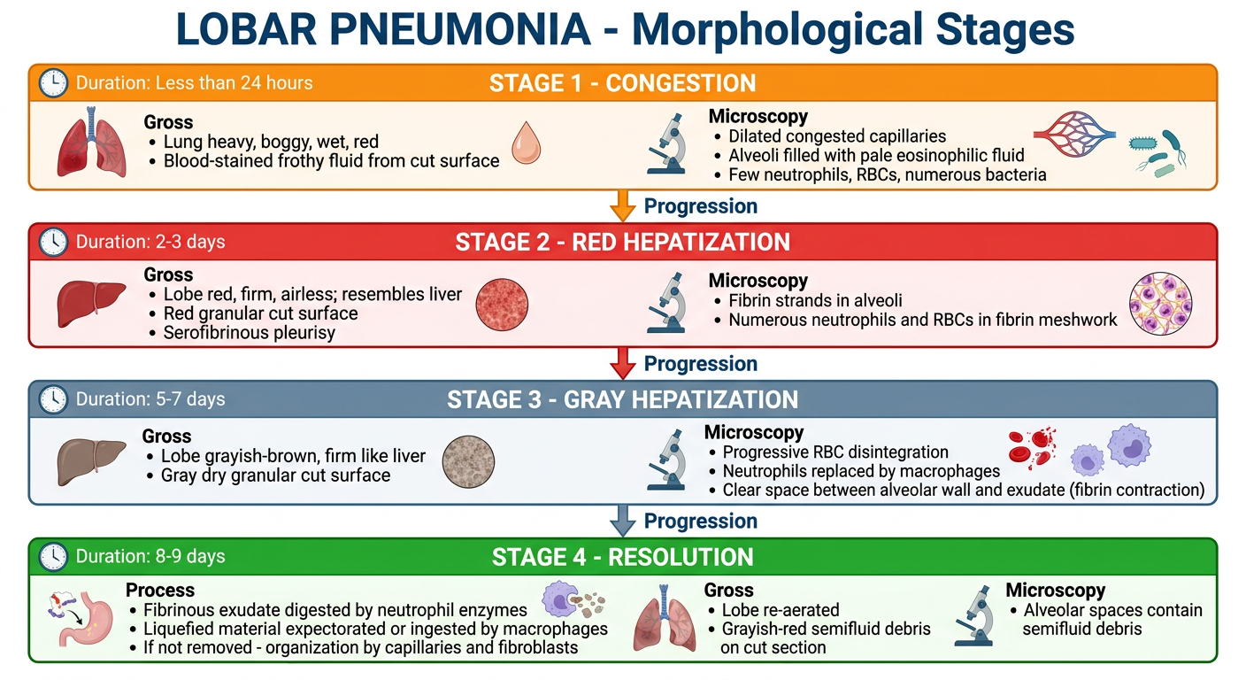
| Stage | Color | Duration |
|---|---|---|
| Congestion | Orange | < 24 hours |
| Red Hepatization | Red | 2-3 days |
| Gray Hepatization | Gray/Blue | 5-7 days |
| Resolution | Green | 8-9 days |

Make a flowchart of classification of pnemonia on badis of given data give it in form of image make it simple to understand and colourful
A colorful, clean, easy-to-read medical classification flowchart titled "CLASSIFICATION OF PNEUMONIA" in bold white text on a dark navy blue header banner at the top. The chart splits into TWO main branches from the title: --- BRANCH 1 (LEFT SIDE): "Based on Clinical Setting" in a bold purple header --- 8 colored boxes arranged vertically or in a tree, each with a distinct bright color, containing: 1. ORANGE box - "Community-Acquired Bacterial" - bullet: S. pneumoniae (most common), H. influenzae, S. aureus, Legionella, Klebsiella, Mycoplasma 2. SKY BLUE box - "Community-Acquired Viral" - bullet: COVID-19 (SARS-CoV-2), RSV, Influenza A & B, Parainfluenza, Adenovirus 3. TEAL box - "Healthcare-Associated" - bullet: S. aureus (MSSA/MRSA), Pseudomonas aeruginosa, S. pneumoniae 4. RED box - "Hospital-Acquired (Nosocomial)" - bullet: MRSA (commonest), Klebsiella, E. coli, Serratia, Pseudomonas 5. YELLOW/GOLD box - "Aspiration Pneumonia" - bullet: Anaerobic oral flora (Bacteroides, Prevotella), S. aureus, Pseudomonas 6. BROWN box - "Chronic Pneumonia" - bullet: Nocardia, Actinomyces, Granulomatous: TB, Histoplasma, Coccidioides 7. DARK RED box - "Necrotizing Pneumonia & Lung Abscess" - bullet: Anaerobic bacteria (most common), S. aureus, K. pneumoniae 8. PINK/MAGENTA box - "Pneumonia in Immunocompromised Host" - bullet: CMV, Pneumocystis jirovecii, MAC, Aspergillus, Candida --- BRANCH 2 (RIGHT SIDE): "Based on Anatomic Distribution" in a bold green header --- 3 colored boxes: A. BRIGHT GREEN box - "Lobar Pneumonia" - Fibrinosuppurative consolidation of large portion or entire lobe. Usually lower lobes. B. CORAL/SALMON box - "Bronchopneumonia" - Patchy scattered solid foci. Consolidation in same or several lobes. C. LAVENDER box - "Interstitial Pneumonia" - Affects interstitial tissue of lungs. Use clear connecting arrows, rounded rectangle boxes, white text labels inside colored boxes, and a professional medical textbook illustration style. The layout is clean, not cluttered, with good spacing.
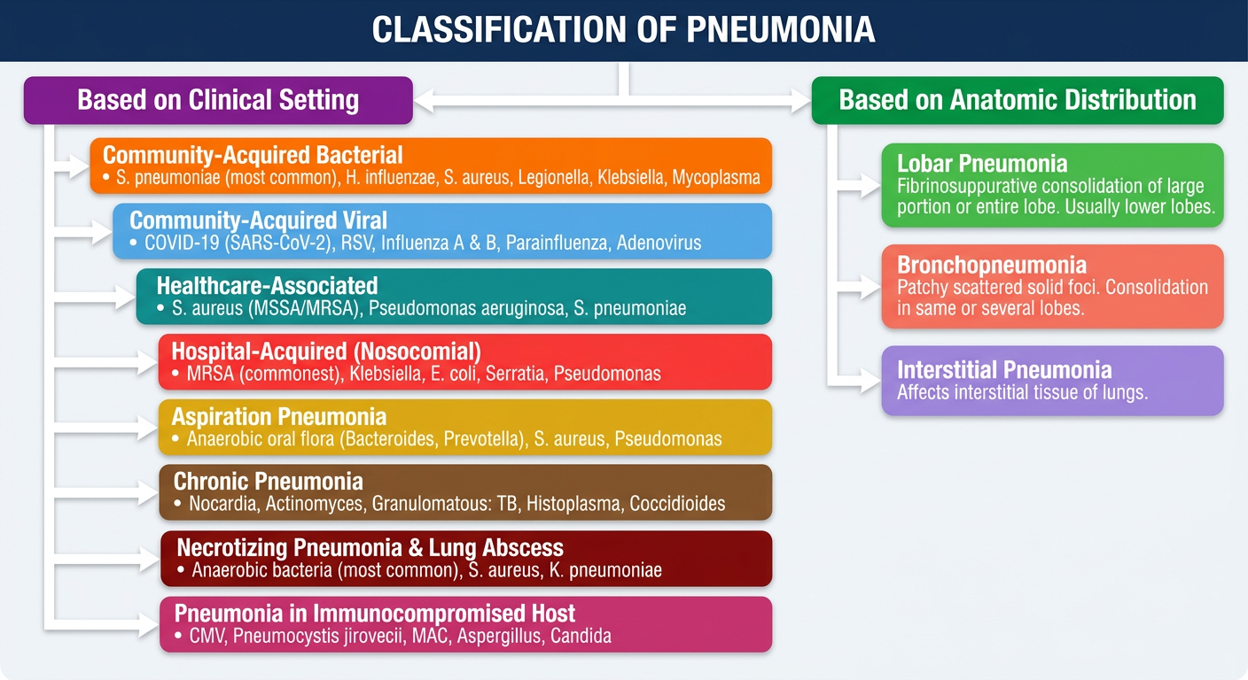
| # | Type | Key Pathogens |
|---|---|---|
| 1 | Community-Acquired Bacterial | S. pneumoniae (MC), H. influenzae, Mycoplasma |
| 2 | Community-Acquired Viral | COVID-19, Influenza A/B, RSV |
| 3 | Healthcare-Associated | MRSA/MSSA, Pseudomonas |
| 4 | Hospital-Acquired (Nosocomial) | MRSA (MC), Klebsiella, E. coli |
| 5 | Aspiration | Anaerobic oral flora |
| 6 | Chronic | TB, Nocardia, Actinomyces, Histoplasma |
| 7 | Necrotizing / Lung Abscess | Anaerobic bacteria (most common) |
| 8 | Immunocompromised Host | PCP, CMV, Aspergillus, Candida |
- Lobar Pneumonia - entire lobe consolidation
- Bronchopneumonia - patchy, scattered consolidation
- Interstitial Pneumonia - affects interstitial tissue

Make fliwchart in form of image and coloyrful
A colorful, clean, easy-to-understand medical flowchart titled "EMPHYSEMA - Morphology" in bold white text on a deep teal header banner at the very top. Below the title, the chart has THREE main vertical sections connected by arrows and dividers: --- SECTION 1 (TOP): "PATHOGENESIS / MECHANISM" in a bold dark blue banner --- A light blue rounded box containing: - "Damage to extracellular matrix by PROTEASE = main role in airway obstruction" - Arrow pointing to: "Loss of elastic tissue in alveolar walls surrounding respiratory bronchioles" - Arrow pointing to: "Small airways can no longer stay open (normally held open by elastic recoil)" - Arrow pointing to: "SMALL AIRWAY OBSTRUCTION" - Side note in yellow sticky box: "Airway infection (bacterial/viral) = does NOT initiate emphysema, but EXACERBATES inflammation and chronic bronchitis" --- SECTION 2 (MIDDLE): "MORPHOLOGY OVERVIEW" in a bold purple banner --- A soft purple rounded box: - "Diagnosis and classification is MAINLY by MACROSCOPIC appearance of lung" - "Best seen on cut surface of formalin-inflated fixed lung" Three colored sub-tags: Irregular emphysema | Distal acinar emphysema | Panacinar emphysema --- SECTION 3 (BOTTOM): Split into two side-by-side colored boxes --- LEFT BOX - "GROSS FEATURES" with bright orange header: - Upper 2/3 of lungs more severely involved - Bullae present in: Irregular & Distal acinar emphysema - Advanced & Panacinar emphysema → VOLUMINOUS LUNGS RIGHT BOX - "MICROSCOPY FEATURES" with bright green header: - Abnormally large alveoli separated by thin septa (no fibrosis) - Destruction of alveolar walls - Loss of attachments of alveoli to outer wall of small airways - Pores of Kohn (interalveolar connections) are ENLARGED Connecting arrows flow top to bottom. Use bright, distinct colors for each section. Clean white background, bold readable text, medical textbook style, professional and easy to study from.
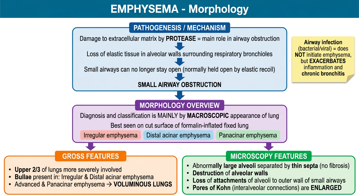
- Protease damages extracellular matrix
- Loss of elastic tissue in alveolar walls
- Small airways collapse - obstruction
- Note: Airway infection only worsens, does not initiate emphysema
- Diagnosis mainly by macroscopic appearance
- 3 types: Irregular, Distal acinar, Panacinar
| Gross | Microscopy |
|---|---|
| Upper 2/3 lungs most affected | Abnormally large alveoli, thin septa |
| Bullae in irregular & distal acinar | Destruction of alveolar walls |
| Panacinar → voluminous lungs | Pores of Kohn enlarged |