
Rahes between middle of thighs which cream is best
best cream for inner thigh rash chafing intertrigo treatment 2025
inner thigh rash intertrigo chafing
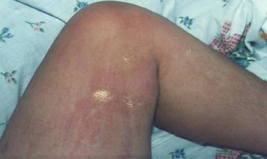
This clinical photograph displays a dermatological condition on the lower limb of a patient, specifically involving the inner thigh and the medial aspect of the knee. The primary finding is a diffuse, salmon-colored to pinkish-red maculopapular eruption. The rash exhibits a mottled and somewhat confluent appearance, with erythema extending across the cutaneous surface. Notably, there are focal areas with a shiny, glistening texture and superficial excoriations, characteristic of pruritic lesions damaged by scratching (lichenification or secondary skin damage). The morphology and 'salmon-colored' hue of the eruption are clinically significant, often associated with systemic inflammatory conditions or specific viral exanthems in dermatology. No distinct vesicles or large pustules are present, though the skin surface appears uneven. This image serves as an educational resource for identifying inflammatory skin patterns and signs of secondary irritation due to pruritus.
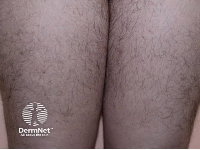
Clinical photography of bilateral anterior-medial thighs at close range, using high-resolution imaging to document healthy skin texture and hair pattern. The image shows the integumentary system in the thigh region with frontal/anterior view and a neutral lighting scheme. Visual features include even skin tone, fine vellus hair density, uniform epidermal surface, and absence of erythema, scaling, plaques, vesicles, ulcers, crusting, edema, or nodules. No focal lesions or pigmentary abnormalities are evident. The regional anatomy corresponds to the anterior medial thigh with subtle skin folds and intertriginous contact areas. There are no signs of inflammation, infection, or trauma in this field of view. This photograph provides a baseline reference for normal cutaneous appearance in the groin/upper thigh area and can be used for comparison with dermatologic conditions such as dermatitis, tinea cruris, candidiasis, intertrigo, psoriasis, pityriasis, or malignant lesions when present. Diagnostic significance lies in establishing a normal skin phenotype for this region, aiding differential diagnosis and clinical correlation in patients presenting with pruritus or rash. Potential uses include medical education, image database curation, and development of computer vision tools for healthy-skin recognition and anomaly detection in dermatology. This baseline image supports reproducible comparisons across clinicians and educational contexts for training.
Rash Between the Inner Thighs (Intertrigo) - Best Creams
Step 1: Identify the Type of Rash
| Signs | Likely Cause |
|---|---|
| Red, moist, itchy skin only | Simple irritant/inflammatory intertrigo |
| White patches, satellite pustules (small bumps around the edge) | Fungal (Candida/yeast) infection |
| Painful, very red, macerated skin | Bacterial infection (Streptococcus) |
| Scaly, ring-shaped border | Tinea cruris (ringworm/fungal) |
Step 2: Choose the Right Cream
For Simple Irritation/Chafing (No Infection)
- Zinc oxide paste - best barrier cream; protects skin from friction and moisture (excellent OTC choice, like Sudocrem or Desitin)
- Petroleum jelly (Vaseline) - forms a protective barrier layer
- Triple paste (zinc oxide + aluminum acetate + petrolatum) - very effective barrier
For Fungal Infection (most common complication)
- Clotrimazole 1% cream (Lotrimin AF) - apply twice daily for 2-4 weeks
- Miconazole 2% cream (Micatin) - apply twice daily
- Ketoconazole 2% cream (Nizoral) - prescription; highly effective
- Nystatin cream - specifically for Candida (yeast) infections
For Inflammation/Itch
- Hydrocortisone 1% cream (OTC) - use short-term only (1-2 weeks); reduces redness and itch
- Do NOT use stronger steroids (like triamcinolone) in skin folds for prolonged periods as they cause skin thinning
For Bacterial Infection
- Mupirocin (Bactroban) ointment - prescription, for bacterial superinfection
- Fusidic acid cream - topical antibiotic
Combination Approach (Most Commonly Prescribed)
Practical Tips to Heal Faster
- Keep the area dry - use a hair dryer on cool setting after bathing
- Wear loose, breathable clothing (cotton underwear)
- Apply cream after drying the area thoroughly
- Use an absorbent powder (like cornstarch-based) after the rash settles, to prevent recurrence
- Avoid potential irritants - scented soaps, deodorant sprays, neomycin-containing creams
When to See a Doctor
- The rash does not improve within 1-2 weeks of OTC treatment
- There is significant pain, warmth, or spreading redness (possible cellulitis)
- You have diabetes or are immunocompromised
- Satellite pustules are present (needs prescription antifungal)
- The rash keeps returning
Disclaimer: This is general medical information. For a proper diagnosis and treatment plan, please consult a doctor or dermatologist, especially if the rash is severe, spreading, or not improving.