
Congestive heart failure
"heart failure"[MeSH Terms] AND "management"[MeSH Terms]
congestive heart failure pathophysiology diagram
heart failure chest X-ray pulmonary edema cardiomegaly
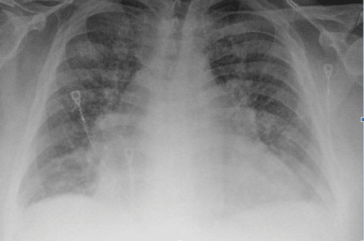
This anterior-posterior (AP) chest X-ray illustrates classic radiological findings of acute congestive heart failure. The primary finding is significant cardiomegaly, with the cardiac silhouette appearing enlarged and globular, occupying more than 50% of the thoracic diameter. Diffuse, bilateral hazy opacities are present throughout both lung fields, consistent with pulmonary edema. These opacifications are most prominent in the perihilar regions and lung bases, obscuring normal pulmonary vasculature and the sharp definition of the diaphragmatic borders. The costophrenic angles appear blunted, suggesting associated pleural effusions. Visible medical hardware includes ECG leads positioned on the chest wall. The image serves as an educational example of how chronic heart failure with reduced ejection fraction manifests as pulmonary venous congestion and interstitial-to-alveolar edema on diagnostic imaging.
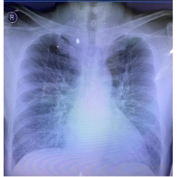
This diagnostic image is a posterior-anterior (PA) chest x-ray radiography demonstrating significant cardiomegaly and signs of pulmonary congestion. The cardiac silhouette is markedly enlarged, with a transverse diameter exceeding 50% of the thoracic width, suggesting multi-chamber enlargement. The lung fields exhibit diffuse, bilateral increased opacities with perihilar haziness and ill-defined vascular markings, characteristic of interstitial pulmonary edema. Cephalization of the pulmonary vessels is visible, further indicating elevated pulmonary venous pressure. These radiological findings are clinically significant for acute heart failure or decompensated cardiac function, likely secondary to a cardiovascular event such as myocardial infarction. The image serves as an educational example of the visual progression from cardiomegaly to impending acute lung edema in a clinical setting.
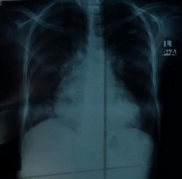
Posteroanterior (PA) view chest X-ray demonstrating clinical features of congestive heart failure and pulmonary edema. The image shows significant cardiomegaly with an enlarged cardiac silhouette. There is evidence of pulmonary venous hypertension, specifically characterized by upper lobe diversion where the vascular markings in the upper lobes are more prominent than those in the lower zones. The hilar regions appear congested with increased density and blurring of the hilar shadows. In the lower lung fields, patchy opacities and areas of increased density are visible, consistent with pulmonary consolidation or alveolar edema. The diaphragmatic angles appear somewhat obscured by the parenchymal density changes. This radiographic presentation is typical for decompensated heart failure, illustrating the redistribution of blood flow and fluid accumulation within the pulmonary parenchyma.
Congestive Heart Failure (CHF)
Definition
Classification
By Ejection Fraction
| Type | EF | Mechanism |
|---|---|---|
| HFrEF (Heart failure with reduced EF) | < 40% | Systolic dysfunction - impaired contraction |
| HFmrEF (mildly reduced) | 40-49% | Mixed |
| HFpEF (preserved EF) | ≥ 50% | Diastolic dysfunction - impaired relaxation/filling |
By NYHA Functional Class
| Class | Description |
|---|---|
| I | No symptoms with ordinary activity |
| II | Slight limitation; symptoms with moderate exertion |
| III | Marked limitation; symptoms with minimal exertion |
| IV | Symptoms at rest; unable to carry on any activity |
Etiology
- Ischemic heart disease / CAD (most common)
- Hypertension (hypertensive cardiomyopathy)
- Valvular heart disease
- Dilated cardiomyopathy (idiopathic, alcohol, viral myocarditis)
- Arrhythmias (tachycardia-mediated cardiomyopathy)
- Thyroid disease (hypo/hyperthyroidism)
- Diabetes, amyloidosis, hemochromatosis, sarcoidosis
- Chemotherapy/cardiotoxins
Pathophysiology
- Sympathetic nervous system activation - Catecholamine release causes vasoconstriction, tachycardia, and increased contractility. Chronically, norepinephrine drives myocyte hypertrophy and apoptosis. - Goldman-Cecil Medicine, p. 3254
- RAAS activation - Renin-angiotensin-aldosterone axis causes sodium and water retention, increasing preload and afterload.
- Ventricular remodeling - Hypertrophy, dilatation, and fibrosis progressively worsen cardiac function.
- Natriuretic peptide release - BNP/ANP are released in response to wall stress; they promote natriuresis and vasodilation (a counter-regulatory response).
Clinical Features
Symptoms
- Dyspnea on exertion (most common)
- Orthopnea - dyspnea when lying flat (requires multiple pillows)
- Paroxysmal nocturnal dyspnea (PND) - awakening with breathlessness
- Fatigue, weakness
- Wheezing ("cardiac asthma")
- Nausea, abdominal distension (right heart failure)
Signs
-
Tachycardia, tachypnea
-
S3 gallop (ventricular filling sound - marker of systolic dysfunction)
-
Crackles/rales at lung bases
-
Elevated JVP (jugular venous pressure)
-
Hepatomegaly (hepatic congestion)
-
Pitting peripheral edema (ankles, legs)
-
Ascites (in severe right heart failure)
-
Displaced apex beat (cardiomegaly)
-
Rosen's Emergency Medicine, p. 3212
Diagnosis
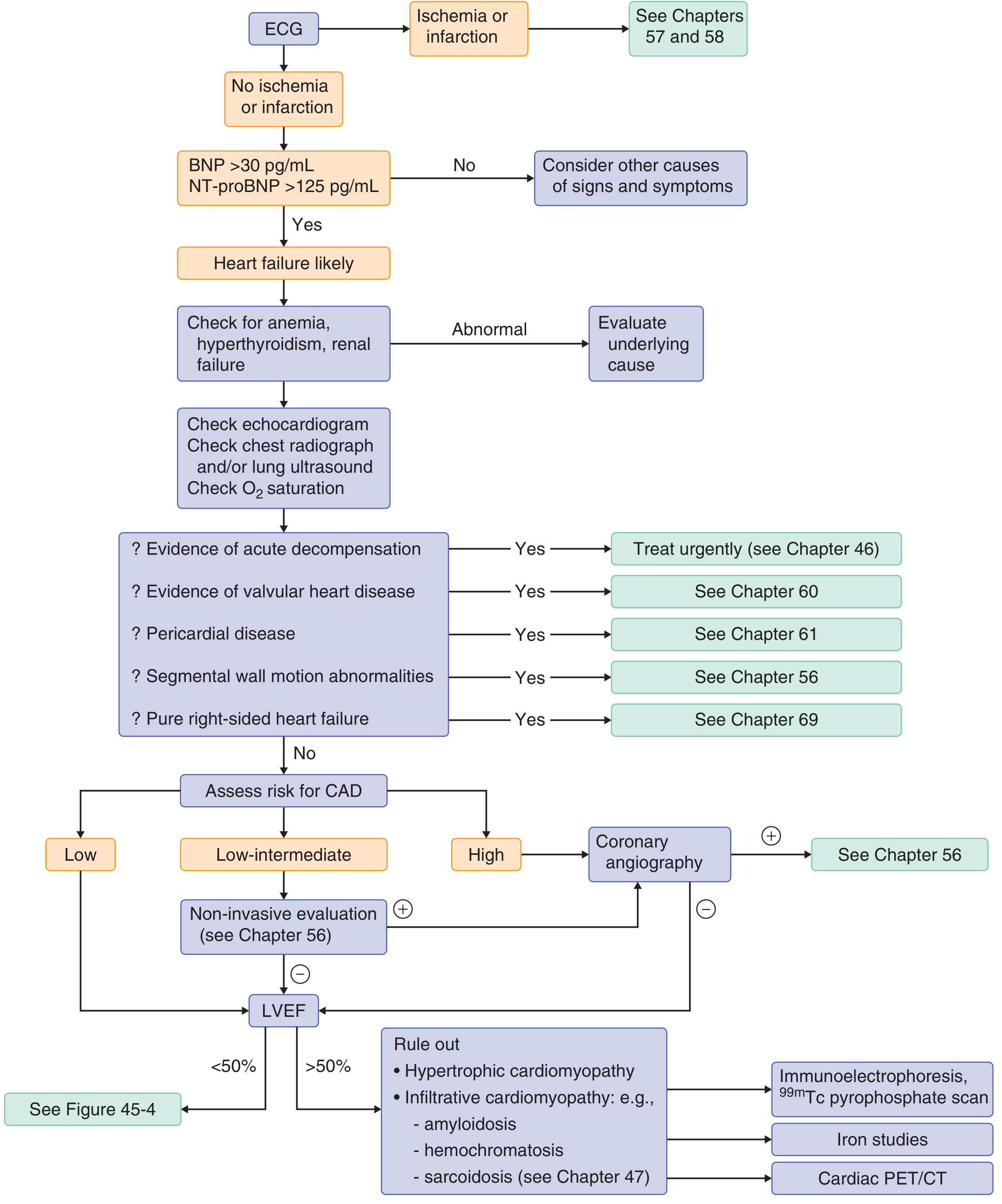
Diagnostic Flowchart
Key Investigations
- Assess rhythm, identify ischemia/prior MI
- Left ventricular hypertrophy (voltage criteria)
- Bundle branch block (determines eligibility for CRT)
- Q waves suggest CAD
- Goldman-Cecil Medicine, p. 2481
- Cardiomegaly (cardiothoracic ratio >50%)
- Cephalization of pulmonary vessels (upper lobe diversion)
- Perihilar "bat-wing" pattern (pulmonary edema)
- Kerley B lines (interstitial edema)
- Bilateral pleural effusions (often right-sided if unilateral)
- BNP < 100 pg/mL or NT-proBNP < 400 pg/mL makes HF unlikely
- BNP > 400 pg/mL or NT-proBNP > 125 pg/mL supports the diagnosis
- BNP between 100-500 pg/mL: indeterminate
- Also useful for monitoring response to treatment
- Note: sacubitril (ARNI) increases BNP but NOT NT-proBNP - use NT-proBNP for monitoring in ARNI-treated patients
- Goldman-Cecil Medicine, p. 2520; Textbook of Family Medicine, p. 777
- Measures LVEF (determines HFrEF vs. HFpEF)
- Identifies wall motion abnormalities, valvular disease, pericardial effusion, cardiomyopathy type
- CBC (detect anemia), BMP (renal function, electrolytes)
- Glucose, lipid profile, HbA1c
- TSH (thyroid disease)
- LFTs (hepatic congestion - elevated transaminases, bilirubin)
- Serum ferritin/iron studies (hemochromatosis)
- Consider: HIV, ANA/rheumatologic panel, serum protein electrophoresis (amyloidosis)
- Goldman-Cecil Medicine, p. 2501
Management
Acute Decompensated Heart Failure
- Supplemental oxygen - target SpO2 >94%
- CPAP/BiPAP - reduces work of breathing and preload; often averts need for intubation
- IV furosemide (loop diuretic) - mainstay for decongestion; 0.5-1 mg/kg in children
- IV morphine (cautiously) - reduces preload and anxiety
- IV vasodilators (nitrates) - reduce preload and afterload in adults
- Inotropes (dobutamine, dopamine) - for cardiogenic shock or low-output states
- Intubation and mechanical ventilation if severe respiratory failure
Chronic Management of HFrEF - "4 Pillars"
| Drug Class | Examples | Starting Dose | Target Dose |
|---|---|---|---|
| ARNI (preferred over ACEi/ARB) | Sacubitril/valsartan | 24/26 mg twice daily | 97/103 mg twice daily |
| ACE Inhibitor (if ARNI not tolerated) | Enalapril, Lisinopril, Ramipril | Low (e.g., enalapril 2.5 mg BD) | Enalapril 10-20 mg BD |
| ARB (if ACEi not tolerated) | Candesartan, Valsartan, Losartan | Low dose | e.g., Candesartan 32 mg OD |
| Beta-Blocker | Bisoprolol, Carvedilol, Metoprolol CR/XL, Nebivolol | Low dose (e.g., bisoprolol 1.25 mg OD) | e.g., Bisoprolol 10 mg OD |
| MRA (mineralocorticoid receptor antagonist) | Spironolactone, Eplerenone | 25 mg OD | 50 mg OD |
| SGLT2 inhibitor | Dapagliflozin, Empagliflozin | 10 mg OD | 10 mg OD |
- Goldman-Cecil Medicine, pp. 3147, 3170, 3074-3083
- Start all drugs at low doses, uptitrate every 2 weeks
- ACEi/ARB must never be combined with ARNI
- Wash-out of 36 hours required when switching from ACEi to ARNI
- Do NOT start beta-blocker during acute decompensation; start only when euvolemic
- Beta-blocker contraindicated in: asthma, 2nd/3rd degree AV block, HR < 60
- ARNI contraindicated in: prior angioedema, SBP < 95 mmHg
Diuretics
- Not mortality-reducing, but essential for symptom relief
- Furosemide (loop diuretic) - first-line
- Titrate to achieve euvolemia (dry weight)
- Monitor electrolytes (hypokalemia, hypomagnesemia) and renal function
Device Therapy
| Device | Indication |
|---|---|
| ICD (implantable cardioverter-defibrillator) | EF ≤ 35%, NYHA II-III, on optimal medical therapy ≥ 3 months |
| CRT (cardiac resynchronization therapy) | EF ≤ 35%, QRS ≥ 150 ms (LBBB morphology), NYHA II-IV |
| CRT-D (combined CRT + ICD) | Meets criteria for both |
| LVAD (left ventricular assist device) | Advanced HF, bridge to transplant or destination therapy |
| Heart transplant | End-stage HF refractory to all therapies |
Atrial Fibrillation in Heart Failure
Prognosis
- 5-year mortality ~50% (worse than many cancers)
- BNP/NT-proBNP are powerful prognostic markers
- Elevated hepatic enzymes (congestion), hyponatremia, and renal dysfunction predict poor outcomes
- Carvedilol is substantially more effective than low-dose short-acting metoprolol for survival
- Multidisciplinary disease management programs (nurse-led, pharmacist-assisted follow-up) significantly reduce hospitalizations and mortality - PMID 41555291
Summary: NYHA-Guided Approach
- Class I-II (mild): Start all 4 drug pillars (ARNI/ACEi, beta-blocker, MRA, SGLT2i) + ICD if EF ≤ 35%
- Class III (moderate): Full 4 pillars + consider CRT if QRS wide; optimize diuretic
- Class IV (severe): Specialist referral; consider LVAD, transplant listing; beta-blocker with caution
- Acute decompensation: Diurese to euvolemia, then optimize chronic therapies