
How blood calcium level is Maintained at normal levels? What is the pathophysiology of this disease? Explain the physiological basis for increased neuromuscular excitability
Reading File
The question covers calcium homeostasis, the pathophysiology of hypoparathyroidism/hypocalcemia, and neuromuscular excitability. Let me search the medical library across all three dimensions in parallel.
Searching sources...
Finding Sources
Finding Sources
Finding Sources
Reading File
Reading File
Finding Sources
Now let me pull the detailed content from Guyton (physiology of calcium regulation + neuromuscular excitability), Frameworks for Internal Medicine (hypocalcemia pathophysiology), and Schwartz's Surgery (hypoparathyroidism):
Reading File
Reading File
Reading File
Let me now get the Guyton physiology content on calcium regulation and the specific neuromuscular excitability mechanism, plus more from Frameworks for Internal Medicine:
Finding Sources
Finding Sources
Finding Sources
Finding Sources
Finding Sources
Reading File
Now let me get the Guyton calcium regulation section and search for images:
Finding Sources
Searching Images
calcium homeostasis PTH parathyroid hormone vitamin D regulation diagram
Searching Images
hypocalcemia neuromuscular excitability tetany Chvostek Trousseau sign
Reading File
Reading File
Excellent - I now have comprehensive textbook content. Let me fetch the key diagram images:
Loading Image
Loading Image
I now have all the information needed for a comprehensive, well-illustrated answer. Let me compose it.
Blood Calcium Homeostasis, Hypocalcemia Pathophysiology, and Neuromuscular Excitability
Part 1: How Blood Calcium Is Maintained at Normal Levels
Normal serum calcium is 8.5-10.5 mg/dL (2.1-2.6 mM). About 50% circulates as free ionized Ca²⁺ (the biologically active fraction), ~40% is bound to albumin, and ~10% is complexed to anions (citrate, phosphate). Only the ionized fraction is regulated. - Harrison's Principles of Internal Medicine 22E
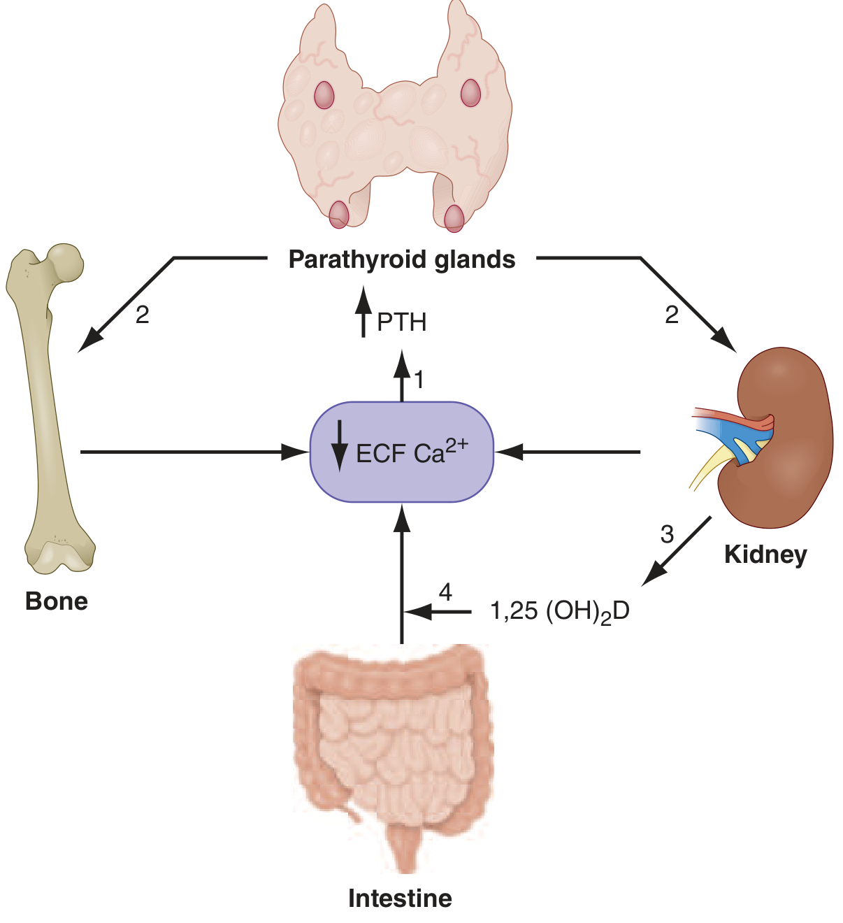
The system integrates signals across four organs: parathyroid glands, bone, kidney, and intestine, orchestrated by two hormones - PTH and 1,25-dihydroxyvitamin D (calcitriol).
The Calcium-Sensing Receptor (CaSR)
The parathyroid glands express a G-protein-coupled calcium-sensing receptor (CaSR) on their chief cells. This receptor continuously monitors ionized Ca²⁺. A fall in Ca²⁺ activates CaSR-mediated signaling that rapidly releases stored PTH from secretory granules. - National Kidney Foundation Primer, 8e
PTH is an 84-amino-acid peptide with a half-life of only 2-4 minutes, allowing rapid fine-tuning of calcium levels.
Actions of PTH (the primary short-term regulator)
| Target Organ | Action | Net Effect |
|---|---|---|
| Bone | Increases osteocyte RANKL → stimulates osteoclast-mediated resorption | Releases Ca²⁺ and PO₄³⁻ into blood |
| Kidney (distal tubule) | Increases Ca²⁺ reabsorption in distal tubule | Reduces urinary Ca²⁺ loss |
| Kidney (proximal tubule) | Stimulates 25(OH)D → 1α-hydroxylase conversion | Increases calcitriol production |
| Kidney (phosphate) | Decreases PO₄³⁻ reabsorption (phosphaturic effect) | Prevents Ca-PO₄ complex formation |
- Frameworks for Internal Medicine; National Kidney Foundation Primer, 8e
Role of Vitamin D (longer-term regulation)
When PTH stimulates renal 1α-hydroxylase, 25-hydroxyvitamin D₃ is converted to the active metabolite 1,25(OH)₂D₃ (calcitriol). The vitamin D pathway itself begins with:
- Skin: UV-B converts 7-dehydrocholesterol → pre-vitamin D₃ → cholecalciferol (D₃)
- Liver: D₃ → 25-hydroxyvitamin D₃ (25(OH)D₃) via CYP2R1
- Kidney: 25(OH)D₃ → 1,25(OH)₂D₃ via 1α-hydroxylase (PTH-dependent)
Calcitriol then acts on the small intestine to upregulate TRPV6 channels and calbindin-D, increasing transcellular Ca²⁺ absorption. It also acts on bone to support mineral mobilization. - Brenner and Rector's The Kidney
The Negative Feedback Loop
When serum Ca²⁺ normalizes or becomes elevated:
- CaSR on parathyroid cells is activated → PTH secretion is suppressed
- Elevated Ca²⁺ also activates the CaSR in the thick ascending limb of the loop of Henle → increases urinary Ca²⁺ excretion
- Calcitriol feeds back directly on the parathyroid gland to suppress PTH gene transcription
In calcium excess, renal synthesis of 24R,25(OH)₂D₃ (a less active metabolite) is favored, further reducing calcium mobilization. - Brenner and Rector's The Kidney
Calcitonin (minor role)
Calcitonin, secreted by thyroid C-cells in response to hypercalcemia, opposes PTH: it inhibits osteoclast activity, decreases intestinal Ca²⁺ absorption, and increases renal Ca²⁺ excretion. Its physiological role in humans is modest. - Lippincott's Biochemistry, 8e
Part 2: Pathophysiology of Hypocalcemia / Hypoparathyroidism
The question implies the "disease" in context is hypoparathyroidism, the most clinically important disorder of this system.
Causes of Hypoparathyroidism
| Category | Specific Cause |
|---|---|
| Surgical (most common) | Inadvertent removal/devascularization of parathyroids during total thyroidectomy or central neck dissection |
| Congenital | DiGeorge syndrome (22q11.2 deletion) - absent parathyroids + absent thymus |
| Autoimmune | Autoimmune polyglandular syndrome type 1 (APS-1) |
| Infiltrative | Heavy metal deposition (iron, copper, aluminium), granulomas |
| Functional | Hypomagnesemia (Mg²⁺ required for PTH secretion and action; severe deficiency blocks both) |
| Resistance to PTH | Pseudohypoparathyroidism (PTH levels are elevated but target organs fail to respond) |
- Schwartz's Principles of Surgery, 11e; Miller's Anesthesia, 10e
The Core Pathophysiological Cascade
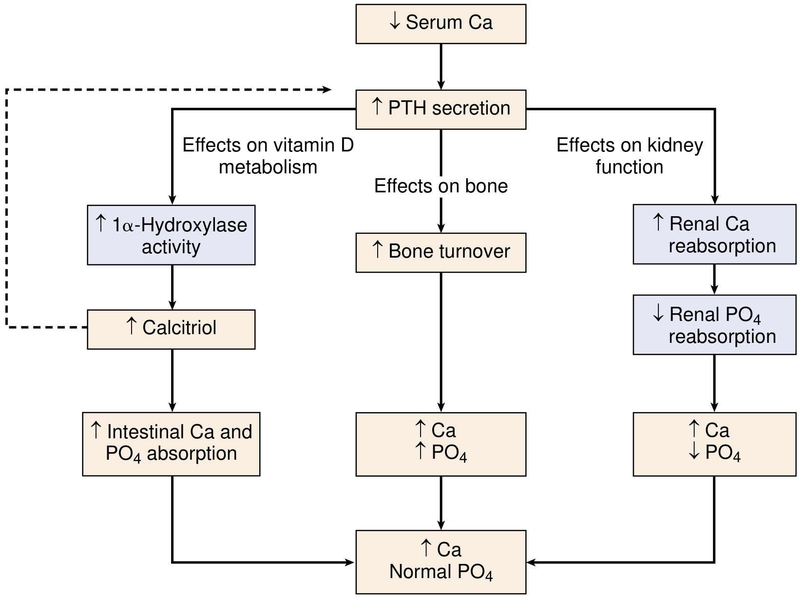
When PTH is absent or deficient:
- No PTH → no bone resorption signal → Ca²⁺ not mobilized from bone
- No PTH → reduced renal Ca²⁺ reabsorption → increased urinary Ca²⁺ loss
- No PTH → no stimulation of renal 1α-hydroxylase → calcitriol levels fall → intestinal Ca²⁺ absorption drops
- No PTH phosphaturic effect → phosphate accumulates in blood (hyperphosphatemia)
- High phosphate + low calcium → further depression of ionized Ca²⁺ (Ca-PO₄ complex formation)
Net result: Low serum Ca²⁺ (hypocalcemia) + high serum PO₄³⁻ (hyperphosphatemia) + low/absent PTH + low calcitriol
In pseudohypoparathyroidism, the biochemical picture is identical but PTH is elevated - the kidneys and bone simply do not respond to it.
Biochemical Hallmarks
| Parameter | Hypoparathyroidism | Pseudohypoparathyroidism |
|---|---|---|
| Serum Ca²⁺ | Low | Low |
| Serum PO₄³⁻ | High | High |
| PTH | Low or absent | Elevated |
| Calcitriol | Low | Low |
Part 3: Physiological Basis for Increased Neuromuscular Excitability
This is the most important consequence of hypocalcemia and has a clear membrane-level explanation.
The Membrane Stabilization Role of Ca²⁺
Extracellular Ca²⁺ ions bind to the outer surface of voltage-gated sodium (Na⁺) channels in nerve and muscle cell membranes. This binding has a charge-screening / stabilizing effect:
- Calcium ions carry a 2+ charge and bind to negatively charged phospholipid head groups and channel proteins on the outer membrane leaflet
- This creates a local positive charge that raises the threshold voltage for Na⁺ channel activation (i.e., makes it harder to trigger an action potential)
- In physiological terms: the threshold potential is shifted to a more positive (less negative) value relative to the resting membrane potential
What Happens in Hypocalcemia
When ionized Ca²⁺ falls:
- The stabilizing effect on Na⁺ channels is lost
- The threshold for action potential generation shifts to a more negative value - closer to the resting membrane potential
- This narrows the "safety gap" between resting potential and threshold
- Even small, sub-threshold stimuli can now trigger action potentials
The result is spontaneous, repetitive firing of nerve and muscle fibers - this is tetany.
"Hypocalcemia increases the excitability of nerve and muscle cells, which can lead to tetany; hypercalcemia decreases neuromuscular excitability." - Frameworks for Internal Medicine
"Acute hypocalcemia results in decreased ionized calcium and increased neuromuscular excitability." - Schwartz's Principles of Surgery, 11e
Clinical Manifestations (in order of increasing severity)
| Severity | Manifestation | Mechanism |
|---|---|---|
| Mild | Perioral and fingertip paresthesias, numbness | Spontaneous sensory fiber firing |
| Moderate | Muscle cramps, carpopedal spasm (Trousseau sign), hyperreflexia | Spontaneous motor fiber firing; enhanced stretch reflex |
| Severe | Laryngospasm, stridor, generalized tetany | Involuntary skeletal/laryngeal muscle tetanic contraction |
| Severe | Seizures, cerebral tetany | Increased CNS neuronal excitability |
| Cardiac | Prolonged QT interval, dysrhythmias, depressed contractility | Altered cardiac repolarization; Ca²⁺-dependent excitation-contraction coupling impaired |
- Miller's Anesthesia, 10e; Frameworks for Internal Medicine
Chvostek's Sign and Trousseau's Sign
- Chvostek's sign: Tapping over the facial nerve anterior to the ear (over the parotid gland) produces ipsilateral facial muscle twitching. This reflects hyperexcitable facial nerve fibers.
- Trousseau's sign: Inflating a sphygmomanometer cuff on the arm above systolic pressure for 3 minutes produces carpal spasm (metacarpophalangeal flexion, finger adduction, wrist flexion). This is the more specific sign. - Frameworks for Internal Medicine
Why Alkalosis Worsens Symptoms
Alkalosis (respiratory or metabolic) reduces the ionized fraction of Ca²⁺ further because:
- H⁺ normally competes with Ca²⁺ for albumin-binding sites
- In alkalosis, fewer H⁺ ions compete → more albumin binding → less free ionized Ca²⁺
- Total serum calcium may be unchanged but ionized fraction falls sharply
This is why hyperventilation (respiratory alkalosis) can precipitate tetany even with borderline total calcium. - Miller's Anesthesia, 10e
Summary
| Topic | Key Points |
|---|---|
| Ca²⁺ maintenance | CaSR detects ionized Ca²⁺; PTH acts on bone (resorption), kidney (Ca²⁺ reabsorption, calcitriol synthesis), and intestine (indirectly via calcitriol); tight negative feedback |
| Pathophysiology | Absent PTH → no bone/renal/intestinal Ca²⁺ recovery + phosphate retention; most common cause is post-surgical |
| Neuromuscular excitability | Ca²⁺ stabilizes Na⁺ channels by charge screening; low Ca²⁺ removes this brake, lowers threshold to firing, causes spontaneous action potentials, tetany |
This is a shared conversation. Sign in to Orris to start your own chat.