Development of face with diagrams
development of the face embryology stages diagram
pharyngeal arches facial prominences frontonasal process embryo
9780323611541_block*.md in /textbooks/9780323611541
craniofacial development neural crest
Development of the Face
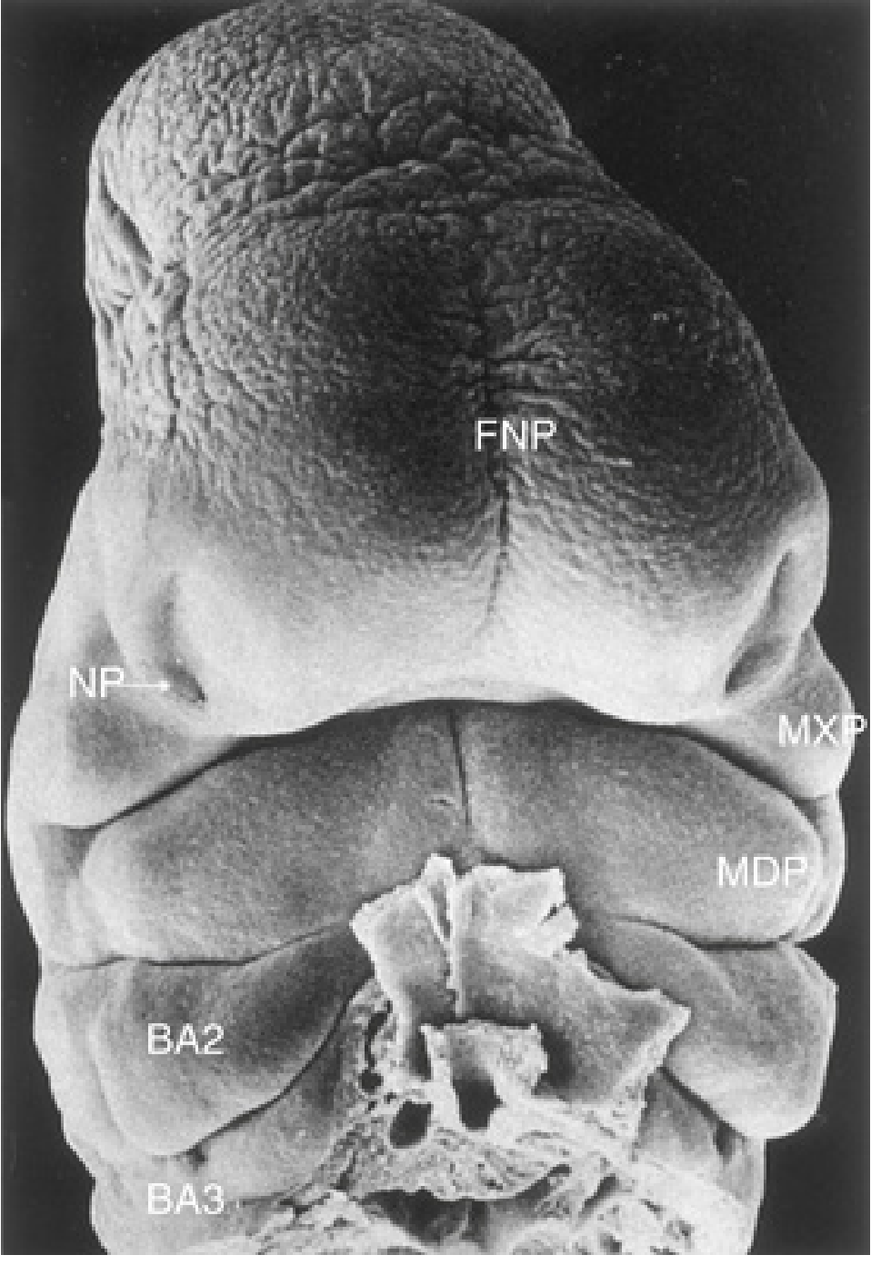
1. The Five Facial Prominences (Week 4)
- 1 frontonasal prominence — surrounds the forebrain; gives rise to the forehead and, later, the nose
- 2 maxillary prominences (paired) — form the lateral boundaries of the stomodeum
- 2 mandibular prominences (paired) — form the caudal boundary of the stomodeum
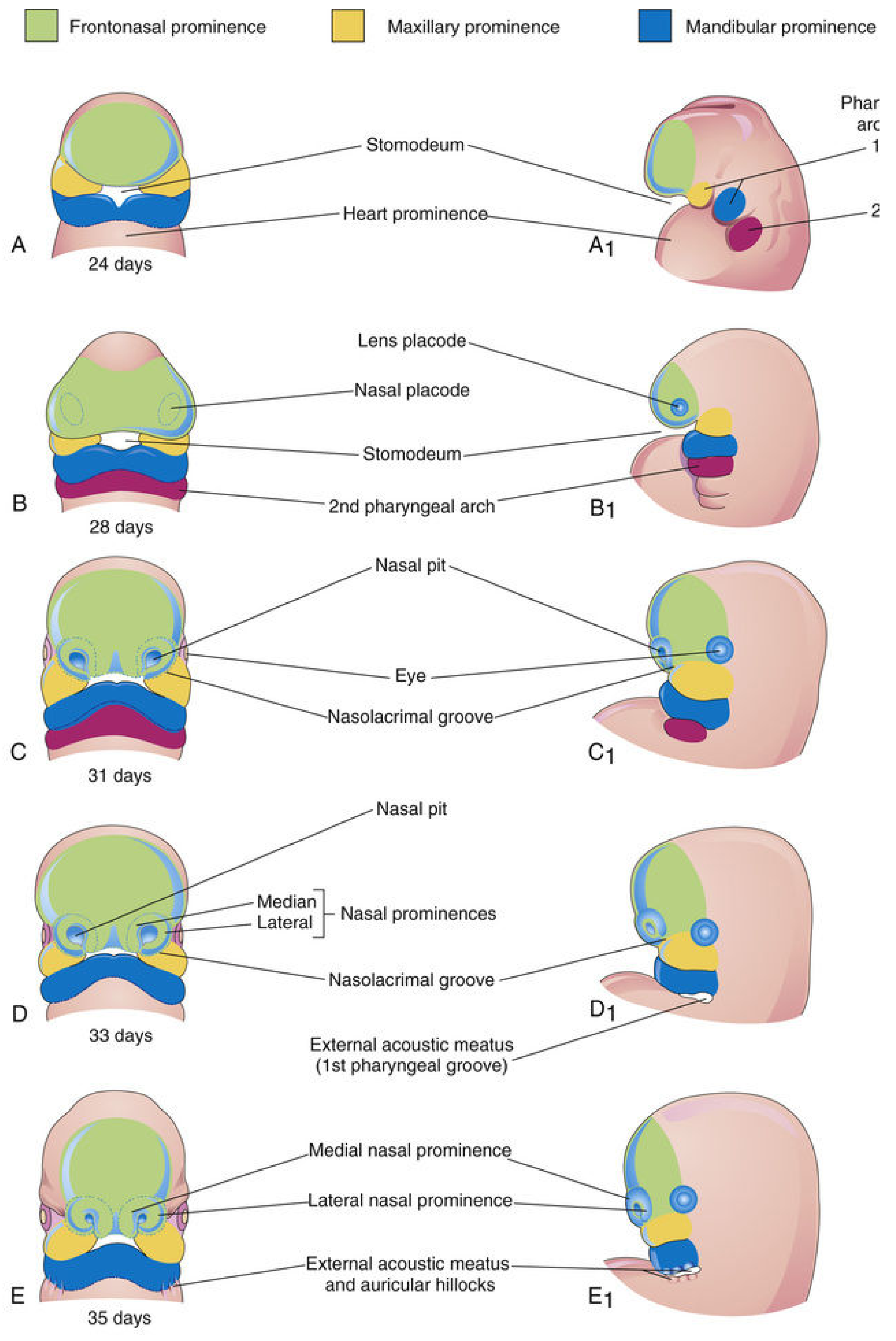
2. Nasal Placodes and Nasal Prominences (End of Week 4 - Week 5)
- The lateral nasal prominences form the alae (sides) of the nose
- The medial nasal prominences will later form the nasal septum, philtrum, and premaxilla
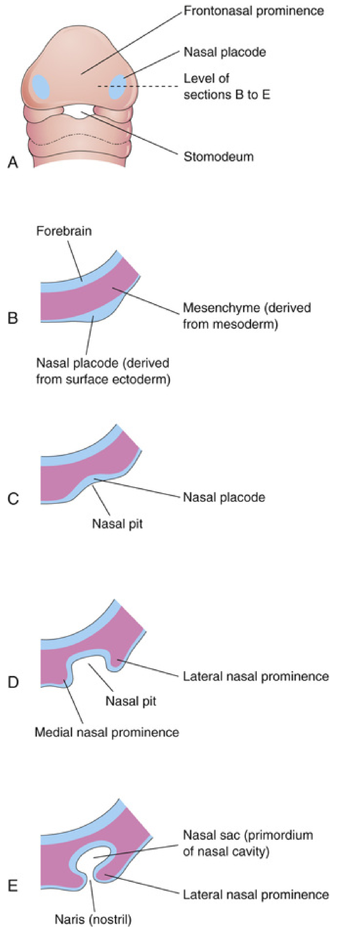
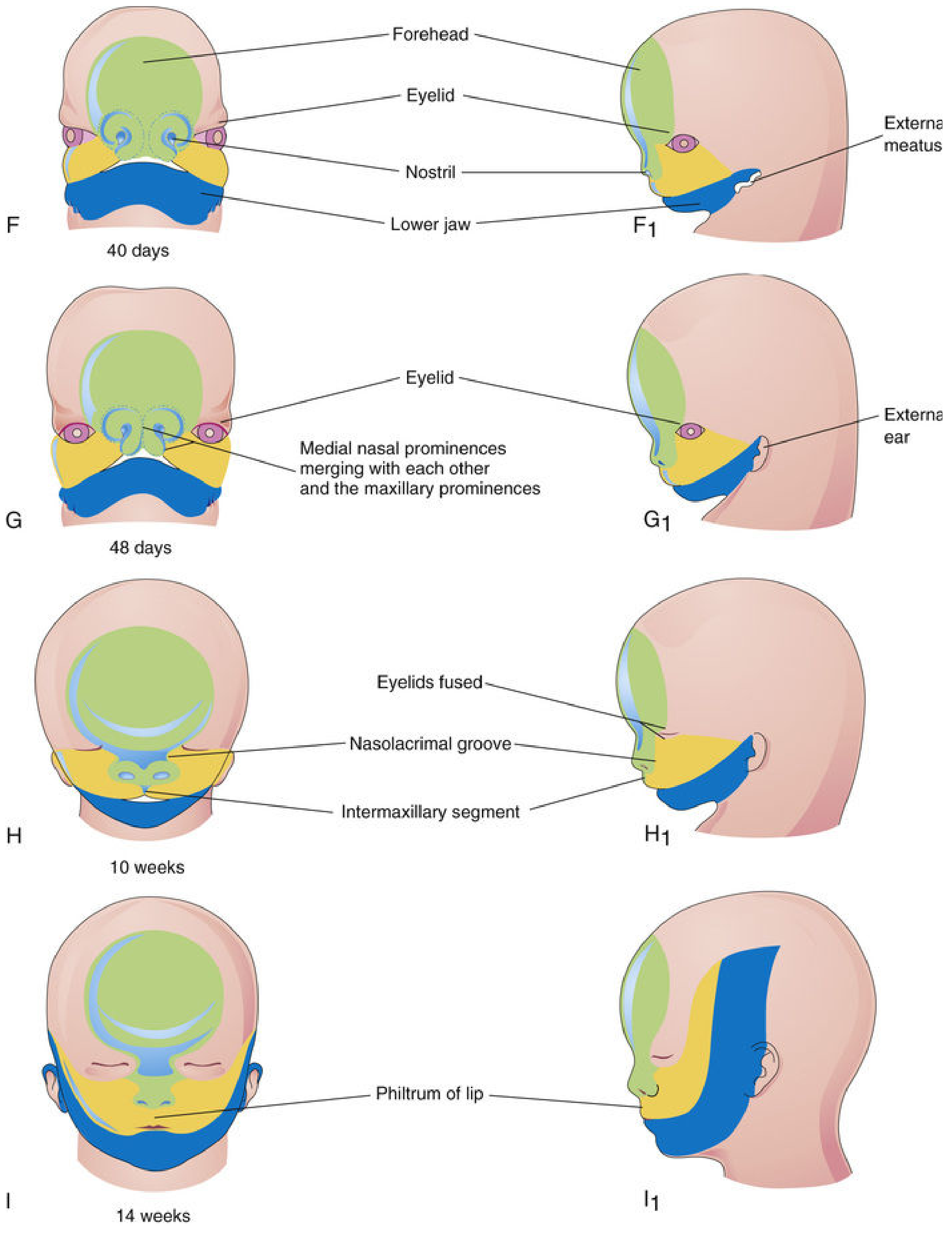
3. Growth and Merging of Prominences (Weeks 5-8)
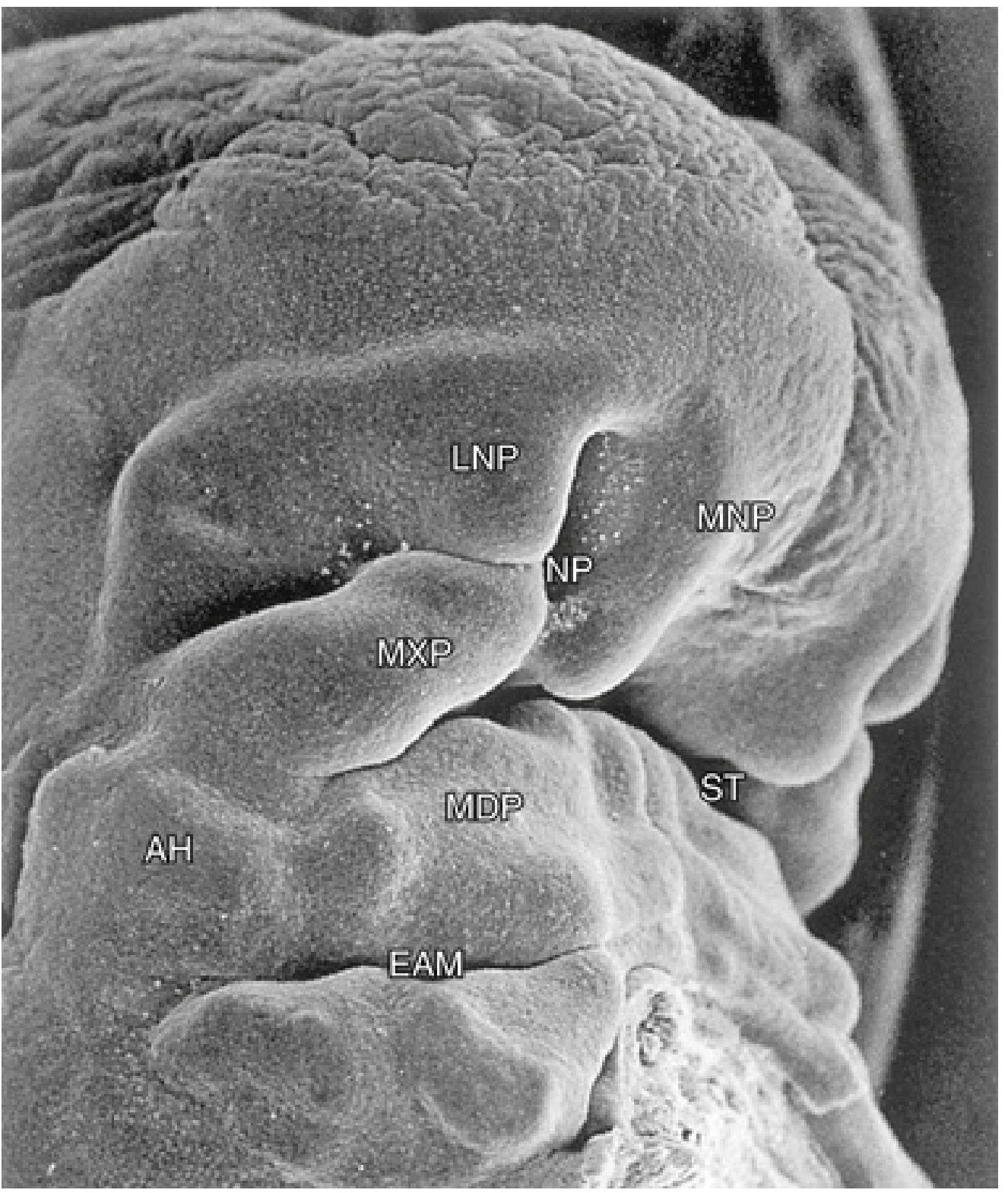
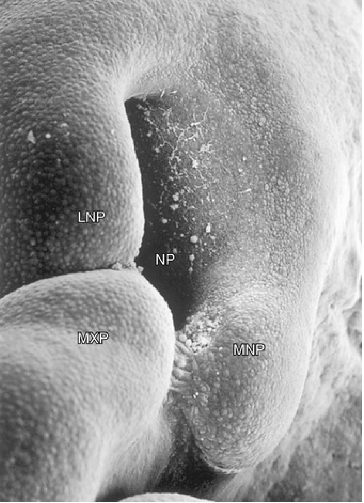
- establishes continuity of the upper jaw and lip
- separates the nasal pits from the stomodeum
- The philtrum (middle part of the upper lip)
- The premaxillary part of the maxilla and its gingiva
- The primary palate
Timeline Summary
| Week | Event |
|---|---|
| 4 (early) | Five facial prominences appear around stomodeum: 1 frontonasal, 2 maxillary, 2 mandibular |
| 4 (late) | Nasal placodes form on frontonasal prominence -> medial & lateral nasal prominences + nasal pits |
| 5 | Maxillary prominences enlarge, grow medially; auricular hillocks appear |
| 6 | Maxillary prominence fuses with lateral nasal prominence (nasolacrimal groove closes) |
| 7-10 | Medial nasal prominences fuse with each other and maxillary prominences -> intermaxillary segment -> philtrum, premaxilla, primary palate |
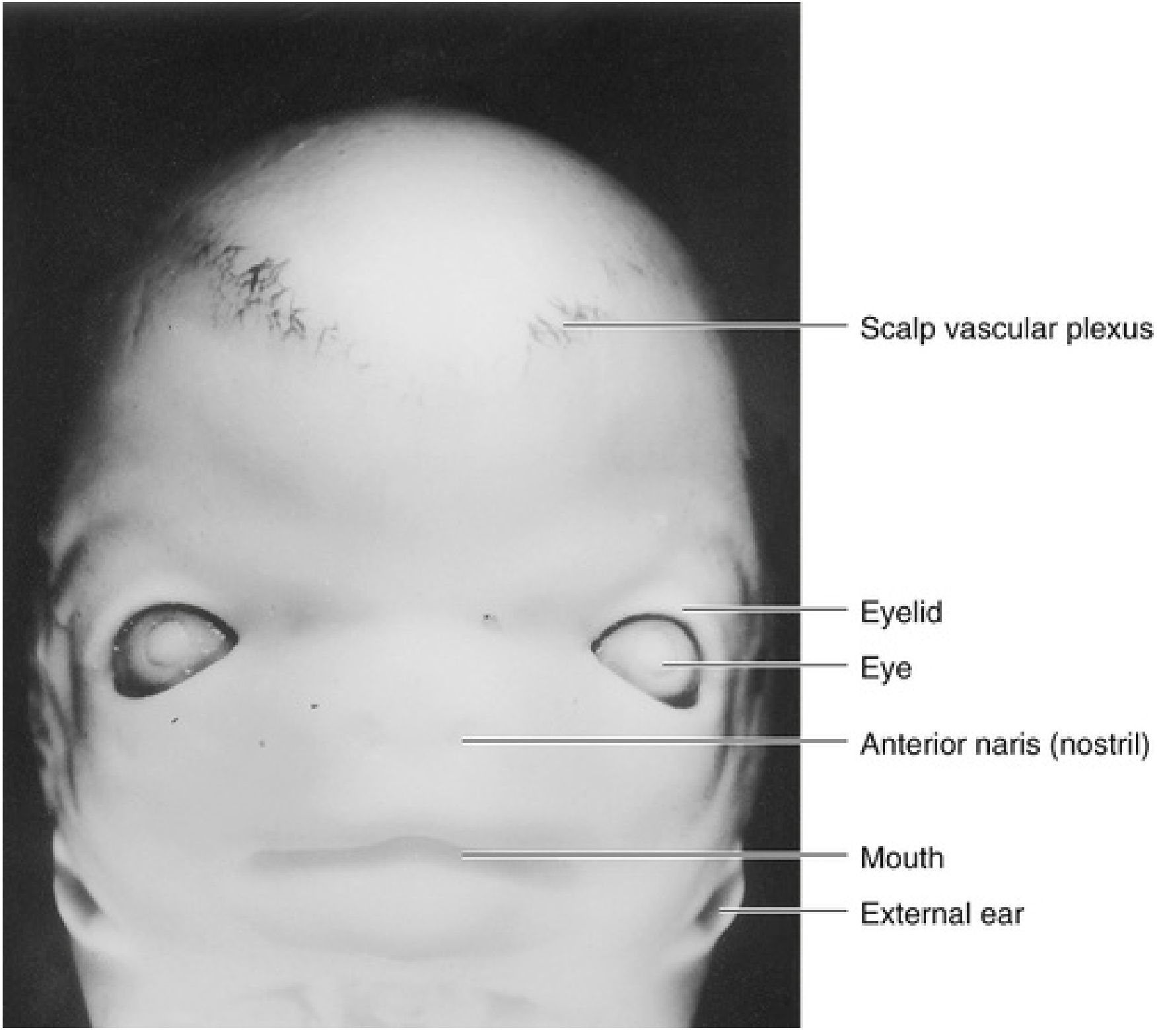
| 8 (end) | Face has recognizably human appearance |
| Fetal period | Refinement of facial proportions (eyes converge, ears ascend) |
Clinical Note
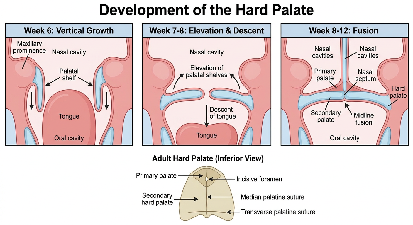
Anomalies and development of hard palate
development of secondary hard palate palatal shelves embryology diagram
cleft palate embryology
cleft palate epidemiology CDC causes treatment
https://www.cdc.gov/birth-defects/about/cleft-lip-cleft-pala…
Development of the Hard Palate and Its Anomalies
What forms the hard palate?
-
Primary palate
- A small triangular part of the adult hard palate anterior to the incisive fossa.
- Derived from the intermaxillary segment, formed by fusion of the two medial nasal prominences.
- Gives rise to the premaxilla, including the region bearing the four incisor teeth.
-
Secondary palate
- Forms the great majority of the hard palate, as well as the soft palate and uvula.
- Derived from the paired lateral palatine processes (palatal shelves) of the maxillary prominences.
- The horizontal bony part becomes the definitive hard palate.
Development: timeline and steps
Week 6: Formation of palatal shelves
- Palatogenesis begins in the sixth week and is normally complete by the twelfth week.
- Two mesenchymal outgrowths, the palatal shelves, arise from the medial aspect of the right and left maxillary prominences.
- Initially, they grow vertically downward on either side of the large developing tongue.
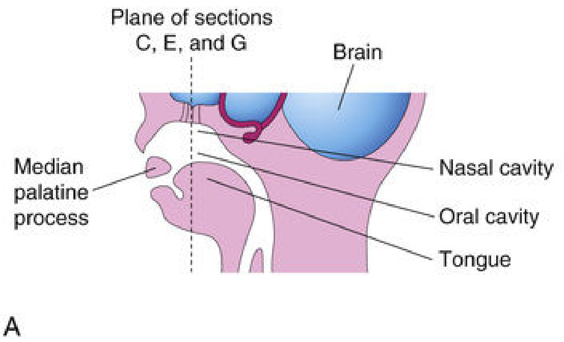
Weeks 7-8: Elevation of palatal shelves
- Growth of the mandible lowers the tongue.
- The palatal shelves elevate from a vertical to a horizontal position above the tongue.
- They grow medially toward each other.
Weeks 8-12: Fusion
- Each other in the midline
- The primary palate anteriorly
- The descending nasal septum superiorly
Ossification of the hard palate
- The hard palate forms by intramembranous ossification.
- There are four main ossification centers:
- One in each maxilla
- One in each palatine bone
- The maxillary and palatine ossification centers appear around the eighth week.
- Incomplete fusion of ossification centers leaves the:
- Median palatine suture
- Transverse palatine suture
Flowchart
Medial nasal prominences
          ↓
Intermaxillary segment
          ↓
Primary palate (anterior to incisive fossa)

Maxillary prominences
          ↓
Paired lateral palatine processes / palatal shelves
          ↓
Initially vertical beside tongue
          ↓
Tongue descends and shelves elevate horizontally
          ↓
Fusion of shelves with each other + primary palate + nasal septum
          ↓
Secondary palate
          ↓
Hard palate anteriorly + soft palate and uvula posteriorly
Cleft palate and other anomalies
1. Cleft palate
Embryological mechanisms
- Failure of palatal shelves to develop
- Failure of shelves to elevate from vertical to horizontal position
- Failure of the shelves to meet in the midline
- Failure of fusion after contact
- Failure of fusion with the primary palate or nasal septum
- Persistent midline epithelial seam
- Mechanical obstruction by a high tongue, such as in mandibular hypoplasia
Classification using the incisive fossa
| Type | Region involved | Embryological basis |
|---|---|---|
| Anterior cleft | Lip, alveolus, primary palate, anterior to incisive fossa | Deficient mesenchyme or failed integration of maxillary prominence and median palatine process |
| Posterior cleft | Secondary palate, hard palate and/or soft palate, posterior to incisive fossa | Defective growth, elevation, apposition, or fusion of palatal shelves |
| Complete cleft palate | Extends from uvula through hard palate to incisive fossa, possibly continuing into alveolus and lip | Combined primary and secondary palatal developmental failure |
| Incomplete cleft palate | Only part of palate involved | Partial failure of palatal fusion |
| Submucous cleft palate | Palatal muscles and bone fail to unite, but mucosa is intact | Defective underlying fusion masked by intact oral and nasal mucosa |
Important clinical forms
Bifid uvula
- Mildest visible form of palatal clefting.
- The uvula is split due to incomplete fusion of its components.
- May indicate an associated submucous cleft palate.
Submucous cleft palate
- Bifid uvula
- Zona pellucida: thin, translucent central soft palate
- Notch at posterior border of hard palate
Pierre Robin sequence
- Characterized by micrognathia, posterior displacement of tongue (glossoptosis), airway obstruction, and frequently cleft palate.
- Small mandible prevents normal lowering of the tongue, obstructing elevation and fusion of the palatal shelves.
Consequences of cleft palate
- Difficulty in sucking and feeding
- Nasal regurgitation of feeds
- Recurrent middle-ear disease and hearing impairment due to Eustachian-tube dysfunction
- Hypernasal speech and articulation difficulties
- Dental malalignment and malocclusion
- Psychosocial effects related to speech, appearance, and repeated treatment
Etiology and risk factors
- Family history and chromosomal or single-gene syndromes
- Maternal smoking
- Alcohol exposure
- Maternal obesity
- Diabetes
- High fever during early pregnancy
- Some medications, particularly selected antiseizure medicines such as valproate and topiramate
- Folate-antagonist drugs and systemic corticosteroids in some settings
Exam points to remember
- Primary palate: from intermaxillary segment, derived from fused medial nasal prominences.
- Secondary palate: from paired palatal shelves of maxillary prominences.
- Hard palate: primary palate plus anterior bony secondary palate.
- Palatal shelves initially lie vertically beside the tongue.
- They elevate horizontally after descent of the tongue, then fuse in the midline.
- Fusion is with each other, primary palate, and nasal septum.
- Palatogenesis: week 6 to week 12; highest susceptibility period is late week 6 to early week 9.
- Cleft anterior to incisive fossa involves primary palate.
- Cleft posterior to incisive fossa indicates secondary-palate defect.
Create a labeled diagram of hard-palate development
A medically accurate, clean labeled educational diagram titled “Development of the Hard Palate”. Create a three-panel embryology sequence using coronal views of the embryonic oral and nasal region. Panel 1: Week 6, paired palatal shelves grow vertically downward from the maxillary prominences on either side of a large central tongue; label nasal cavity, palatal shelf, tongue, maxillary prominence, oral cavity. Panel 2: Week 7 to 8, tongue descends and the palatal shelves elevate to a horizontal position above it; use directional arrows and label elevation of palatal shelves and descent of tongue. Panel 3: Week 8 to 12, horizontal shelves fuse in the midline, anteriorly with the primary palate and superiorly with the nasal septum; label midline fusion, nasal septum, primary palate, secondary palate, hard palate, oral cavity and nasal cavities. Include a small bottom schematic of the final adult hard palate with the primary palate anterior to the incisive foramen, secondary hard palate posterior to it, median palatine suture, and transverse palatine suture. Use precise readable English labels, arrows, and a simple textbook-style anatomical cross-sectional diagram.