
Holoprosencephaly in detail with pictures
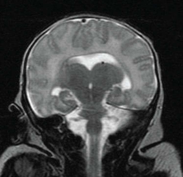
holoprosencephaly types pathology clinical features 2024
holoprosencephaly cyclopia proboscis facial malformations
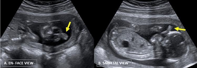
Two B-mode grayscale two-dimensional antenatal ultrasound images demonstrate fetal facial anomalies in a fetus at approximately 16 weeks gestation. (A) The 'en face' view shows a cross-sectional representation of the fetal face with visible orbits and a prominent midline soft tissue projection. (B) The 'sagittal' view provides a side profile clearly illustrating the forehead, nasal region, and oral cavity, with a distinct tubular or cylindrical structure extending outwards from the midline of the forehead. This structure, marked by yellow arrows in both views, is a proboscis, a hallmark facial dysmorphism often associated with holoprosencephaly spectrum disorders such as ethmocephaly or cyclopia. These images are critical for the prenatal diagnosis of midline craniofacial defects and the assessment of associated central nervous system malformations.
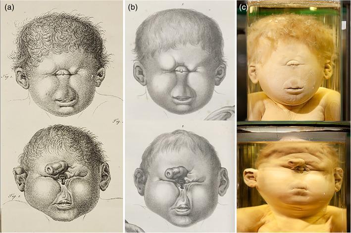
This composite image illustrates severe craniofacial malformations, specifically holoprosencephaly manifestations, comparing historical medical art with preserved pathology specimens. The image is divided into three columns: (a) 19th-century copperplate engravings, (b) lithographs, and (c) color photographs of preserved wet specimens. The top row depicts cyclopia, characterized by a single, centrally located eye (synophthalmia) in a single orbital cavity and an absent nose (arrhinia) with a visible philtrum and mouth. The bottom row demonstrates a more complex cephalic malformation where a single orbital opening is situated below a tubular, fleshy appendage known as a proboscis, which is located in the supraorbital region. These comparisons highlight the evolution of medical illustration from detailed artistic interpretation to modern specimen preservation. The visual focus is on midline facial defects, dysmorphic ears, and the anatomical variations of the proboscis and eye fusion, providing a historical and clinical perspective on teratological study and dysmorphology.
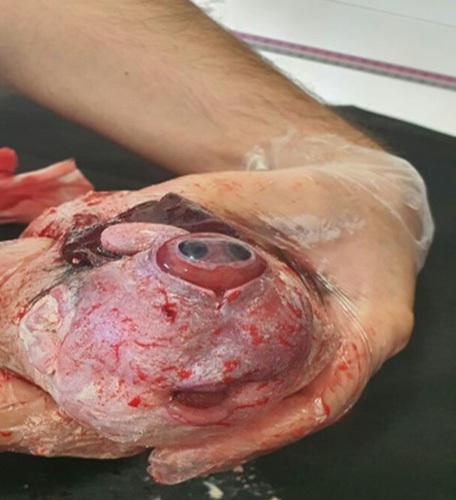
Clinical photograph of a neonatal specimen demonstrating severe craniofacial malformations consistent with holoprosencephaly. The central feature is cyclopia, characterized by a single median orbit located in the midline of the face. The orbit contains a fused or partially divided ocular globe with visible iris and pupil structures, partially covered by rudimentary eyelids. Superior to the orbit, a cylindrical skin-covered appendage known as a proboscis is present. The nose is absent (arrhinia). Inferior to the median eye, a malformed oral cavity is visible, showing a small mouth opening and a glimpse of the tongue. The surrounding facial skin displays significant discoloration, congestion, and visible superficial vasculature. Superior to the proboscis, there is evidence of exposed neural tissue or severe cranial defect, possibly related to anencephaly or a significant skull vault deficiency. This image serves as a clinical example of extreme midline defects arising from the failure of the prosencephalon to divide during embryonic development.
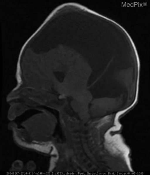
**Imaging Modality:** Sagittal T1-weighted Magnetic Resonance Imaging (MRI).
alobar holoprosencephaly monoventricle fused thalami brain
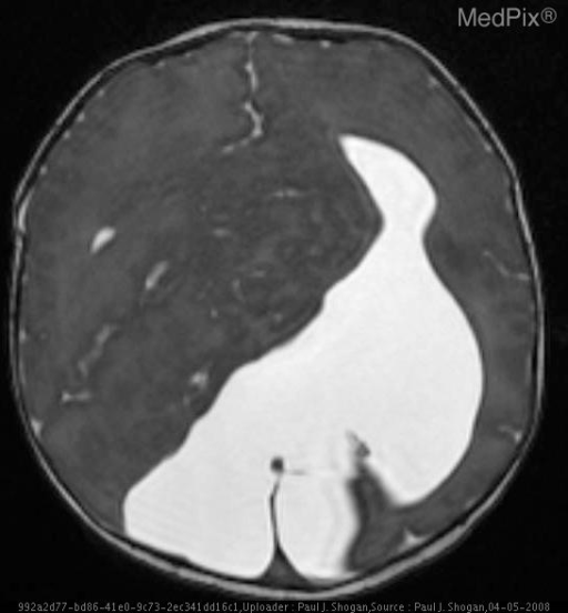
**Imaging Modality:** Axial T2-weighted Magnetic Resonance Imaging (MRI) of the brain.
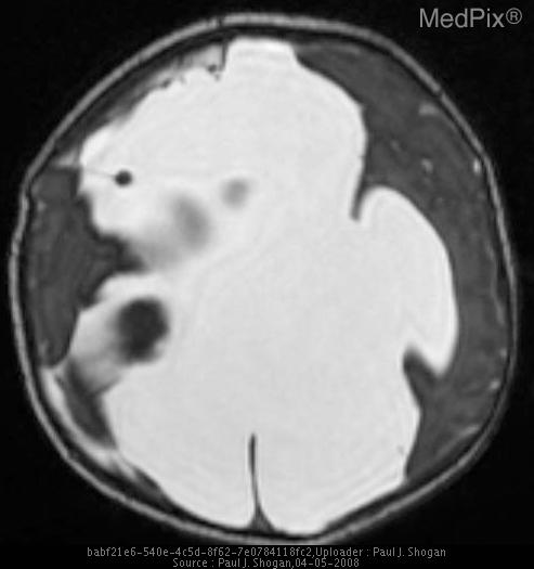
**Imaging Modality:** T2-weighted axial Magnetic Resonance Imaging (MRI) of the brain.
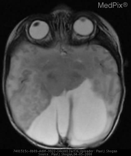
**Imaging Modality:** Axial T2-weighted MRI of the brain.
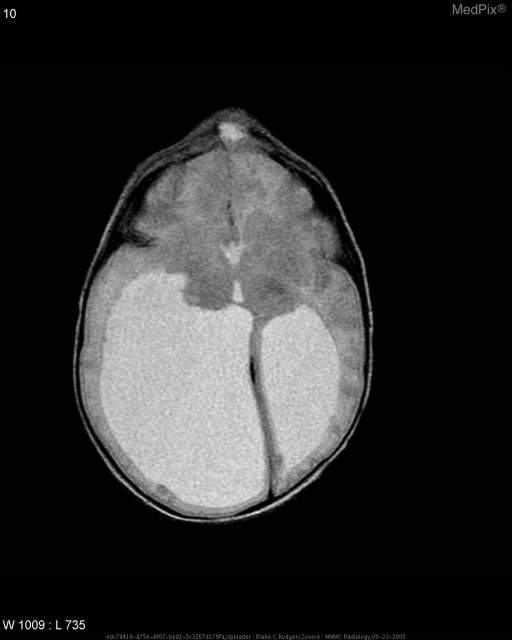
**Imaging Modality:** Non-contrast Computed Tomography (CT) scan of the head.
Create a comprehensive summary document with all HPE classifications