
Structures enclosed in axillarybsheath
Reading File
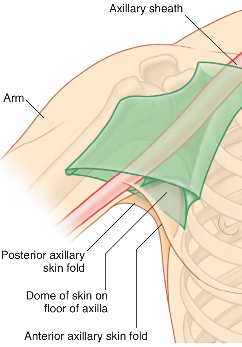
The axillary sheath is a fascial sleeve derived from the prevertebral fascia that encloses the axillary vessels and the brachial plexus. The key structures inside it are the axillary artery, axillary vein, and the three cords (and branches) of the brachial plexus.
Searching sources...
Finding Sources
Finding Sources
Reading File
Reading File
Reading File
Now let me fetch the axillary sheath diagram for visual support:
Loading Image
Finding Sources
Here is a complete, textbook-based answer:
Structures Enclosed in the Axillary Sheath
The axillary sheath is a sleeve-like extension of the prevertebral (deep cervical) fascia. It arises from the anterior and middle scalene muscles in the lower neck and follows the neurovascular bundle as it passes into the axilla, enclosing the following structures:
Contents of the Axillary Sheath
| Structure | Details |
|---|---|
| Axillary artery | Continuation of the subclavian artery; the subclavian artery is also initially enclosed proximally as the sheath forms |
| Axillary vein | Lies medial to the axillary artery within the sheath |
| Brachial plexus (cords and terminal/collateral branches) | Surrounds the axillary artery inside the sheath |
These three - axillary artery, axillary vein, and brachial plexus - are the primary structures enclosed.
"The axillary artery, axillary vein, and brachial plexus lie within the sleeve-like covering of fascia termed the axillary sheath."
- Gray's Anatomy for Students
"The prevertebral fascia in an anterolateral position extends from the anterior and middle scalene muscles to surround the brachial plexus and subclavian artery as these structures pass into the axilla. This fascial extension is the axillary sheath."
- Gray's Anatomy for Students
Origin and Formation
The sheath is an extension of the prevertebral fascia (not the deep fascia of the arm). Proximally, it encloses the subclavian artery and roots/trunks of the brachial plexus as they emerge between the scalene muscles. Distally, it becomes the sheath around the axillary vessels and the cords/branches of the brachial plexus.
Clinical Relevance: Axillary Nerve Block
Because the axillary sheath forms a continuous fascial compartment, injecting local anesthetic into the sheath at the axilla (guided by palpating the axillary artery pulse with the arm abducted and externally rotated) distributes the anesthetic to all branches of the brachial plexus simultaneously - producing a brachial plexus nerve block for procedures on the hand, wrist, and forearm.
A tourniquet may be applied 2-3 cm distal to pectoralis major insertion to limit distal spread of the anesthetic and improve proximal diffusion.
Potential complications include:
- Direct needle injury to brachial plexus branches
- Axillary artery puncture
- Inadvertent intra-arterial injection of local anesthetic
(Sources: Gray's Anatomy for Students; Fischer's Mastery of Surgery, 8th ed.; Pye's Surgical Handicraft, 22nd ed.)
Memory Aid
"AVB in the sheath" - Axillary artery, Vein, Brachial plexus.
This is a shared conversation. Sign in to Orris to start your own chat.