
Hey, Please give some idea for vitamins physiology models.
Reading File
Vitamins physiology covers fat-soluble (A, D, E, K) and water-soluble (B-complex, C) vitamins - their absorption, transport, mechanisms of action, deficiency states, and toxicities. Let me search the library for rich, textbook-level content on this.
Searching sources...
Finding Sources
Finding Sources
Finding Sources
Good results. Now let me pull content from the most physiology-focused sources in parallel.
Reading File
Reading File
Reading File
Excellent content retrieved. Now let me get more detail on individual vitamins - especially the B-complex mechanisms and Vitamin D pathway - from Harper's Biochemistry and Basic Medical Biochemistry.
Reading File
Reading File
I now have rich, textbook-level content from multiple authoritative sources. Let me also fetch the thiamin molecular image from Harper's to include in the response.
Loading Image
Finding Sources
I now have comprehensive material to provide a thorough, well-organized answer. Here it is:
Vitamin Physiology - Key Concepts & Models
Vitamins are micronutrients that the body cannot synthesize in adequate quantities and must obtain from the diet. They are divided into fat-soluble (A, D, E, K) and water-soluble (B-complex, C) groups, each with distinct absorption mechanisms, physiological roles, and deficiency states.
1. ABSORPTION MODELS
Fat-Soluble Vitamins (A, D, E, K) - Lipid Micelle Model
Fat-soluble vitamins are processed exactly like dietary lipids:
- Incorporated into micelles in the intestinal lumen
- Transported to the apical membrane of enterocytes
- Diffuse across the membrane → packaged into chylomicrons
- Extruded into lymph → delivered to systemic circulation via the thoracic duct
Vitamin A specifically uses membrane protein STRA6 for retinol uptake, while carotenoids enter via SR-B1, CD36, and NPC1L1. Inside the cell, retinol binds CRBP2 and is re-esterified by LRAT before chylomicron packaging.
- Costanzo Physiology 7th Edition, p. 3098
- Yamada's Textbook of Gastroenterology, p. 189
Water-Soluble Vitamins - Na⁺-Cotransport Model
Most water-soluble vitamins are absorbed via Na⁺-dependent cotransport in the small intestine.
Vitamin B12 (cobalamin) - the exception: This has a unique, multi-step model:
- Pepsin in the stomach releases dietary B12 from food
- Free B12 binds R proteins (haptocorrin) secreted in saliva
- Pancreatic proteases in the duodenum degrade R proteins → B12 transfers to intrinsic factor (IF), a glycoprotein from gastric parietal cells
- The B12-IF complex resists protease digestion and travels to the ileum, where specific receptors mediate absorption
Clinical consequence: After gastrectomy, loss of parietal cells = no intrinsic factor → B12 malabsorption → pernicious anemia. Oral B12 is useless - it must be given by injection.
- Costanzo Physiology 7th Edition, p. 3103-3107
2. VITAMIN D ACTIVATION CASCADE
Vitamin D follows a two-step hydroxylation model before becoming biologically active:
Dietary Vitamin D3 (Cholecalciferol) — INACTIVE
         ↓  Liver (cytochrome P-450 enzyme)
25-Hydroxycholecalciferol (25-OHD) — INACTIVE (major circulating form)
         ↓  Kidney proximal tubule (1α-hydroxylase)
1,25-Dihydroxycholecalciferol (Calcitriol) — ACTIVE
Active Calcitriol actions:
-
Induces synthesis of calbindin D-28K (vitamin D-dependent Ca²⁺-binding protein) in intestinal epithelial cells → promotes Ca²⁺ absorption
-
Acts on kidney and bone for calcium homeostasis
-
The renal 1α-hydroxylation step is lost in CKD → declining calcitriol levels
-
Costanzo Physiology 7th Edition, p. 3112
-
Harrison's Principles of Internal Medicine 22E, p. 822-828
3. B-VITAMIN COENZYME MODELS (Harper's Biochemistry)
Each B vitamin functions as a coenzyme or coenzyme precursor in specific metabolic pathways:
| Vitamin | Active Coenzyme | Key Metabolic Role | Deficiency Syndrome |
|---|---|---|---|
| B1 (Thiamin) | Thiamin diphosphate (TDP) | Pyruvate dehydrogenase, α-ketoglutarate DH, branched-chain keto acid DH; transketolase in pentose phosphate pathway | Beriberi (dry/wet), Wernicke-Korsakoff syndrome |
| B2 (Riboflavin) | FMN, FAD | Mitochondrial respiratory chain, fatty acid oxidation, citric acid cycle | Cheilosis, glossitis, seborrheic dermatitis (ariboflavinosis) |
| Niacin (B3) | NAD, NADP | Oxidation/reduction across metabolism; ADP-ribosylation for DNA repair; cyclic ADP-ribose signaling | Pellagra (3 Ds: Dermatitis, Diarrhea, Dementia) |
| B6 (Pyridoxine) | Pyridoxal phosphate | Transamination, decarboxylation of amino acids; heme synthesis | Seborrheic dermatitis, microcytic anemia, convulsions |
| B12 (Cobalamin) | Methylcobalamin, adenosylcobalamin | Methionine synthesis, methylmalonyl-CoA → succinyl-CoA | Megaloblastic anemia, subacute combined degeneration of spinal cord |
| Folate (B9) | Tetrahydrofolate (THF) | One-carbon transfers; nucleotide synthesis; cell division | Megaloblastic anemia; neural tube defects |
| Biotin | Biocytin | Carboxylation reactions (pyruvate carboxylase, acetyl-CoA carboxylase) | Alopecia, dermatitis, CNS abnormalities |
| Pantothenic acid | Coenzyme A (CoA) | Acyl group transfer in fatty acid and carbohydrate metabolism | Fatigue, GI symptoms, neurological symptoms |
Thiamin - Structure
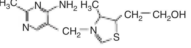
- Harper's Illustrated Biochemistry 32nd Ed., p. 553-554
Niacin note: Niacin is not strictly a vitamin - it can be synthesized from the essential amino acid tryptophan (60 mg Trp = 1 mg niacin). Its NAD is also the substrate for ADP-ribosylation (DNA repair) and produces cyclic ADP-ribose, which mobilizes intracellular calcium in response to hormones/neurotransmitters.
4. VITAMIN K - CARBOXYLATION MODEL
Vitamin K acts as cofactor for γ-carboxylase, which converts glutamate (Glu) residues to γ-carboxyglutamate (Gla) in clotting factors and other proteins.
Proteins dependent on this reaction:
- Coagulation factors: II (prothrombin), VII, IX, X (all require Gla for Ca²⁺ binding and membrane interaction)
- Anticoagulant proteins: Protein C, Protein S
- Bone proteins: Osteocalcin, matrix Gla protein - bind calcium, causing a conformational change to interact with membrane phospholipids
- Gas6: Controls apoptosis and differentiation in nervous system
Osteocalcin levels in circulation serve as an index of vitamin D status.
- Harper's Illustrated Biochemistry 32nd Ed., p. 321-323
5. VITAMIN A - VISUAL CYCLE MODEL
Vitamin A (retinol) is essential for:
- Vision (rhodopsin cycle in rods)
- Cell differentiation and proliferation (via retinoic acid nuclear receptors RAR/RXR)
- Immune integrity
The retinol is stored in large quantities in the liver. Mutations in LRAT (the esterification enzyme) cause Leber congenital amaurosis and retinitis pigmentosa; mutations in RBP (retinol-binding protein) cause night blindness.
6. VITAMIN E - ANTIOXIDANT MODEL
Vitamin E (α-tocopherol) acts as a lipid-soluble chain-breaking antioxidant that scavenges free radicals and protects cell membranes from lipid peroxidation.
- Uptake: via NPC1L1 and SR-BI at the apical enterocyte membrane
- Secretion: via ABCA1 (apoA1 pathway) and chylomicrons (apoB pathway)
- Deficiency causes: reduced RBC lifespan, spinocerebellar ataxia, loss of deep tendon reflexes, myopathies, night blindness
Ataxia with isolated vitamin E deficiency (AVED) is caused by mutations in the α-tocopherol transfer protein (α-TTP), preventing vitamin E incorporation into hepatic lipoproteins.
- Yamada's Textbook of Gastroenterology, p. 194-198
7. VITAMIN C - COLLAGEN SYNTHESIS MODEL
Vitamin C (ascorbic acid) is the cofactor for prolyl hydroxylase and lysyl hydroxylase - enzymes that hydroxylate proline and lysine residues in procollagen. This hydroxylation is essential for:
- Cross-linking of collagen fibers (structural integrity)
- Stability of the triple helix
Scurvy (vitamin C deficiency) results from defective collagen formation, leading to:
-
Subcutaneous hemorrhage, perifollicular hemorrhages
-
Swollen, bleeding gums
-
Poor wound healing
-
Aching bones and joints
-
Basic Medical Biochemistry - A Clinical Approach 6e
Quick Reference - Deficiency Models
| Vitamin | Classic Deficiency Mnemonic/Model |
|---|---|
| A | Night blindness → Xerophthalmia → Bitot's spots |
| D | Rickets (children) / Osteomalacia (adults) |
| E | Spinocerebellar ataxia, areflexia |
| K | Bleeding diathesis (PT prolonged) |
| B1 | Beriberi (wet = cardiac; dry = neural) / Wernicke-Korsakoff |
| B2 | Ariboflavinosis - "cheilosis, corneal vascularization" |
| B3 | Pellagra - "4 Ds: Dermatitis, Diarrhea, Dementia, Death" |
| B6 | Sideroblastic anemia, peripheral neuropathy |
| B9 | Megaloblastic anemia + neural tube defects |
| B12 | Megaloblastic anemia + subacute combined degeneration |
| C | Scurvy - perifollicular hemorrhage, corkscrew hairs |
These models cover the major physiological frameworks used to understand vitamins: absorption (micelle vs. Na⁺-cotransport), activation cascades (Vitamin D), coenzyme function (B-vitamins), antioxidant protection (E), carboxylation (K), and biosynthetic cofactor roles (C). Each provides a distinct teaching model that integrates biochemistry, physiology, and clinical medicine.
This is a shared conversation. Sign in to Orris to start your own chat.