
Maternal pelvis
Reading File
Finding Sources
Finding Sources
Searching Images
diagram maternal pelvis obstetric pelvic inlet midpelvis outlet landmarks
Searching PubMed
maternal pelvis obstetric
Searching the Web
maternal pelvis obstetric anatomy pelvic inlet midpelvis outlet authoritative
Reading File
Reading File
Reading File
Maternal pelvis
The maternal pelvis is the bony passage through which the fetus passes during vaginal birth. It consists of two hip bones, the sacrum, and coccyx.
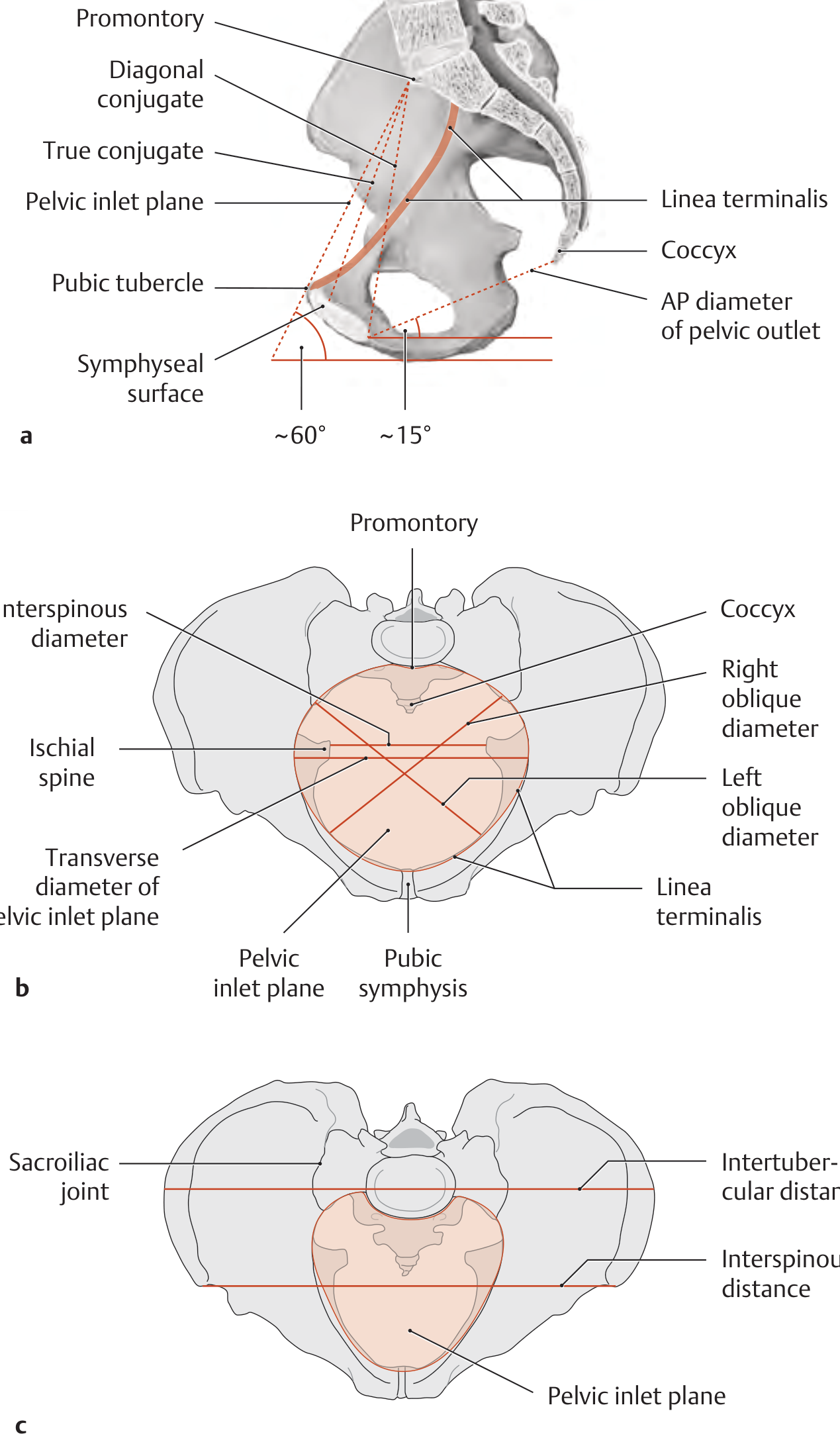
Divisions
-
False (greater) pelvis
- Above the pelvic brim
- Supports the enlarging uterus in pregnancy
- Not part of the bony birth canal
-
True (lesser) pelvis
- Below the pelvic brim
- Forms the bony birth canal
- Has three clinically important planes: inlet, cavity/midpelvis, and outlet
1. Pelvic inlet
The inlet is bounded by:
- Posteriorly: sacral promontory
- Laterally: linea terminalis
- Anteriorly: upper border of symphysis pubis
Important diameters
- Obstetric conjugate: shortest anteroposterior diameter, from sacral promontory to the posterior surface of the symphysis pubis. About 11 cm.
- Diagonal conjugate: sacral promontory to inferior border of symphysis pubis. About 12.5-13 cm and can be estimated clinically.
- Transverse diameter: about 13 cm.
Engagement occurs when the fetal biparietal diameter has passed the pelvic inlet. General Anatomy and Musculoskeletal System (THIEME Atlas of Anatomy), p. 161.
2. Midpelvis or pelvic cavity
- Extends from the inlet to the level of the ischial spines.
- The ischial spines are key obstetric landmarks.
- The interspinous diameter is the narrowest transverse diameter of the pelvis, usually about 10-11 cm.
- Fetal station is assessed in relation to the ischial spines:
- Station 0: at the spines
- Negative stations: above
- Positive stations: below
3. Pelvic outlet
Boundaries:
- Anteriorly: lower border of symphysis pubis and pubic arch
- Laterally: ischiopubic rami and ischial tuberosities
- Posteriorly: sacrotuberous ligaments and tip of coccyx
The anteroposterior outlet diameter is approximately 9 cm, but can increase by about 2 cm as the coccyx moves backward during birth. General Anatomy and Musculoskeletal System (THIEME Atlas of Anatomy), p. 161.
Pelvic types
| Type | Features | Obstetric relevance |
|---|---|---|
| Gynecoid | Rounded inlet, wide subpubic arch, non-prominent ischial spines | Most favorable for vaginal birth |
| Android | Heart-shaped inlet, narrow anterior pelvis | May cause deep transverse arrest or persistent occipitoposterior position |
| Anthropoid | Oval inlet with long AP diameter | Occipitoposterior position is more common |
| Platypelloid | Broad transverse inlet and short AP diameter | Engagement may be difficult |
Obstetric importance
The fetal head must negotiate changing pelvic dimensions: the inlet is relatively widest transversely, while the outlet has a relatively greater anteroposterior dimension. This contributes to the fetal head’s internal rotation during normal labor. The pelvis is also not completely rigid: the symphysis pubis, sacroiliac joints, and sacrococcygeal joint gain some mobility in pregnancy under hormonal influence. Berek & Novak’s Gynecology, p. 150.
This is a shared conversation. Sign in to Orris to start your own chat.