
Pharyngeal arches
pharyngeal arches embryology diagram derivatives
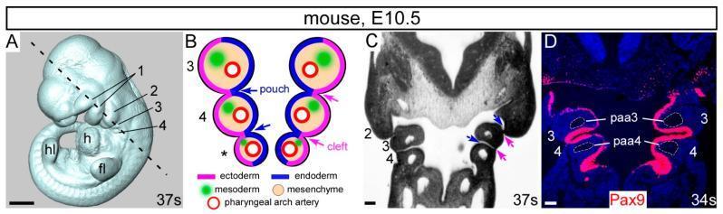
This composite educational image illustrates the developmental anatomy of the pharyngeal arches in a mouse embryo at embryonic day 10.5 (E10.5). (A) A 3D whole-mount embryo reconstruction showing anatomical landmarks including the forelimb bud (fl), hindlimb bud (hl), heart (h), and the sequential pharyngeal arches (1-4). (B) A schematic diagram of a coronal section detailing the embryological layers: outer ectoderm (pink), internal endoderm (blue) forming pouches, and a mesenchymal core containing mesoderm (green) and pharyngeal arch arteries (red circles). (C) A corresponding histological coronal section showing the physical arrangement of arches 2, 3, and 4. (D) An immunofluorescence-stained coronal section showing Pax9 expression (red signal) localized within the pharyngeal endoderm. The image highlights the specific positioning of the 3rd and 4th pharyngeal arch arteries (paa3, paa4) within their respective arches. This material demonstrates key concepts in embryology, specifically the rostral-caudal development of the pharyngeal apparatus and the molecular markers of the pharyngeal endoderm essential for cardiovascular and craniofacial morphogenesis.
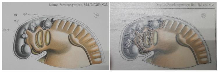
This comparative anatomical diagram illustrates the embryological development of the pharyngeal arches, using a stage 33 lungfish embryo as a model for vertebrate morphogenesis. The image is presented in two panels: the left displays a shaded specimen with key landmarks labeled, including 'Gl. Pl.' (Glandular Plate), 'M.H.' (Midbrain/Mittelhirn), and 'Ggl. max. md.' (Maxillomandibular ganglion). The right panel utilizes a historic transparent overlay method to demonstrate the migration of neural crest cells, represented by small black ovals. These cells are concentrated along the cephalic region and the dorsal-ventral axis of the pharyngeal arches, which appear as segmented bulges. The illustration highlights the pathways of migratory cells from the neural tube into the pharyngeal apparatus, which eventually contribute to the development of the aorticopulmonary septum, cardiac outflow tract, and craniofacial structures. This comparison is an educational resource for developmental biology and embryology, focusing on neural crest cell distribution and the evolution of vertebrate circulatory systems.
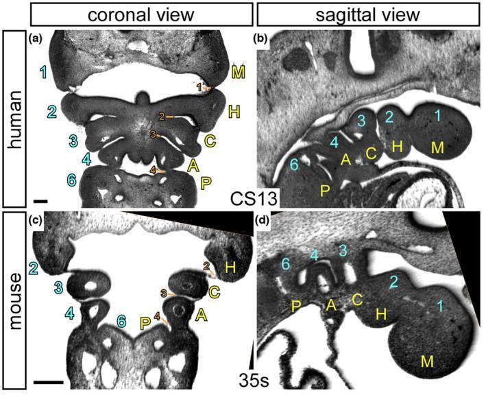
High-resolution episcopic microscopy (HREM) images illustrating the comparative embryological anatomy of the pharyngeal region in a human embryo (Carnegie Stage 13) and a mouse embryo (35 somites). The plate includes coronal (transverse) and sagittal sections, highlighting the segmental organization of pharyngeal arches and pouches. The images contrast the traditional numerical nomenclature (1, 2, 3, 4, 6) with a proposed descriptive naming system: Mandibular (M), Hyoid (H), Carotid (C), Aortic (A), and Pulmonary (P). In the human coronal view (a), bilateral symmetry of the arches is visible, while the sagittal view (b) clearly depicts the cranial-to-caudal sequence of these structures. Pharyngeal pouches are numerically labeled (1-4, 6) in orange, representing endodermal outpocketings between the arches. This visual resource is designed for developmental biology and embryology education, emphasizing that there are five distinct arches formed during normal amniote development, which has significant implications for understanding congenital cardiac malformations and the derivatives of the pharyngeal apparatus.
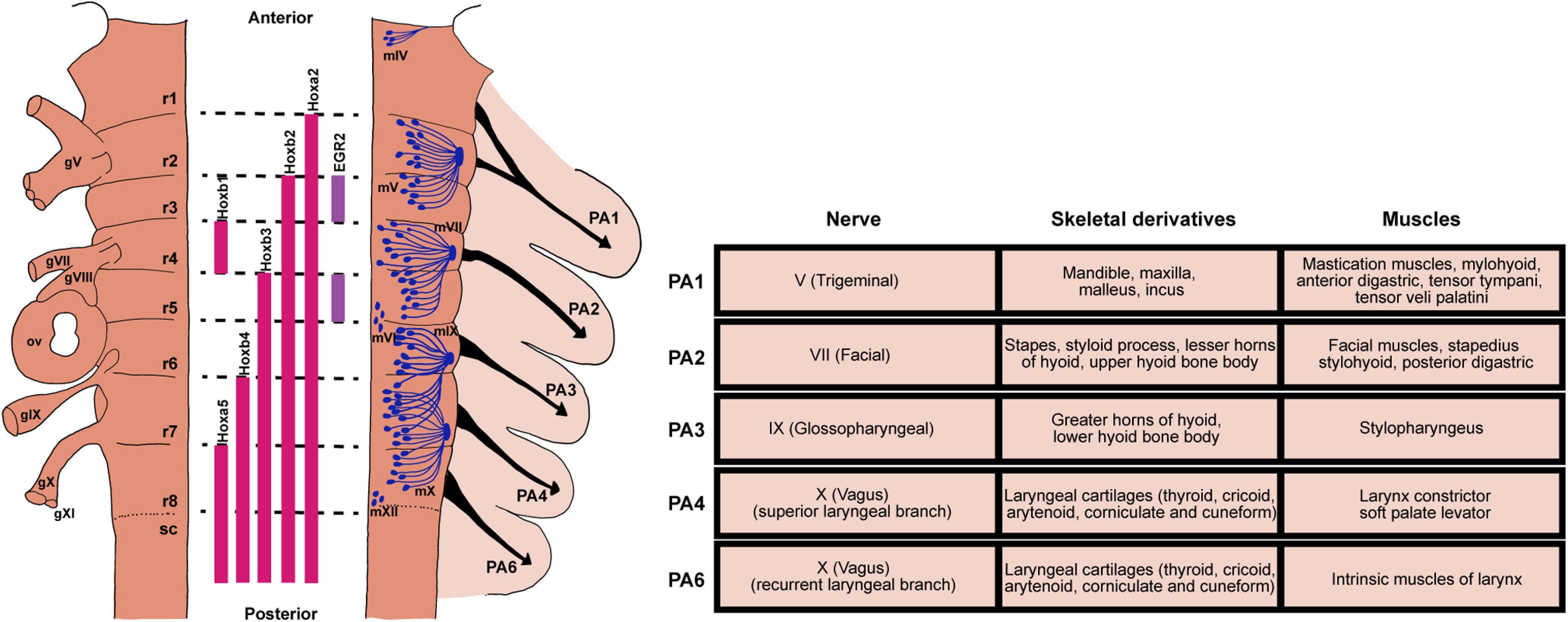
This medical illustration depicts the embryological development of the vertebrate hindbrain and pharyngeal arches (PAs). The left panel presents a schematic of the segmented hindbrain, divided into eight rhombomeres (r1–r8). It highlights the nested expression patterns of Hox genes (Hoxa2, Hoxb2, Hoxb3, Hoxb4, Hoxa5) and transcription factors like EGR2 (Krox-20) that define segmental identity. Laterally, it shows the spatial relationship with cranial sensory ganglia (gV, gVII–gXI) and the otic vesicle (ov). The distribution of motor nuclei (mV, mVII, mIX, mX, mXII) and neural crest cell migration toward specific PAs (PA1, PA2, PA3, PA4, PA6) are also mapped. The right panel features a detailed comparison table summarizing the derivatives of each pharyngeal arch. It lists the associated cranial nerves (Trigeminal V, Facial VII, Glossopharyngeal IX, and Vagus X branches), skeletal derivatives (e.g., mandible, stapes, hyoid, laryngeal cartilages), and specific muscular developments (e.g., muscles of mastication, facial expression, and larynx). This infographic serves as a comprehensive educational guide for understanding cranial nerve innervation and musculoskeletal embryology.
Pharyngeal (Branchial) Arches
Overview
- The Developing Human (Moore), p. 463-487
- Langman's Medical Embryology, p. 306-312
Structure of Each Arch
| Component | Origin | Role |
|---|---|---|
| Cartilage / Skeletal core | Neural crest cells | Gives rise to bones, cartilage, ligaments |
| Artery | Lateral mesoderm + 2nd heart field (arches 3-6) | Connects truncus arteriosus to dorsal aorta |
| Muscle primordium | Paraxial (myogenic) mesoderm | Forms specific head/neck muscles |
| Nerve | Cranial nerve for that arch | Always innervates derivatives of its own arch |
- The Developing Human, p. 463
Derivatives of Each Arch

Arch 1 - Mandibular Arch (CN V - Trigeminal)
- Proximal end → Malleus + Incus (middle ear ossicles)
- Middle part regresses; perichondrium → anterior ligament of malleus + sphenomandibular ligament
- Ventral end acts as a template; mandible forms by intramembranous ossification of surrounding mesenchyme (not from the cartilage directly)
- Maxillary process mesenchyme → premaxilla, maxilla, zygomatic bone, part of temporal bone
- Muscles of mastication (temporalis, masseter, medial + lateral pterygoids)
- Mylohyoid, anterior belly of digastric
- Tensor tympani, tensor veli palatini
- Note: sensory supply to face skin from all 3 branches of CN V (V1, V2, V3)
Arch 2 - Hyoid Arch (CN VII - Facial)
- Stapes (middle ear ossicle)
- Styloid process of temporal bone
- Stylohyoid ligament
- Upper part of body and lesser cornu of hyoid
- Muscles of facial expression
- Stapedius, stylohyoid, posterior belly of digastric
- Auricular muscles
Arch 3 (CN IX - Glossopharyngeal)
- Lower part of body and greater cornu of hyoid
- Stylopharyngeus (the only muscle)
Arch 4 (CN X superior laryngeal branch - Vagus)
- Thyroid cartilage (superior cornu)
- Fuses with arch 6 derivatives
- Cricothyroid, levator veli palatini
- Constrictors of the pharynx
Arch 6 (CN X recurrent laryngeal branch - Vagus)
- Cricoid, arytenoid, corniculate, cuneiform cartilages
- Intrinsic muscles of the larynx
- Striated muscles of the esophagus
Summary Table
| Arch | Nerve | Skeletal Derivatives | Muscles |
|---|---|---|---|
| 1 (Mandibular) | CN V3 | Malleus, incus, mandible, maxilla, zygomatic, temporal | Mastication, mylohyoid, ant. digastric, tensor tympani, tensor veli palatini |
| 2 (Hyoid) | CN VII | Stapes, styloid process, stylohyoid lig., lesser cornu + upper body of hyoid | Facial expression, stapedius, stylohyoid, post. digastric |
| 3 | CN IX | Greater cornu + lower body of hyoid | Stylopharyngeus |
| 4 | CN X (superior laryngeal) | Thyroid cartilage, superior cornu | Cricothyroid, levator veli palatini, pharyngeal constrictors |
| 6 | CN X (recurrent laryngeal) | Cricoid, arytenoid, corniculate, cuneiform cartilages | Intrinsic laryngeal muscles, striated esophagus |
- The Developing Human, Table 9.1, p. 467
- Langman's Medical Embryology, p. 308-309
Pharyngeal Pouches (Internal - Endodermal)
| Pouch | Derivatives |
|---|---|
| 1 | Tympanic (middle ear) cavity; pharyngotympanic (Eustachian) tube |
| 2 | Palatine tonsils; tonsillar fossa |
| 3 | Inferior parathyroid gland (dorsal wing); Thymus (ventral wing) |
| 4 | Superior parathyroid gland; ultimobranchial body → parafollicular (C) cells of thyroid |
Memory tip: Pouches 3 and 4 are "crossed" - pouch 3 gives the inferior parathyroid (which migrates further caudally with the thymus) and pouch 4 gives the superior parathyroid.
- Langman's Medical Embryology, Table 17.2, p. 310
Pharyngeal Grooves (External - Ectodermal)
- There are 4 pairs of ectodermal grooves between the arches.
- Only the 1st groove contributes to adult structures → becomes the external acoustic meatus (ear canal).
- Grooves 2-4 are buried by the overgrowth of the 2nd arch, forming the cervical sinus, which is normally obliterated.
Pharyngeal Membranes
- Form where the ectoderm of grooves meets the endoderm of pouches.
- Only the 1st membrane persists → becomes the tympanic membrane (eardrum).
Clinical Correlates
Branchial (Cervical) Sinus / Fistula / Cyst
- Most result from failure of the 2nd groove and cervical sinus to obliterate.
- Opens along the anterior border of the sternocleidomastoid in the inferior third of the neck.
- Bilateral in ~10% of affected neonates.
- The Developing Human, p. 479
First Pharyngeal Arch Syndrome
- Caused by insufficient migration of neural crest cells into the first arch in the 4th week.
- Two main forms:
- Treacher Collins syndrome (mandibulofacial dysostosis): autosomal dominant, mutation in TCOF1 gene (encodes "treacle" protein involved in rRNA biogenesis); malar hypoplasia, down-slanting palpebral fissures, lower eyelid colobomas, deformed external ears.
- Pierre Robin sequence: micrognathia → posterior tongue displacement → bilateral cleft palate.
- The Developing Human, p. 486-487
DiGeorge Syndrome (22q11.2 deletion)
- Defective development of 3rd and 4th pharyngeal pouches.
- Results in: absent thymus + absent parathyroids → T-cell immunodeficiency + hypoparathyroidism (hypocalcemia) + cardiac outflow tract defects.