
Bells palsy
Bell's palsy treatment corticosteroids antivirals
Bell's palsy facial nerve palsy clinical features
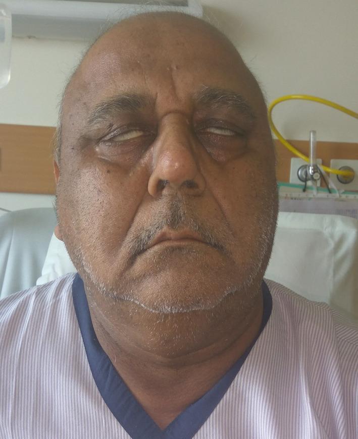
This clinical photograph shows the frontal view of an elderly male patient exhibiting signs of bilateral lower motor neuron facial nerve palsy (Bell's palsy). A prominent finding is Bell's phenomenon, where the patient's eyeballs rotate upward and outward when attempting to close the eyes, leaving the sclera visible due to incomplete eyelid closure (lagophthalmos). The facial features appear relatively symmetrical but mask-like, with a loss of normal expression lines. The mouth is slightly open with evidence of drooling at the commissures, consistent with orbicularis oris muscle weakness. This presentation is highly suggestive of cranial nerve VII involvement, often associated with systemic conditions such as Guillain-Barr syndrome, sarcoidosis, or leptomeningeal metastasis. The image is an educational resource for demonstrating neurological deficits and the physical examination findings of bilateral facial paralysis.
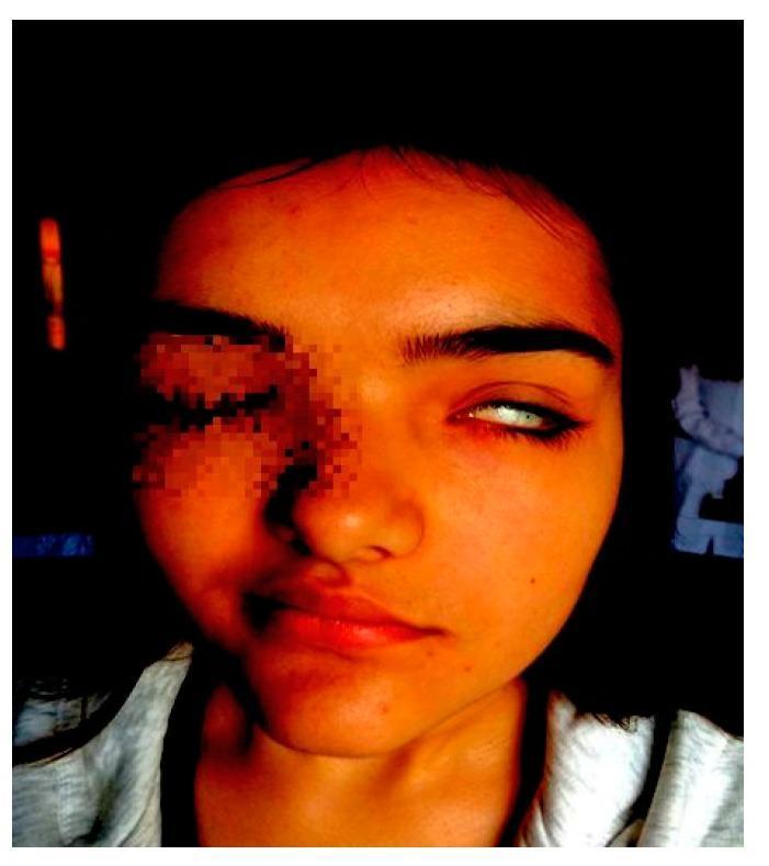
This clinical photograph of a pediatric patient demonstrates classic features of left-sided peripheral facial nerve palsy (Bell's palsy), commonly associated with Melkersson-Rosenthal Syndrome in this context. The image illustrates significant facial asymmetry: the left eye exhibits lagophthalmos (incomplete closure) with notable scleral show (Bell's phenomenon), where the eyeball rolls upward as the patient attempts to close the eye. On the lower face, there is a distinct obliteration of the left nasolabial fold and a compensatory right-sided deviation of the labial commissure, resulting from the loss of muscle tone on the paralyzed left side. These findings represent a House-Brackmann grade 4 dysfunction. The image serves as a visual diagnostic tool for identifying lower motor neuron facial weakness, characterized by the involvement of both the upper and lower facial muscles on the affected side.
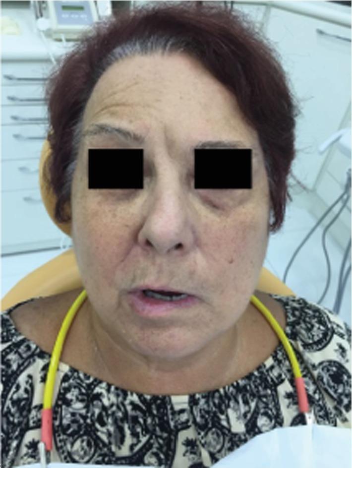
A frontal clinical photograph of an elderly female patient demonstrating symptoms of unilateral facial nerve paralysis, specifically Bell's palsy, in a pretreatment state. The image illustrates significant facial asymmetry localized to the left side. Key clinical findings include the flattening of the left forehead wrinkles, a slight drooping of the left lower eyelid (suggestive of orbicularis oculi weakness), and a pronounced drooping of the left labial commissure (mouth corner) compared to the right side. The nasolabial fold on the left appears effaced or less defined than on the right. Additional visible skin features consistent with the patient's age include solar lentigines (hyperpigmented age spots) on the cheeks and forehead, and dynamic rhytids (wrinkles) on the unaffected right side of the forehead. This visual serves as an educational example of House-Brackmann Grade V facial paralysis, where resting asymmetry is prominent and motor function is severely impaired.
Bell palsy corticosteroids
Bell's Palsy
Definition & Epidemiology
- Incidence: ~23-25 per 100,000/year; roughly 1 in 60 persons will develop it during a lifetime
- Affects men and women roughly equally; occurs at all ages
- Risk factors: pregnancy (especially last trimester and first 2 weeks postpartum), diabetes mellitus, possibly hypertension
- Recurrence rate: ~7-8% of cases, average interval ~10 years
Pathophysiology
- HSV-1 (herpes simplex virus type 1): HSV DNA has been found in the endoneurial fluid surrounding the facial nerve and in the geniculate ganglion - HSV-1 reactivation is the leading cause in the majority of cases
- VZV (varicella-zoster virus): implicated in up to one-third of cases and is the second most common cause; when it causes vesicular lesions, the syndrome is called Ramsay Hunt syndrome rather than Bell's palsy
- Other viruses including SARS-CoV-2 and HIV seroconversion have also been implicated
Clinical Features
| Feature | Details |
|---|---|
| Onset | Abrupt; 50% reach maximal weakness within 48 h, nearly all within 3-4 days |
| Pattern | Unilateral; affects both upper AND lower face (LMN pattern - key to distinguish from UMN lesion) |
| Forehead involvement | YES - cannot raise eyebrow, cannot wrinkle forehead (differentiates from stroke) |
| Eye closure | Lagophthalmos (incomplete closure); Bell's phenomenon (eye rolls up on closure attempt) |
| Pain | Retroauricular/postauricular pain often precedes paralysis by 1-2 days |
| Taste | Impaired unilateral taste (chorda tympani involvement) - usually resolves by 2nd week |
| Hyperacusis | Due to stapedius muscle paralysis (branch of CN VII) |
| Nasolabial fold | Effaced on affected side |
| Drooling | Due to orbicularis oris weakness |
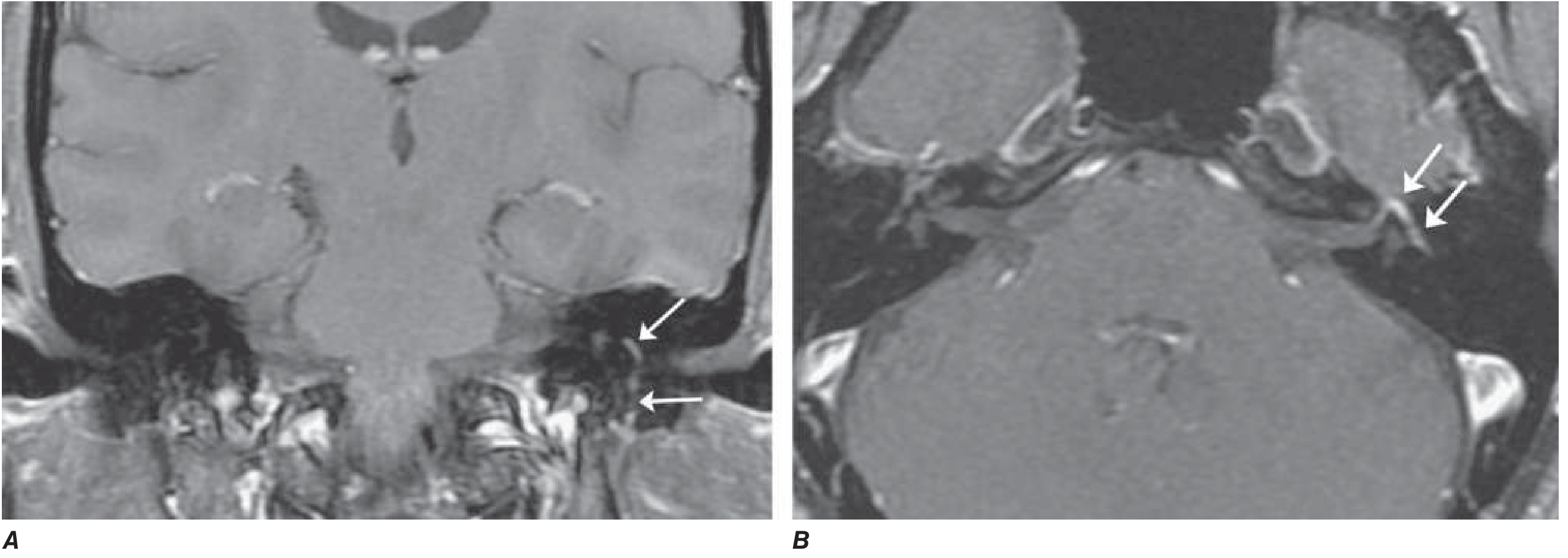
MRI Findings
Clinical Photographs
Differential Diagnosis
- Ramsay Hunt syndrome (VZV reactivation): severe facial palsy + vesicular eruption in external auditory canal ± ear pain ± CN VIII involvement
- Lyme disease (Borrelia burgdorferi): can cause unilateral or bilateral facial palsy; in endemic areas, accounts for ≥10% of facial palsy cases
- Sarcoidosis: can cause bilateral facial palsy; check ACE level and chest imaging
- Guillain-Barré syndrome: bilateral facial palsy possible; check CSF
- HIV seroconversion: CN VII involvement
- Diabetes, Sjögren's syndrome, amyloidosis, leprosy
- Carcinomatous meningitis (LMN pattern possible)
- Melkersson-Rosenthal syndrome: recurrent facial paralysis + facial/labial edema + fissured tongue
- Stroke/UMN lesion: forehead SPARED (upper face has bilateral cortical representation); lower face only affected; often with arm/leg weakness or aphasia
- Parotid tumors or acoustic neuroma: typically slower onset
Workup
- Typical abrupt-onset unilateral facial palsy
- No risk factors pointing to another cause
- No vesicles in external auditory canal (would suggest Ramsay Hunt)
- Otherwise normal neurological exam
- ESR/CRP, fasting glucose (diabetes)
- Lyme serology (if endemic area)
- HIV serology
- ACE level + chest X-ray (sarcoidosis)
- Lumbar puncture (Guillain-Barré)
- Gadolinium-enhanced MRI (facial nerve enhancement)
Prognosis
- ~70-80% recover completely within 1-2 months
- ~85% achieve near-normal function overall
- Most favorable prognostic signs:
- Incomplete paralysis in the first week
- Early recovery of some motor function within 5-7 days
- Return of taste in the first week
- If denervation on EMG by day 10: expect long delay (months), incomplete recovery
- Recurrence: ~7-8%; average interval ~10 years
Treatment
1. Corticosteroids (first-line, strongest evidence)
- Evidence: increases return of facial function from 63% to 83% at 3 months (Goldman-Cecil); modestly shortens recovery and improves functional outcome
- Must be started early (within 72 hours of onset for best effect)
- A 2024 systematic review and meta-analysis (PMID 37940744) compared oral vs. intravenous steroids; oral route is standard first-line
- A 2025 systematic review (PMID 40741786) noted statistical fragility in some steroid-antiviral trial conclusions, worth noting for severe cases
2. Antivirals (adjunct, case-dependent)
- Antiviral monotherapy: not recommended - no benefit alone
- Combined with steroids: evidence is mixed. Most large RCTs (Sullivan et al.) show no added benefit of valacyclovir (1000 mg/day x 5-7 days) or acyclovir (400 mg 5x/day x 10 days) when added to steroids in general Bell's palsy
- Exceptions - give antivirals:
- Vesicular lesions present (ear canal, palate) - suggests Ramsay Hunt, where antivirals ARE indicated
- Severe/complete facial palsy (some evidence of additive benefit with valacyclovir + prednisolone in this subgroup - Hato et al.)
- HIV or immunocompromised
3. Eye care (mandatory in incomplete eyelid closure)
- Artificial tears during the day
- Lubricating eye ointment at night
- Paper tape/eye patch to close eyelid during sleep
- Consider ophthalmology referral if corneal exposure is significant
4. No role for surgical decompression
- Evidence does not support surgical decompression; may be harmful
5. Supportive/rehabilitation
- Physiotherapy and facial muscle massage
- For permanent paralysis: cosmetic surgical procedures to restore facial symmetry
Key Distinguishing Points: LMN vs UMN Facial Palsy
| Feature | LMN (Bell's palsy) | UMN (Stroke) |
|---|---|---|
| Forehead | Involved (cannot wrinkle) | Spared (bilateral cortical supply) |
| Eye closure | Impaired | Usually preserved |
| Lower face | Weak | Weak |
| Taste/Hyperacusis | Possible | Absent |
| Other CN deficits | Rare | Possible |
| Limb weakness/aphasia | Absent | Often present |
Recent evidence note: A 2025 systematic review (PMID 40741786) highlights statistical fragility in steroid-antiviral combination trials for Bell's palsy - the benefit of adding antivirals to steroids may be less robust than previously thought, though this does not change current first-line steroid recommendations.
Cinacalcet
cinacalcet secondary hyperparathyroidism CKD
cinacalcet calcimimetic hyperparathyroidism
Cinacalcet
Drug Class & Overview
Chemical Structure
- A 3-trifluoromethyl group (shown in red below)
- A naphthalene moiety (shown in blue)
- One chiral center with R-absolute configuration - the R-enantiomer is the pharmacologically active form
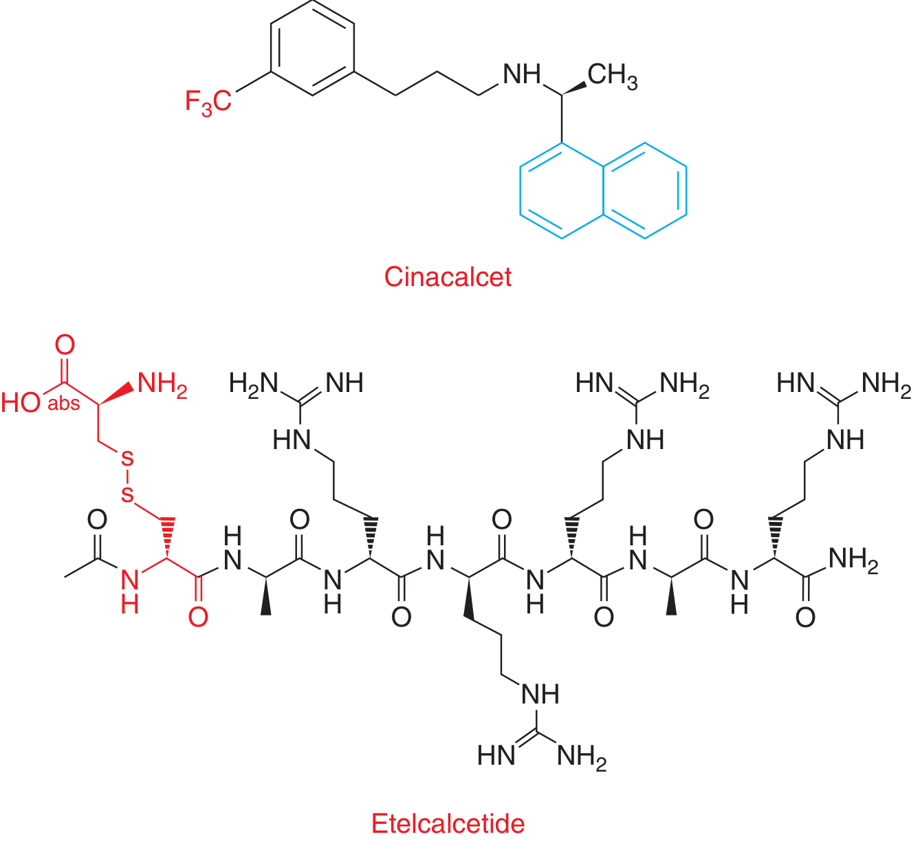
Mechanism of Action
CaSR Pharmacology
| Type | Description | Examples |
|---|---|---|
| Type I calcimimetics | Full CaSR agonists; activate CaSR directly without cofactors | Inorganic di/trivalent cations, aminoglycosides, polylysine |
| Type II calcimimetics | Positive allosteric modulators; require Ca²⁺ to be present; lower the Ca²⁺ threshold for CaSR activation | Cinacalcet, etelcalcetide |
- CaSR is expressed on parathyroid chief cells (and also kidney, thyroid C-cells, bone)
- Cinacalcet binds to the transmembrane domain of CaSR (allosteric site), distinct from the Ca²⁺ binding site
- This increases CaSR sensitivity to extracellular Ca²⁺ - i.e., it lowers the set-point for Ca²⁺-mediated PTH suppression
- Result: PTH secretion is suppressed at prevailing (or even low-normal) calcium concentrations
- Downstream: serum PTH falls → serum Ca²⁺ falls → serum phosphate falls
Pharmacokinetics (ADME)
| Parameter | Cinacalcet | Etelcalcetide (comparison) |
|---|---|---|
| Route | Oral | IV only |
| Absorption | First-order; Tmax 2-6 h | Linear PK after IV |
| Peak PTH effect | 2-4 h after dose | - |
| Volume of distribution | ~1000 L (extraordinarily large) | - |
| Metabolism | Hepatic: CYP3A4, CYP2D6, CYP1A2 | Not a CYP substrate |
| Elimination | Renal 85%, biliary 15% | Eliminated by dialysis |
| Half-life | 30-40 h | 3-5 days (in HD patients) |
| Food effect | Take with evening meal - improves absorption, reduces GI side effects | N/A |
Approved Indications
- Secondary hyperparathyroidism (SHPT) in CKD patients on dialysis - primary use
- Hypercalcemia in parathyroid carcinoma - reduces unresectable hypercalcemia
- Hypercalcemia in primary hyperparathyroidism (PHPT) - for patients who are NOT surgical candidates
Important: Cinacalcet is NOT recommended in CKD patients not on dialysis - in pre-dialysis CKD, it causes marked hypocalcemia (62% develop serum Ca²⁺ < 8.4 mg/dL vs. 6% on placebo), creating a safety concern. (Brenner & Rector's The Kidney)
Dosing
| Indication | Starting Dose | Titration | Maximum |
|---|---|---|---|
| SHPT in CKD-dialysis | 30 mg once daily | Every 2-4 weeks | 180 mg/day |
| Parathyroid carcinoma | 30 mg twice daily | Titrate to normalize Ca²⁺ | 90 mg four times daily |
| Primary HPT (non-surgical) | 30 mg twice daily | Titrate to normalize Ca²⁺ | 90 mg twice daily |
Clinical Evidence: The EVOLVE Trial
- Primary outcome: Composite of death, nonfatal MI, hospitalization for unstable angina, heart failure, or peripheral vascular event
- ITT result: Nonsignificant 7% reduction in primary outcome (HR 0.93; 95% CI 0.85-1.02; p = 0.11)
- Age-adjusted result: Nominally significant reduction (HR 0.88; 95% CI 0.79-0.97; p = 0.008)
- Key confound: Cinacalcet group was older (median 55 vs. 54 years) AND a substantial proportion of placebo patients crossed over to cinacalcet commercially
- Positive findings despite primary endpoint miss:
- Significantly reduced parathyroidectomy rates
- Significantly reduced calciphylaxis rates
- 16% lower hazard of non-atherosclerotic cardiovascular events
- Lowers FGF23, which independently correlates with CV death reduction
Adverse Effects
| Adverse Effect | Details |
|---|---|
| Hypocalcemia | Most important; do NOT start if Ca²⁺ < 8.4 mg/dL |
| Nausea/vomiting | Very common; improved by taking with evening meal; may require switching agent |
| Seizures | Risk increased with severe hypocalcemia; extra caution in seizure disorders |
| Adynamic bone disease | If PTH drops too low (< 100 pg/mL); reduce/stop dose if PTH < 150 pg/mL |
| QT prolongation | Via hypocalcemia |
- Serum Ca²⁺ and phosphate: within 1 week of starting/dose change
- PTH: within 4 weeks of starting/dose change
- Stop or reduce dose if PTH < 150 pg/mL
Drug Interactions
| Interaction Type | Examples | Clinical Impact |
|---|---|---|
| CYP3A4 inhibitors (raise cinacalcet levels) | Ketoconazole, erythromycin, itraconazole | Caution - dose adjustment needed |
| CYP2D6 substrates (cinacalcet inhibits CYP2D6) | Most beta-blockers, flecainide, vinblastine, TCAs | Dose adjustment of co-medications needed |
| Ca²⁺ homeostasis drugs | Vitamin D analogues, bisphosphonates, calcitonin, glucocorticoids, cisplatin, gallium | Additive hypocalcemia risk |
Cinacalcet vs. Etelcalcetide (IV Calcimimetic)
| Feature | Cinacalcet | Etelcalcetide |
|---|---|---|
| Route | Oral | IV (end of HD session) |
| Adherence | Lower (oral, daily) | Higher (given by staff 3x/week) |
| Drug interactions | Multiple CYP interactions | None (not a CYP substrate) |
| GI adverse effects | Nausea/vomiting common | Similar GI side effects |
| GI bleeding risk | Lower | Increased risk of upper GI bleeding |
| CKD not on dialysis | Not recommended | Not recommended |
Place in Therapy
SHPT Management Ladder (CKD on Dialysis):
1. Dietary phosphate restriction
2. Phosphate binders (sevelamer preferred)
3. Active vitamin D analogues (calcitriol, paricalcitol)
   → BUT: raise Ca and PO₄ → worsen vascular calcification
4. Cinacalcet (or etelcalcetide)
   → Lowers PTH + Ca + PO₄ simultaneously
   → Especially preferred when:
      - Hypercalcemia or hypercalcemic tendency
      - Hyperphosphatemia
      - High risk for vascular calcification
5. Parathyroidectomy
   → For refractory cases (iPTH > 1000, failed medical Rx,
      calciphylaxis, persistent hypercalcemia)
- Patient has marginal or frank hypercalcemia (vitamin D analogues would worsen it)
- Patient has hyperphosphatemia (cinacalcet lowers phosphate too)
- As an adjunct to low-dose active vitamin D (cinacalcet upregulates CaSR expression, increasing vitamin D efficacy)
- To avoid or delay parathyroidectomy
- Parathyroid carcinoma (controlling hypercalcemia in unresectable disease)
- PHPT in surgical non-candidates