

This image is not clear can you make it clear with parts marking
A clear, high-resolution medical anatomy poster titled "The Male Reproductive System" showing 8 detailed diagrams with crisp, readable labels: 1) Anterior View of bladder, prostate, penis showing Fundus of bladder, Urethral crest, Prostatic utricle, Openings of ejaculatory ducts, Urogenital diaphragm, Bulbourethral gland, Crus of penis, Bulb of penis, Corpus cavernosum, Corpus spongiosum, Glans penis, Prepuce, Navicular fossa; 2) Sagittal Section of pelvic organs showing Sacrum, Urinary bladder, Ureter, Seminal vesicle, Ejaculatory duct, Prostate gland, Rectum, Pubic symphysis, Suspensory ligament of penis, Testis, Epididymis; 3) Cross-Section of Penis showing Tunica albuginea, Corpus cavernosum, Corpus spongiosum, Deep artery of penis, Buck's fascia, Dorsal vein and artery; 4) Posterior View of bladder showing Ampulla of ductus deferens, Seminal vesicle, Spermatic cord, Pampiniform plexus; 5) Spermatogenesis microscopic diagram showing Sertoli cell, Primary spermatocyte, Secondary spermatocyte, Spermatid, Spermatozoon; 6) The Testis showing Epididymis head body tail, Seminiferous tubule, Rete testis, Efferent ductules, Tunica albuginea; 7) The Prostate zones showing Central zone, Peripheral zone, Transitional zone, Fibromuscular stroma; 8) Vasculature and Innervation showing Testicular artery, Dorsal nerve of penis, Internal pudendal artery. All labels are large, black, clearly printed with leader lines. White background. Professional medical illustration style.
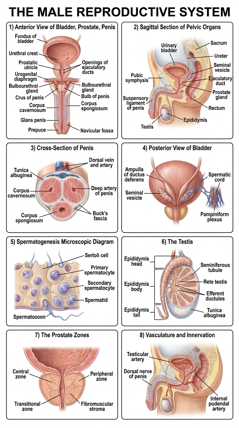
1. Anterior View (Bladder, Prostate, Penis)
- Fundus of bladder, Opening of ureter, Interureteric fold, Trigone of bladder
- Urethral crest, Prostatic utricle, Openings of ejaculatory ducts, Openings of prostatic ducts
- Male Urethra divisions: Prostatic urethra -> Membranous urethra -> Bulbous urethra -> Spongy (Cavernous) urethra -> Pendulous (Penile) urethra
- Bulbourethral (Cowper's) gland & duct, Urogenital diaphragm, Crus of penis, Bulb of penis
- Deep artery of penis, Urethral lacunae, Buck's fascia, Corpus cavernosum, Corpus spongiosum
- Navicular fossa, Corona of glans penis, Glans penis, Prepuce, External urethral opening
2. Pelvic Organ - Sagittal Section
- Sacrum, Peritoneum, Urinary bladder, Ureter, Ampulla of ductus deferens
- Seminal vesicle, Ejaculatory duct, Prostate gland, Rectovesical pouch, Rectum
- Pubic symphysis, Suspensory ligament of penis, Perineal membrane
- Corpus cavernosum, Corpus spongiosum, Glans penis, Epididymis, Testis
- Levator ani muscle, Internal & External anal sphincters, Anococcygeal ligament
3. Cross-Section of the Penis
- Subcutaneous median dorsal vein, Dartos fascia (superficial), Buck's fascia (deep)
- Deep dorsal vein, Dorsal artery & nerve of penis
- Tunica albuginea, Corpus cavernosum, Corpus spongiosum
- Deep artery of penis, Intercavernous septum, Penile urethra
4. Posterior View
- Median umbilical ligament, Deep & Superficial inguinal rings, Spermatic cord
- Cremasteric fascia & muscle, Prostate, Ampulla of ductus deferens, Seminal vesicle
- Ejaculatory duct, Pampiniform plexus, Tunica vaginalis (visceral & parietal layers)
- Internal spermatic fascia, External spermatic fascia, Epididymis, Aberrant ductule
5. Spermatogenesis (Microscopic)
- Sertoli's cell, Spermatogonium, Primary spermatocyte
- Secondary spermatocyte, Spermatid, Spermatozoon
6. The Testis
- Ductus deferens, Efferent ductules, Appendix of epididymis, Appendix of testis
- Epididymis (Head, Body, Tail), Seminiferous tubule, Septa of testis
- Lobules of testis, Rete testis, Mediastinum testis, Tunica albuginea
7. The Prostate (Zones)
- Central zone, Peripheral zone, Transitional zone, Fibromuscular stroma
- Periurethral gland region, Preprostatic sphincter, Ejaculatory duct, Distal striated sphincter
8. The Perineum
- Bulbospongiosus muscle, Ischiocavernosus muscle, Superficial transverse perineal muscle
- Perineal membrane, Posterior scrotal artery & nerve, Perineal artery & nerve
- Inferior rectal artery & nerve, Internal pudendal artery, Dorsal nerve of penis
9. Vasculature & Innervation
- Cremasteric, Testicular, Deferential arteries & veins
- Superior, Middle & Inferior vesical arteries, Internal pudendal artery
- Prostatic venous plexus, Pampiniform plexus, Dorsal artery & vein of penis
- Autonomic nerves, Dorsal nerve of penis, Anterior scrotal nerve
- Genital branch of genitofemoral nerve
I asking you to create the clarity pic will you
A highly detailed, crystal clear medical anatomy illustration poster of "THE MALE REPRODUCTIVE SYSTEM" on white background. Professional textbook quality. Include these clearly labeled sections with large readable black text and precise leader lines: TOP LEFT - Anterior View: detailed coronal cross-section of penis, prostate, bladder. Label: Fundus of bladder, Trigone of bladder, Opening of ureter, Prostatic urethra, Membranous urethra, Bulbous urethra, Spongy urethra, Urogenital diaphragm, Bulbourethral gland, Crus of penis, Bulb of penis, Corpus cavernosum, Corpus spongiosum, Deep artery of penis, Glans penis, Prepuce, External urethral opening, Scrotum TOP RIGHT - Sagittal Section of male pelvis: Label: Sacrum, Urinary bladder, Ureter, Seminal vesicle, Prostate gland, Ejaculatory duct, Rectum, Pubic symphysis, Corpus cavernosum, Corpus spongiosum, Testis, Epididymis, Levator ani muscle, Internal anal sphincter, External anal sphincter MIDDLE LEFT - Cross-section of penis (circular diagram): Label: Skin, Dartos fascia, Buck's fascia, Deep dorsal vein, Dorsal artery, Dorsal nerve, Tunica albuginea, Corpus cavernosum, Deep artery of penis, Corpus spongiosum, Urethra MIDDLE CENTER - Posterior view of bladder and testes: Label: Urinary bladder, Ureter, Ampulla of ductus deferens, Seminal vesicle, Ejaculatory duct, Prostate, Spermatic cord, Epididymis, Testis, Pampiniform plexus BOTTOM LEFT - Spermatogenesis diagram and Testis structure: Label: Sertoli cell, Spermatogonium, Primary spermatocyte, Secondary spermatocyte, Spermatid, Spermatozoon, Seminiferous tubule, Epididymis head body tail, Rete testis, Tunica albuginea BOTTOM RIGHT - Vasculature and Innervation: Label: Testicular artery, Cremasteric artery, Dorsal nerve of penis, Internal pudendal artery, Pampiniform plexus, Anterior scrotal nerve RIGHT SIDE - The Prostate zones: Label: Central zone, Peripheral zone, Transitional zone, Fibromuscular stroma, Prostatic urethra All anatomical drawings are highly realistic, colorful, detailed. Labels use clean sans-serif font, black text, clear leader lines. Medical school poster style.
400, message: Got more than 4194304 bytes when reading: b'data: {"candidates": [{"content": {"role": "model","parts": [{"inlineData": {"mimeType": "image/png"...'.
We encountered an error while processing your request. Please try again or reach out to us for help.
I asking you to create the clarity pic will you
Crystal clear professional medical anatomy poster of THE MALE REPRODUCTIVE SYSTEM. White background. Multiple labeled diagrams arranged neatly. Highly detailed realistic anatomical illustrations with sharp black label text and clean leader lines. Section 1 - ANTERIOR VIEW of penis and bladder (top left): shows bladder, prostate, full penis in coronal section. Labels: Fundus of bladder, Trigone, Prostatic urethra, Membranous urethra, Bulbous urethra, Spongy urethra, Urogenital diaphragm, Bulbourethral gland, Corpus cavernosum, Corpus spongiosum, Glans penis, Prepuce, External urethral meatus, Scrotum, Crus of penis, Bulb of penis. Section 2 - SAGITTAL SECTION of male pelvis (top right): full side-view cutaway. Labels: Sacrum, Urinary bladder, Seminal vesicle, Prostate gland, Rectum, Pubic symphysis, Corpus cavernosum, Testis, Epididymis, Levator ani, Anal sphincters. Section 3 - CROSS-SECTION OF PENIS (middle left, circular): Labels: Skin, Dartos fascia, Bucks fascia, Deep dorsal vein, Dorsal artery, Dorsal nerve, Corpus cavernosum, Tunica albuginea, Corpus spongiosum, Penile urethra, Deep artery of penis. Section 4 - POSTERIOR VIEW of bladder and testes (center): Labels: Bladder, Ureter, Ampulla of vas deferens, Seminal vesicle, Ejaculatory duct, Prostate, Spermatic cord, Pampiniform plexus, Epididymis, Testis. Section 5 - TESTIS internal structure (bottom left): Labels: Tunica albuginea, Seminiferous tubules, Rete testis, Efferent ductules, Epididymis head body tail, Vas deferens, Septa, Lobules. Section 6 - SPERMATOGENESIS diagram (bottom left inset): Labels: Sertoli cell, Spermatogonium, Primary spermatocyte, Secondary spermatocyte, Spermatid, Spermatozoon. Section 7 - PROSTATE ZONES (right middle): Labels: Central zone, Peripheral zone, Transitional zone, Fibromuscular stroma, Prostatic urethra, Ejaculatory duct. Section 8 - VASCULATURE AND INNERVATION (bottom right): Labels: Testicular artery, Dorsal nerve of penis, Internal pudendal artery, Pampiniform plexus, Anterior scrotal nerve, Autonomic nerves. Medical school textbook illustration. Very sharp. Large readable labels.
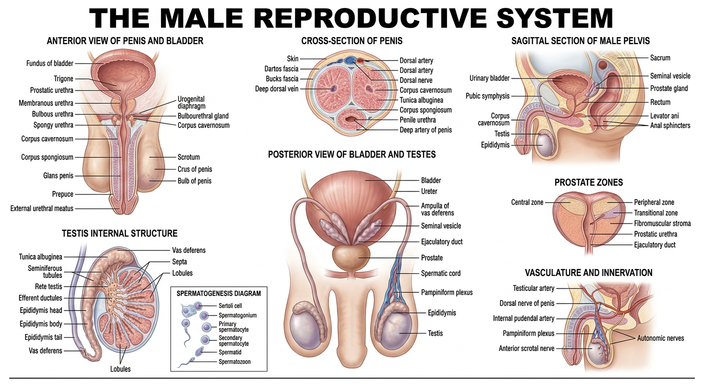
- Anterior View - bladder, prostate, penis with urethra divisions
- Sagittal Section - full pelvic cutaway showing organ relationships
- Cross-Section of Penis - all internal layers labeled
- Posterior View - seminal vesicles, vas deferens, epididymis, testes
- Testis Structure - internal anatomy with seminiferous tubules
- Spermatogenesis - step-by-step sperm cell development
- The Prostate Zones - central, peripheral, transitional zones
- Vasculature & Innervation - blood supply and nerve pathways