

1000123382.jpg
Identify this condition in male groin region The patient is having itching and rashes Along with something sticky substance being secreted
analyse_media
Reading File
Based on the image analysis and symptoms, the most likely diagnosis is Tinea Cruris (fungal infection / "Jock Itch"), though I also note features consistent with Seborrheic Dermatitis or Intertrigo. Let me search the textbooks and recent literature in parallel.
Searching sources...
Finding Sources
Searching PubMed
tinea cruris groin fungal infection treatment
Good - multiple textbooks confirm Tinea Cruris. Let me now read the detailed clinical findings and get the relevant textbook image.
Reading File
Reading File
Excellent - I now have comprehensive information. Let me also fetch the textbook image for visual reference and get the treatment section.
Loading Image
Reading File
I now have all the information needed for a thorough answer. Let me also quickly get the treatment section for Tinea Cruris.
Reading File
I now have all the information needed. Here is a complete clinical assessment:
Diagnosis: Tinea Cruris ("Jock Itch")
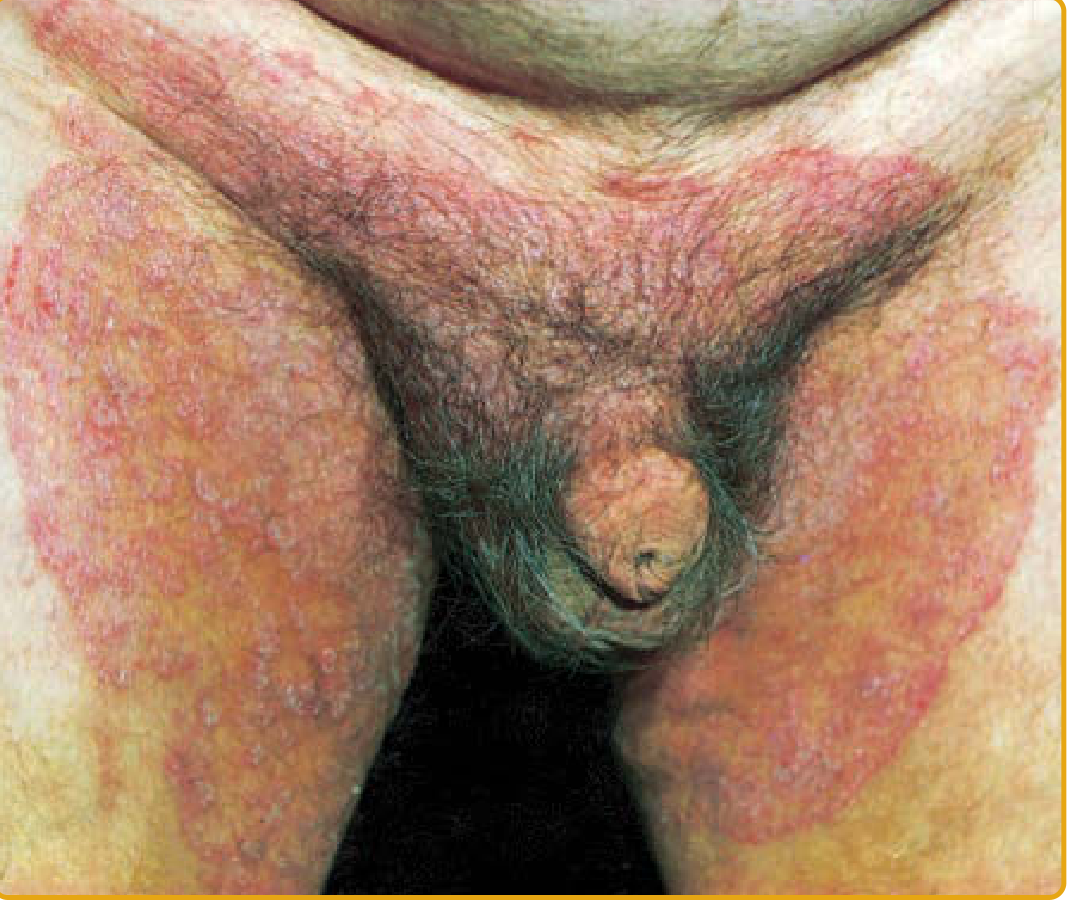
The image shows a localized, pinkish-red (erythematous) patch with slight scaling in the male pubic/groin region, consistent with Tinea Cruris - the second most common dermatophytosis worldwide. The symptoms you describe (itching, rash, sticky/moist discharge) are classic.
Below is a textbook reference image for comparison:
What Is It?
Tinea cruris is a dermatophyte fungal infection of the groin, genitalia, pubic area, perineal, and perianal skin. It is caused primarily by:
- Trichophyton rubrum (most common; coalesces with extension toward pubic/buttock/abdominal areas)
- Epidermophyton floccosum (shows central clearing with involvement of genitocrural crease)
- Less commonly: T. interdigitale, T. verrucosum
It is 3 times more common in men than women, and adults are affected more frequently than children. - Fitzpatrick's Dermatology, p. 2975
Why These Symptoms?
| Symptom | Explanation |
|---|---|
| Itching (pruritus) | Classic feature - caused by fungal invasion of the stratum corneum and host inflammatory response |
| Rash / erythema | Well-marginated annular plaques with a slightly raised, scaly border extending from inguinal fold to inner thigh |
| Sticky/moist discharge | Maceration from moisture and occlusion in the warm groin environment; can indicate secondary bacterial superinfection |
The "sticky substance" likely represents maceration (skin breakdown from chronic moisture) or early secondary infection/weeping, which is worsened by occlusion (tight underwear, synthetic fabrics, sweating).
How It Spreads
- Direct contact (person-to-person or via fomites like shared towels)
- Autoinfection from tinea pedis (athlete's foot) - fungus can spread from feet to groin when putting on clothes. Check for scaling between toes.
- Exacerbated by occlusion, humidity, and heat - Tintinalli's Emergency Medicine, p. 1694
Differential Diagnosis
According to Fitzpatrick's Dermatology (Table 160-18), conditions to differentiate include:
| Condition | Key Distinguishing Feature |
|---|---|
| Erythrasma | Coral-red fluorescence under Wood lamp (caused by Corynebacterium) |
| Cutaneous Candidiasis | Involves scrotum (tinea cruris typically SPARES scrotum); satellite pustules around edges |
| Intertrigo | No raised border; pure moisture/friction; no hyphae on KOH |
| Inverse Psoriasis | Smooth, non-scaling red plaques; psoriasis elsewhere on body |
| Seborrheic Dermatitis | Greasy, yellowish scales; usually also on scalp/face |
| Contact Dermatitis | History of new irritant/allergen; no annular pattern |
Diagnosis Confirmation
A KOH (potassium hydroxide) preparation of skin scrapings from the leading edge of the lesion will show branching hyphae under microscopy. This is the gold standard bedside test. - Tintinalli's EM, p. 1694
Treatment
Topical antifungals (first-line, applied twice daily for 2-4 weeks):
| Drug | Brand | Availability |
|---|---|---|
| Clotrimazole 1% cream | Lotrimin | OTC - first choice |
| Miconazole 2% cream/powder | Zeasorb AF | OTC |
| Ketoconazole 2% cream | Nizoral | Prescription |
| Econazole 1% cream | Spectazole | Prescription - preferred if maceration/weeping present (has antibacterial properties) |
Given the sticky/moist discharge in this case, Econazole would be the preferred topical agent.
Oral antifungals (for extensive, refractory, or recurrent cases):
- Terbinafine 250 mg once daily x 1-2 weeks
- Itraconazole 100 mg once daily x 2 weeks
- Fluconazole 150 mg weekly x 2-4 weeks
Supportive Measures
- Keep the area clean and dry - pat dry thoroughly after bathing
- Wear loose-fitting, breathable cotton underwear
- Apply antifungal powder (miconazole or clotrimazole) daily to prevent recurrence
- If tinea pedis (athlete's foot) is also present, treat the feet simultaneously and apply socks before underwear to prevent autoinfection
- Do NOT apply plain steroid creams (hydrocortisone) alone - this will worsen fungal infection
- Avoid sharing towels or clothing
When to See a Doctor Urgently
Seek prompt medical care if:
- Area becomes warm, swollen, or produces pus (possible cellulitis/abscess)
- Fever develops
- No improvement after 4-6 weeks of topical antifungals
- Rash spreads rapidly
Disclaimer: This is educational information only. Please consult a dermatologist or physician for formal diagnosis and treatment, especially given the presence of discharge which may indicate secondary infection requiring specific management.
What are the signs of secondary bacterial infection in tinea cruris?
Secondary bacterial infection complicating tinea cruris occurs when skin barrier breakdown from fungal invasion and scratching allows bacteria (usually Staphylococcus aureus or Streptococcus) to invade. Key signs include increased pain/tenderness, purulent discharge, crusting, warmth, and swelling beyond the original rash border.
Searching sources...
Finding Sources
Finding Sources
Searching PubMed
tinea cruris secondary bacterial infection superinfection
Let me search more broadly across dermatology textbooks for bacterial superinfection in skin/fungal infections.
Finding Sources
Finding Sources
Finding Sources
Let me search across dermatology and general medicine textbooks for signs of bacterial superinfection in skin conditions and intertrigo/maceration topics.
Finding Sources
Finding Sources
Finding Sources
Finding Sources
Finding Sources
Reading File
Searching the Web
signs secondary bacterial infection tinea cruris superinfection clinical features treatment
I have enough information for a thorough, well-sourced answer. Here it is:
Signs of Secondary Bacterial Infection in Tinea Cruris
Secondary bacterial infection is a recognized complication of tinea cruris. The fungal infection disrupts the skin barrier, and persistent scratching creates open entry points for bacteria - most commonly Staphylococcus aureus and Group A Streptococci. According to StatPearls (NCBI), "secondary infections may result in inflammation and discomfort," and the PMC comprehensive review states "secondary bacterial infection should be treated with oral antibiotics."
Local Signs (at the rash site)
| Sign | What to Look For |
|---|---|
| Increased pain/tenderness | Shifts from itch-dominant to pain-dominant; even light touch is painful - the clearest indicator |
| Purulent (pus) discharge | Yellow, green, or cloudy exudate draining from the lesion; distinct from the clear/serous moisture of simple maceration |
| Yellow or honey-colored crusting | Thick golden crusts overlying the rash (classic sign of S. aureus impetiginization) |
| Pustules | Small pus-filled bumps (pustules) appearing within or around the original plaque |
| Increased erythema / redness | Redness that is deeper, darker, and extends well beyond the original fungal rash border |
| Warmth | The skin feels notably hot to the touch compared to surrounding normal skin |
| Swelling (edema) | Puffiness or induration of the surrounding skin, suggesting deeper tissue involvement |
| Rapid border expansion | The infected area grows quickly in size - bacterial spread is faster than fungal |
| Skin breakdown / ulceration | Open sores or erosions, especially in macerated folds |
| Foul odor | A distinctly unpleasant smell from the exudate (bacterial metabolism byproducts) |
Systemic Signs (indicates deeper / spreading infection)
These suggest progression to cellulitis or systemic sepsis - require urgent medical attention:
- Fever (> 38°C / 100.4°F)
- Chills or rigors
- Enlarged, tender lymph nodes in the groin (inguinal lymphadenopathy)
- Red streaks extending from the rash toward the lymph nodes (lymphangitis - a medical emergency)
- Malaise, fatigue, or feeling generally unwell
- Rapid heart rate (tachycardia)
How It Differs From Uncomplicated Tinea Cruris
| Feature | Tinea Cruris Alone | With Secondary Bacterial Infection |
|---|---|---|
| Discharge | None, or thin/clear maceration | Thick, yellow/green pus |
| Pain | Itch predominates | Burning + pain predominates |
| Crusting | Fine, dry white scale | Thick, honey-colored/yellow crusts |
| Borders | Well-defined, raised scaly edge | Blurred, spreading, edematous |
| Systemic symptoms | Absent | May be present (fever, lymphadenopathy) |
| Odor | Mild/musty | Foul |
A Special Complication: Majocchi's Granuloma
When bacterial infection or skin trauma drives fungal organisms deeper into the dermis/subcutaneous tissue, StatPearls notes that Majocchi's granuloma can develop - an uncommon but serious complication where dermatophytes disseminate into subcutaneous tissue following skin breakdown. This presents as deep nodules or plaques and requires systemic antifungal therapy.
Risk Factors That Increase the Likelihood of Secondary Infection
- Diabetes mellitus (impaired immunity and skin healing)
- Obesity (more friction, deeper skin folds, more moisture)
- Immunocompromise (HIV, steroid use, chemotherapy)
- Prolonged scratching (breaks the skin barrier)
- Inappropriate topical steroid application (suppresses local immunity, allows organisms to thrive)
- Poor hygiene or prolonged moisture exposure
Treatment When Secondary Bacterial Infection Is Present
- Topical antifungal + antibacterial combination: Econazole (preferred - has intrinsic antibacterial properties) or combine a topical antifungal with a topical antibiotic (e.g., mupirocin) - Tintinalli's Emergency Medicine, p. 1694
- Oral antibiotics if more than superficial: Flucloxacillin/dicloxacillin for S. aureus; Amoxicillin-clavulanate as a broader choice; if MRSA suspected, trimethoprim-sulfamethoxazole or doxycycline
- Continue antifungal treatment - do NOT stop; both infections must be treated simultaneously
- Wound care: Gentle cleansing with mild antiseptic (chlorhexidine), keep dry
- Oral antifungal (terbinafine, itraconazole) if the infection is extensive or in an immunocompromised patient
Seek urgent care if you notice fever, spreading redness, red streaks, severe pain, or systemic symptoms - these can indicate cellulitis or necrotizing fasciitis, which are medical emergencies requiring IV antibiotics and possibly surgical debridement.
This is a shared conversation. Sign in to Orris to start your own chat.