
Electromyography
Electromyography (EMG) is a broad topic - could mean the basic overview, the testing procedure/technique, clinical indications, or how to interpret findings (e.g., for a specific condition). Let me narrow this down so I can give you the most useful answer.
Asking for Preferences
What would you like to know about EMG?
Reading File
Finding Sources
Searching the Web
electromyography EMG overview definition uses
Reading File
Reading File
Reading File
I have enough to give a solid overview. I'll skip a PubMed check since this is a well-established, non-controversial physiologic/diagnostic concept with no fast-moving evidence base.
Electromyography (EMG) - Overview
What it is
Electromyography is an electrodiagnostic technique that records the electrical activity of skeletal muscle, most commonly using a needle (concentric) electrode inserted directly into the muscle, though surface electrodes over the skin can also be used for some applications. It assesses the properties of the motor unit (a motor neuron and all the muscle fibers it innervates), and is typically paired with nerve conduction studies (NCS) as part of a combined electrodiagnostic exam - the NCS component measures how nerves conduct impulses, while EMG measures the muscle's electrical response - Goldman-Cecil Medicine, p. 275; Firestein & Kelley's Textbook of Rheumatology.
The four phases examined during needle EMG
- Insertional activity - the brief burst of electrical activity that occurs in the first second after the needle is inserted into muscle
- Spontaneous activity - electrical activity recorded at rest (should normally be silent)
- Voluntary activity - motor unit potentials generated during active muscle contraction
- Recruitment pattern - how electrical activity changes as contraction intensity increases (i.e., how many/which motor units are recruited)
- Goldman-Cecil Medicine, p. 275
Key abnormal findings and what they suggest
| Finding | Typical cause |
|---|---|
| Prolonged insertional activity | Acute denervation or active inflammatory myopathy |
| Fibrillations / positive sharp waves | Acute denervation, active inflammatory myopathy |
| Fasciculations | Chronic neuropathy, motor neuron disease |
| Myotonic discharges | Myotonic disorders, acid maltase deficiency |
| Large-amplitude, long-duration, polyphasic ("neuropathic") potentials | Chronic neuropathy, anterior horn cell disease |
| Small-amplitude, short-duration, polyphasic ("myopathic") potentials | Chronic myopathy, neuromuscular junction disorders |
| Reduced recruitment | Chronic neuropathic disorders |
| Rapid/early recruitment | Chronic myopathies |
- Goldman-Cecil Medicine, p. 275-276
Why it's clinically useful
- EMG helps localize the source of weakness - anterior horn cell (e.g., motor neuron disease), nerve root (radiculopathy), plexus (plexopathy), a single peripheral nerve (mononeuropathy), or multiple nerves (polyneuropathy) - versus a primary muscle disorder.
- It distinguishes active inflammatory myopathies (dermatomyositis, polymyositis, inclusion body myositis) from chronic/structural myopathies, and is sensitive (though nonspecific) for detecting muscle pathology in general - Firestein & Kelley's Textbook of Rheumatology, p. 4435.
- It's used to guide muscle biopsy site selection in suspected myositis, since inflammation is often patchy - the biopsy is typically taken from the contralateral, non-EMG-tested muscle because needle EMG itself can cause histologic artifact that confounds biopsy interpretation - Firestein & Kelley's Textbook of Rheumatology, p. 4435-4436.
- Combined with repetitive nerve stimulation, it can diagnose neuromuscular junction disorders: myasthenia gravis shows a decrementing response with repetitive stimulation, while presynaptic myasthenic syndromes (e.g., Lambert-Eaton) show an incrementing response with fast repetitive stimulation - Goldman-Cecil Medicine, p. 274.
Important caveat: no single EMG finding is pathognomonic (diagnostic on its own) for any specific disease - results must be interpreted alongside the clinical picture, nerve conduction data, and often muscle biopsy or serology - Goldman-Cecil Medicine, p. 275.
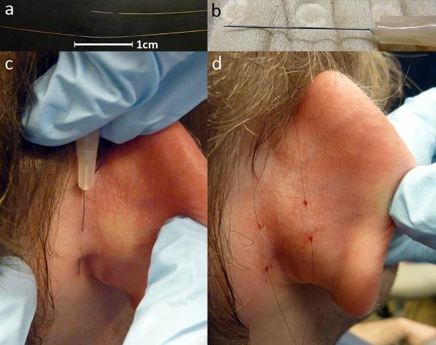
Here's a real clinical photo set illustrating fine-wire/needle electrode placement used for EMG-type recordings:
Let me know if you'd like more detail on the actual testing procedure/patient experience, or on interpreting EMG in a specific condition (e.g., carpal tunnel syndrome, ALS, myopathy work-up).
This is a shared conversation. Sign in to Orris to start your own chat.