
Hypersensitivity of teeth
"dentin hypersensitivity"[MeSH Terms] AND treatment
"dentin hypersensitivity" AND management
dentin hypersensitivity dentinal tubules hydrodynamic mechanism diagram
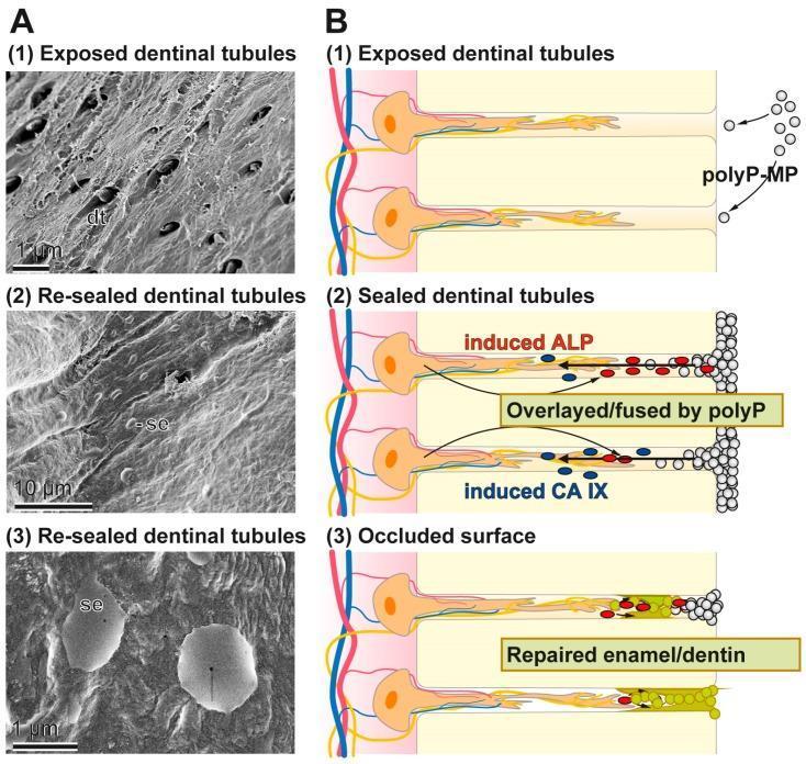
This composite educational image illustrates the process of dentinal tubule occlusion using polyP-based microparticles, relevant to treating dental hypersensitivity and caries. Section A presents three scanning electron microscopy (SEM) images showing the progression from (1) 'Exposed dentinal tubules' with visible dark openings (dt), to (2) 'Re-sealed dentinal tubules' showing a smoothed surface (c-se), and (3) a high-magnification view of an 'Occluded surface' featuring globular mineral deposits (se). Section B contains a corresponding three-stage pathophysiology diagram. It depicts: (1) polyP-microparticles (polyP-MP) entering open tubules; (2) the biochemical interaction where polyP induces alkaline phosphatase (ALP) and carbonic anhydrase IX (CA IX), leading to fusion; and (3) the final state of 'Repaired enamel/dentin' where tubules are fully occluded. The diagram highlights the transition from exposed odontoblastic processes to a mineralized, sealed state. This material is designed for dental education focusing on biomaterial-driven tooth repair and mineralization mechanisms.
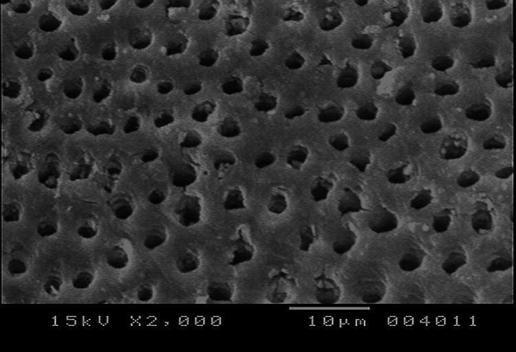
Scanning Electron Microscope (SEM) image at 2000x magnification showing the ultrastructure of human dentin. The diagnostic image displays a high density of dentinal tubules distributed across the calcified tissue surface. These tubules appear as roughly circular or elliptical dark openings against a lighter, textured intertubular dentin matrix. According to the 10 μm scale bar, the tubule diameters average approximately 1-2 μm. While many tubules demonstrate patent (open) lumens characteristic of a control or untreated sample, some exhibit partial occlusion by debris or mineral deposits. The image is used in dental research and endodontics to study dentin hypersensitivity, tubule occlusion treatments, and the structural integrity of tooth surfaces. The organization follows a relatively regular, porous pattern, serving as a baseline for comparing the efficacy of desensitizing agents or bonding materials in clinical dentistry.
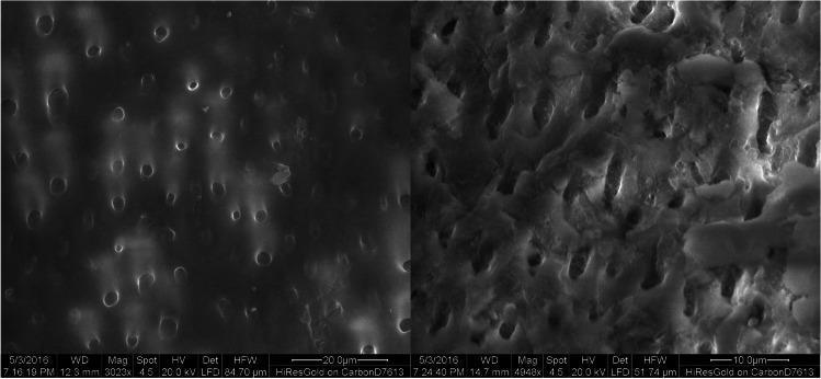
This diagnostic image consists of two high-magnification scanning electron microscopy (SEM) micrographs showing the surface of human dentin. The micrographs serve as a control group to demonstrate the normal anatomical morphology of dentin after the removal of the smear layer. The primary visual feature is the presence of numerous open dentinal tubules, which appear as dark, circular to oval orifices distributed relatively evenly across a smooth intertubular dentin matrix. The tubule openings are unobstructed, clearly defined, and show no signs of mineralization, sealing, or debris. This state of patent tubules is clinically significant in dental research related to dentin hypersensitivity and the efficacy of desensitizing agents or laser treatments designed to occlude these pathways. The left panel shows a wider field of view at approximately 3000x magnification (20m scale bar), while the right panel provides a higher magnification view (10m scale bar) emphasizing the structural integrity of the peritubular and intertubular dentin.
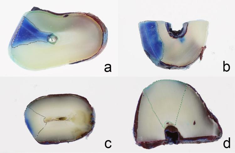
This composite of four macroscopic clinical photographs (labeled a–d) illustrates a comparative study of dentinal tubule occlusion efficiency using methylene blue dye penetration in human dentin specimens. Each panel shows a cross-section of a tooth specimen with a dotted line demarcating the treatment area. (a) Control group: Shows extensive, deep, triangular-shaped blue dye penetration through the dentin, indicating open tubules. (b) Desensitizer paste (TP/DP) group: Displays a moderate, less defined region of dye penetration localized toward the specimen periphery. (c) CO2 laser-treated group: Exhibits restricted, linear dye penetration along a structural crack, suggesting partial tubule sealing. (d) Combined laser and desensitizer paste group: Shows minimal, nearly punctate dye penetration, demonstrating the highest efficacy in sealing dentinal tubules. The progression from (a) to (d) visually represents the synergistic effect of physical and chemical occlusion methods in treating dentin hypersensitivity by reducing permeability. The specimens appear as yellowish-white ivory-colored sections with peripheral reddish-brown enamel/cementum margins.
Hypersensitivity of Teeth (Dentin Hypersensitivity)
Definition
Epidemiology
- Prevalence: 8-57% in the general population (varies widely with assessment method)
- Most common in the 3rd-4th decades of life
- Affects women slightly more than men
- Most commonly involves buccal cervical surfaces of canines and premolars
- Molar-Incisor Hypomineralization (MIH) is a significant risk factor - a recent systematic review and meta-analysis (Santos et al., 2024) confirmed significantly higher dentin hypersensitivity rates in MIH patients
Mechanism - The Hydrodynamic Theory (Brannstrom, 1966)
- Dentin is exposed via gingival recession, enamel erosion, abrasion, or abfraction
- Dentinal tubules become patent - open channels from the pulp to the surface
- A stimulus (cold, heat, osmotic, air) causes rapid fluid movement within the tubules
- This fluid movement activates A-delta nerve fibers (mechanoreceptors) at the pulp-dentin junction
- Result: sharp, short pain
Etiology and Predisposing Factors
1. Gingival Recession (Exposure of root dentin/cementum)
- Periodontal disease
- Aggressive tooth brushing
- Periodontal surgery
- Orthodontic tooth movement
2. Loss of Enamel / Cementum
- Erosion: dietary acid (citrus, carbonated drinks), gastric acid (GERD, bulimia)
- Abrasion: abrasive toothpastes, hard toothbrushes
- Attrition: bruxism / parafunctional habits
- Abfraction: eccentric loading causing cervical stress fractures
3. Iatrogenic Factors
- Scaling and root planing
- Bleaching agents (hydrogen/carbamide peroxide)
- Air abrasion procedures
- Restorative procedures (loss of smear layer)
Clinical Features
| Feature | Description |
|---|---|
| Character | Sharp, shooting, short-duration pain |
| Duration | Seconds (not persistent) |
| Triggers | Cold > hot, sweet/sour, air, touch |
| Location | Usually cervical / buccal surfaces of premolars and canines |
| Vitality test | Positive (tooth is vital) |
| Percussion | Negative (no periapical pathology) |
Differential Diagnosis (must exclude before diagnosing DH)
- Cracked tooth syndrome (pain on release of biting)
- Fractured restoration
- Caries (sustained pain, carious cavity visible)
- Pulpitis (spontaneous, prolonged pain)
- Periapical pathology (tenderness to percussion)
- Galvanic pain (two dissimilar metals in contact)
Assessment Tools
- Schiff Cold Air Sensitivity Scale (SCASS): 0-3 scale using air-blast
- Visual Analogue Scale (VAS): patient-reported pain intensity
- Yeaple probe: quantified tactile stimulus
- These should be combined for research purposes
Management
Home-Use (Patient-Applied) Agents
| Agent | Mechanism | Example |
|---|---|---|
| Potassium nitrate (5%) | Depolarizes nerve fibers (nerve desensitization) | Sensodyne Original |
| Strontium chloride (10%) | Tubule occlusion (strontium precipitate) | Sensodyne Classic |
| Stannous fluoride | Tubule occlusion + fluoride mineralization | Oral-B Pro-Expert |
| Sodium fluoride | Calcium fluoride smear layer; remineralization | Various |
| Arginine-calcium carbonate | Physically occludes tubules with Ca/carbonate | Colgate Sensitive Pro-Relief |
In-Office Treatments
| Agent / Procedure | Mechanism |
|---|---|
| Fluoride varnishes (5% NaF) | Tubule occlusion via CaF2 deposits |
| Oxalate-based agents | Calcium oxalate crystals seal tubule orifices |
| Adhesive bonding resins | Mechanically seal tubules with resin tags |
| Glass ionomer cement / composite | Physical coverage of exposed dentin |
| Laser therapy (Nd:YAG, Er:YAG, diode) | Tubule obliteration by micro-melting dentin |
| Bioglass / bioactive glass | Ion exchange releases Ca/P to form hydroxyapatite |
Emerging Approaches
- Photothermal mineralizing nanoparticles: A 2025 study (Huang et al.) demonstrated photothermal-activated nanoparticles that promote dentin remineralization to seal tubules
- PIEZO1 mechanoreceptors: Recent research (2025) is exploring PIEZO1-mediated mechanotransduction in dental pain signaling as a future therapeutic target
- PolyP microparticles: Polyphosphate-based biomaterials that induce ALP-mediated mineralization and seal tubules (shown in SEM image above)
Surgical Management (for severe recession)
- Gingival grafting (connective tissue graft, free gingival graft)
- Coronally advanced flap
- Indicated when significant recession is the root cause and conservative therapy fails
Prevention
- Use a soft toothbrush and correct brushing technique (Bass method)
- Avoid abrasive toothpastes
- Wait 30-60 minutes after consuming acid before brushing
- Manage GERD if present
- Custom occlusal splint for bruxism
- Regular use of fluoride/potassium nitrate toothpaste
Key Points Summary
- DH is diagnosed by exclusion - rule out caries, pulpitis, cracked tooth first
- The hydrodynamic theory (Brannstrom) is the accepted mechanism
- Cold stimuli are the most common trigger
- Arginine-calcium carbonate and fluoride varnishes are among the most evidence-supported in-office treatments
- If conservative therapy fails, consider dentin bonding agents or gingival surgery
- Treat the underlying cause (acid erosion, recession, bruxism) alongside symptomatic relief