
Obstetrics anaesthesia in brief
obstetric anesthesia epidural spinal labor delivery diagram
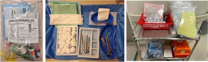
A three-panel clinical photograph displaying the organized medical equipment and kits prepared for obstetric care in a COVID-19 isolation setting. The left panel shows a sealed, transparent 'COVID-19 Epidural Set' containing essential anesthesia supplies such as syringes, tubing, and medications with a clear inventory label. The center panel displays a 'Vaginal Delivery Set' laid out on a sterile blue drape, featuring absorbent underpads, sterile green drapes, gauze, cotton wool balls in a divided tray, and various surgical instruments including clamps, scissors, and kidney basins. The right panel depicts a two-tiered stainless steel delivery trolley; the top shelf holds a red basket with emergency medications and syringes, alongside packaged linens, while the bottom shelf contains a perimortem caesarean section set and a distinctive orange emergency supply box. This setup illustrates the specialized triage and preparedness protocols for managing labor and obstetric emergencies (e.g., postpartum hemorrhage, eclampsia) in a high-risk infectious disease environment.
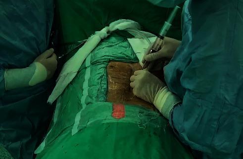
A clinical photograph captures the administration of epidural anesthesia prior to a spinal surgical procedure. The patient is positioned prone on an operating table, with the lumbar region exposed within a sterile surgical field created by green surgical drapes. The exposed skin is discolored brownish-orange from the application of a topical antiseptic solution, such as povidone-iodine. A clinician, wearing sterile blue surgical gowns and gloves, is shown performing the procedure. The clinician is seen holding a syringe connected to an epidural needle, which is inserted into the interlaminar space of the lumbar vertebrae. This visual illustrates the 'loss-of-resistance' technique used for identifying the epidural space. White surgical tape and markers indicate specific vertebral segments or the midline. The image demonstrates standard intraoperative anesthetic preparation and regional anesthesia delivery in the context of orthopedic or neurosurgical spine interventions.
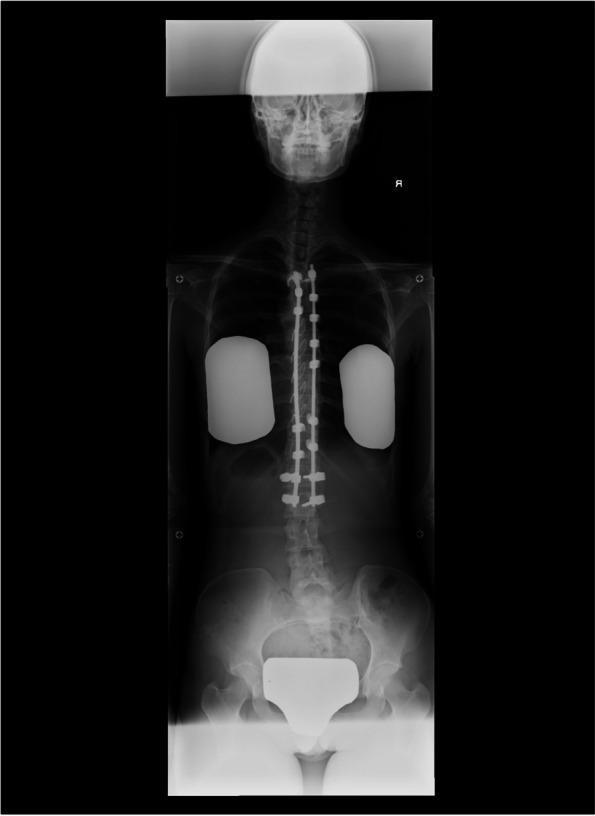
A full-body anteroposterior (AP) X-ray of a pregnant patient with a history of adolescent idiopathic scoliosis (AIS). The image highlights extensive posterior spinal instrumentation consisting of dual longitudinal metal rods and multiple bilateral pedicle screws. The instrumentation spans from the upper thoracic spine (Th3) to the mid-lumbar region (L2). The primary educational focus is on the structural alignment achieved post-fixation surgery and its impact on subsequent obstetric care. Notable features include the visualization of the skull, rib cage, and pelvis, with radiopaque shielding placed over the lung fields and the pelvic region to minimize radiation exposure to the fetus. The alignment of the cervical and lower lumbar vertebrae (below L2) is visible, which is clinically relevant for assessing potential sites for neuraxial anesthesia (epidural or spinal) during labor. The image demonstrates the anatomical constraints and modified spinal landmarks often encountered in patients with a history of corrective spinal fusion.
Obstetric Anaesthesia - A Comprehensive Overview
1. Physiologic Changes of Pregnancy Relevant to Anaesthesia
Airway & Pulmonary Changes
- Capillary engorgement of nasal, oropharyngeal, and laryngeal mucosa occurs throughout pregnancy - worsened by preeclampsia, second-stage pushing, and tocolytic therapy. Always use small ETT (6.0-6.5 mm) and have a difficult airway plan ready.
- Mallampati score increases progressively throughout labor.
- Breast engorgement may impair laryngoscopy - use a short-handled laryngoscope.
- Minute ventilation increases by 50% (tidal volume up 45%, small RR increase) driven by progesterone.
- FRC falls by 30% (RV down 25%, ERV down 20%) - this dramatically reduces the apnea-safe window during induction; preoxygenation is mandatory.
- Progesterone causes bronchodilation; FEV1/FVC ratio is unchanged.
| Variable | Change | Magnitude |
|---|---|---|
| Plasma volume | ↑ | 40-50% |
| Total blood volume | ↑ | 40% |
| Haemoglobin | ↓ | 11-12 g/dL (dilutional) |
| Fibrinogen | ↑ | ~100% |
| FRC | ↓ | ~30% |
| Minute ventilation | ↑ | ~50% |
| Oxygen consumption | ↑ | ~20% |
| MAC (volatile agents) | ↓ | ~30-40% |
Cardiovascular Changes
- Cardiac output rises 40-50% by term (increased HR + stroke volume).
- Systemic vascular resistance falls (progesterone-mediated vasodilation).
- Aortocaval compression by the gravid uterus when supine: causes decreased venous return, hypotension, and fetal compromise. Always use left lateral tilt (>15° wedge under right hip) after 20 weeks.
- Blood pressure normally falls in mid-pregnancy and returns to baseline at term.
Gastrointestinal Changes
- Lower oesophageal sphincter tone reduced by progesterone.
- Gastric emptying delayed; intragastric pressure elevated by gravid uterus.
- All obstetric patients are treated as having a full stomach regardless of time of last oral intake.
- Aspiration prophylaxis: sodium citrate 0.3M (15-30 mL orally), ranitidine/omeprazole, metoclopramide 10 mg IV.
Haematologic Changes
- Hypercoagulable state: fibrinogen doubles, factors VII, VIII, X, XII elevated.
- Platelets may fall slightly. Minimum platelet count for neuraxial: typically >70,000-80,000/μL.
- Pseudocholinesterase activity decreases by ~25% - prolonged succinylcholine effect.
Neurological Changes
- MAC for inhalational agents reduced by 30-40% (progesterone and endorphin effects).
- Epidural and spinal drug requirements are reduced (engorged epidural veins reduce epidural space volume; increased sensitivity of nerve roots).
2. Placental Transfer of Drugs
- The driving force for placental drug transfer is the concentration gradient of free (unbound) drug between maternal and fetal blood.
- Lipid solubility, low molecular weight (<500 Da), low protein binding, and un-ionized form all favor placental transfer.
- Fetal blood is more acidic (pH ~7.35) than maternal blood - ion trapping of weak bases (e.g., local anaesthetics, opioids) can occur in fetal distress (fetal acidosis), increasing fetal drug levels.
- Nearly all parenteral opioids and sedatives readily cross the placenta. Regional techniques are preferred to minimize fetal drug exposure.
- Neuromuscular blockers: highly ionized, large molecules - do NOT cross in clinically significant amounts.
3. Pain Pathways in Labour
| Stage | Pain Type | Dermatomes Involved |
|---|---|---|
| First stage (latent) | Visceral (uterine contractions + cervical dilation) | T11-T12 |
| First stage (active) | Visceral (radiates to lumbosacral, gluteal, thighs) | T10-L1 |
| Second stage | Somatic (perineal distension) + visceral | T10-S4 |
- Visceral afferents travel with sympathetic fibers via the uterovaginal plexus → inferior hypogastric plexus → T10-L1 nerve roots.
- Perineal pain (second stage) is carried by the pudendal nerve (S2-S4).
4. Labour Analgesia
Non-Pharmacologic Methods
- Psychoprophylaxis (Lamaze), hydrotherapy, TENS, intradermal water injections, acupuncture.
Systemic Medication
- Opioids (IV/IM): pethidine (meperidine), morphine, fentanyl, remifentanil PCA. All cross placenta and can cause neonatal respiratory depression.
- Remifentanil PCA: increasingly popular; ultra-short acting but requires close monitoring.
- Nitrous oxide (50% N2O/O2 - Entonox): self-administered during contractions; moderate analgesia, minimal fetal effect.
- Ketamine (sub-dissociative IV doses 0.25 mg/kg): useful supplement in second stage.
Neuraxial Analgesia (Gold Standard)
- Initiated when patient requests it; does not increase operative delivery rate with dilute LA-opioid mixtures.
- Catheter placed at L2-L3 or L3-L4.
- Test dose: bupivacaine 5 mg or lidocaine 45 mg + epinephrine 15 μg (monitors for intrathecal or intravascular placement).
- Typical maintenance: bupivacaine 0.0625-0.125% + fentanyl 2-3 μg/mL or sufentanil 0.3-0.5 μg/mL.
- PCEA (patient-controlled epidural analgesia) allows parturient control.
- Spinal component: intrathecal fentanyl 25 μg ± bupivacaine 2.5 mg → rapid onset ("walking epidural").
- Epidural catheter: maintained for prolonged analgesia, escalation to surgical block.
- Especially useful for severe early labor pain or when delivery is imminent.
5. Anesthesia for Caesarean Section
Preoperative Preparation
- Aspiration prophylaxis as above.
- Large-bore IV access (at least 16G), baseline BP monitoring.
- Left lateral tilt maintained throughout.
- Surgical level T4 dermatome required (to upper sternum/"nipple line").
Neuraxial Anaesthesia (Preferred)
- Hyperbaric 0.75% bupivacaine 12-13.5 mg (1.6-1.8 mL) - provides 90-120 min surgical anaesthesia.
- Additives: fentanyl 10-20 μg (rapid, short-acting adjuvant) or morphine 0.1-0.15 mg (12-18 hours postoperative analgesia).
- Advantages: simple, fast, reliable, dense block; small drug dose.
- Disadvantages: fixed duration, cannot be extended, higher rate of hypotension.
- Slower onset, larger drug volumes; can be titrated and extended.
- 2% lidocaine + epinephrine (1:200,000) OR 3% 2-chloroprocaine - typically 15-25 mL in divided doses over 5-10 min.
- Useful when pre-existing labor epidural can be converted: add bicarbonate to alkalinize solution, add fentanyl, increase concentration.
- Test dose essential; incremental dosing in 5 mL aliquots.
- Spinal provides rapid dense block; epidural allows extension if surgery prolonged.
- Especially useful for complex or anticipated long surgeries.
General Anaesthesia for CS
- Pre-oxygenation for minimum 3 min (or 4 vital capacity breaths at 100% O2).
- Rapid sequence induction (RSI) with cricoid pressure.
- Induction: propofol 2 mg/kg or thiopental 4-5 mg/kg (if available).
- Succinylcholine 1.5 mg/kg (remember reduced pseudocholinesterase; if contraindicated, rocuronium 1.2 mg/kg with sugammadex reversal available).
- Intubation with cuffed ETT (6.0-6.5 mm).
- Maintain with low-dose volatile agent (0.5-1 MAC) + N2O/O2 (50:50) until delivery; avoid >1 MAC to prevent uterine atony.
- After delivery: increase analgesia (opioids), IV oxytocin.
- Extubate awake (reversed, following commands) in left lateral position.
6. Anesthetic Complications
Hypotension (Most Common)
- Occurs in up to 80% of spinal anaesthesia for CS without prophylaxis.
- Mechanism: sympathetic blockade → vasodilation + venodilation → decreased preload and afterload.
- Management: left uterine displacement, IV fluid co-loading (1-1.5 L crystalloid or colloid), phenylephrine infusion (preferred over ephedrine as it maintains uteroplacental blood flow better); phenylephrine IV bolus 40-120 μg. Ephedrine if maternal bradycardia present.
Pulmonary Aspiration (Mendelson Syndrome)
- Risk exacerbated by delayed gastric emptying and reduced LOS tone.
- pH <2.5 and volume >25 mL is high risk.
- Prevention: aspiration prophylaxis + RSI + cricoid pressure.
- Treatment: supportive; bronchodilators, CPAP/intubation if needed.
Total Spinal Anaesthesia
- Intrathecal injection of epidural dose → rapid onset of very high block.
- Features: sudden hypotension, bradycardia, apnoea, unconsciousness.
- Management: immediate intubation + IPPV, vasopressors (ephedrine/epinephrine), atropine, CPR if needed.
Local Anaesthetic Systemic Toxicity (LAST)
- From intravascular injection of epidural agent (bupivacaine most cardiotoxic).
- Features: early CNS (tinnitus, perioral numbness, seizures) → cardiac arrhythmias, arrest.
- Management: stop LA injection, call for help, seizure control (midazolam/thiopental), lipid emulsion therapy (20% intralipid 1.5 mL/kg IV bolus then 0.25 mL/kg/min), CPR if needed.
Post-Dural Puncture Headache (PDPH)
- From accidental dural puncture with epidural needle (large bore, 17-18G → incidence ~70-80% if untreated).
- Characteristics: postural, bilateral frontal/occipital, worse sitting/standing, better lying flat; may have neck stiffness, photophobia, diplopia (CN VI palsy).
- Incidence with spinal needles: 1-2% with pencil-point (Whitacre/Sprotte) needles; higher with cutting-tip (Quincke).
- Management:
- Conservative (24-48h): bed rest, hydration, caffeine (300-500 mg), paracetamol, NSAIDs.
- Definitive: Epidural blood patch (EBP) - 15-20 mL autologous blood into epidural space; >90% success rate.
High/Total Neuraxial Block
- Excessive spread of spinal block → C3-C5 involvement → diaphragmatic paralysis.
- Management: immediate intubation + ventilation, vasopressors.
Maternal Mortality
- Leading causes (US CDC 2017): cardiovascular disease (14%), sepsis (13%), cardiomyopathy (12%), haemorrhage (11%), embolism (10%), stroke (8%), hypertensive disorders (7%).
- Anaesthesia-related deaths: only 0.4% of maternal deaths - markedly reduced by shift to neuraxial anaesthesia.
7. High-Risk Conditions
Preeclampsia / Eclampsia
- Avoid aortocaval compression; invasive BP monitoring for severe features.
- Neuraxial anaesthesia is preferred if platelet count allows (check platelets before every block; typically avoid if <70,000-80,000).
- Airway edema is often severe - use smaller ETT.
- Severe hypertension: treat with labetalol, hydralazine, or nicardipine before induction.
- Magnesium sulfate (anticonvulsant): potentiates neuromuscular blockers - reduce dose of NMBDs and monitor carefully.
Obstetric Haemorrhage
- Haemorrhage is the leading cause of maternal mortality worldwide.
- Causes: uterine atony (most common), retained placenta, obstetric lacerations, placenta previa/accreta, uterine rupture.
- Management: large-bore IV access, blood products, oxytocin, ergometrine, carboprost, tranexamic acid; early recourse to general anaesthesia if haemodynamically unstable.
- Neuraxial anaesthesia is generally contraindicated in haemodynamically unstable patients.
Cardiac Disease in Pregnancy
- Cardiovascular conditions now account for >25% of pregnancy-related deaths in the US.
- Decisions regarding neuraxial vs. GA depend on specific lesion:
- Stenotic lesions (mitral stenosis, aortic stenosis): poorly tolerate tachycardia and decreased SVR.
- Regurgitant lesions: generally benefit from the decreased SVR of neuraxial block.
- High-risk lesions (Eisenmenger's, severe pulmonary hypertension, Marfan's): require multidisciplinary team and invasive monitoring.
Obesity
- Higher incidence of gestational HTN, preeclampsia, gestational DM, difficult airway, OSA.
- Neuraxial placement technically more difficult; consider early insertion.
- Accelerated desaturation at induction; careful positioning for RSI.
Diabetes Mellitus
- Gastroparesis may worsen aspiration risk.
- Glucose monitoring throughout labor.
- Insulin requirements change dramatically after delivery.
8. Nonobstetric Surgery During Pregnancy
- Incidence: ~1-2% of pregnancies require surgery.
- Highest risk: 1st trimester (organogenesis), 3rd trimester (preterm labour risk).
- No specific anaesthetic agent is definitively contraindicated or proven safe in all trimesters.
- No anesthetic technique is preferred over another as long as: oxygenation and BP are maintained, hyperventilation is avoided (respiratory alkalosis reduces uteroplacental blood flow), and left uterine displacement is used after 20 weeks.
- Avoid N2O in first trimester (inhibits methionine synthase).
- Fetal monitoring during/after surgery in viable gestations.
9. Neonatal Resuscitation (Apgar Score)
| Score | 0 | 1 | 2 |
|---|---|---|---|
| Appearance | Blue/pale all over | Blue extremities, pink body | Pink all over |
| Pulse | Absent | <100 bpm | ≥100 bpm |
| Grimace | None | Grimace | Cry/cough/sneeze |
| Activity (tone) | Limp | Some flexion | Active movement |
| Respiration | Absent | Weak/irregular | Strong cry |
- Assessed at 1 and 5 minutes.
- Score 7-10: normal; 4-6: moderate depression; 0-3: severe depression.
- Intrauterine asphyxia during labour is the most common cause of neonatal depression seen in the delivery room.
- Initial resuscitation: warmth, tactile stimulation, airway positioning, suction if needed; oxygen; PPV with bag-mask; chest compressions + epinephrine if HR <60 bpm.
10. Key Clinical Pearls (Summary)
- All obstetric patients = full stomach regardless of fasting time; aspiration prophylaxis mandatory.
- Left lateral tilt after 20 weeks to prevent aortocaval compression.
- FRC is reduced by 30% - preoxygenate thoroughly; desaturation is rapid.
- MAC is reduced 30-40% in pregnancy.
- Neuraxial drug requirements are reduced due to engorged epidural veins and increased nerve root sensitivity.
- Spinal (hyperbaric bupivacaine 0.75%) is first choice for elective CS.
- Phenylephrine infusion is preferred vasopressor for spinal hypotension (preserves uteroplacental flow).
- GA requires RSI with cricoid pressure; use cuffed ETT 6.0-6.5 mm.
- PDPH is treated definitively with epidural blood patch.
- Bupivacaine is more cardiotoxic than lidocaine; treat LAST with 20% intralipid.
- Magnesium potentiates NMBDs in preeclamptic patients.
- Haemorrhage is the leading cause of maternal mortality worldwide; anaesthesia accounts for only 0.4%.