
Explain hyperthermia malignant pathophysiology
"malignant hyperthermia"[MeSH Terms] AND pathophysiology
Malignant Hyperthermia - Pathophysiology
Overview
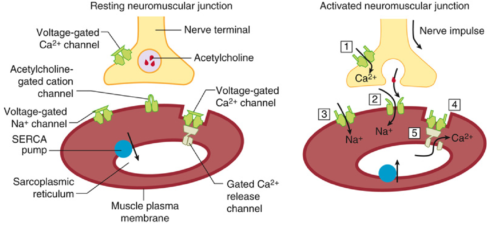
Step 1 - Normal Excitation-Contraction Coupling (The Baseline)
- A nerve impulse reaches the motor end plate and triggers acetylcholine (ACh) release.
- ACh binds nicotinic receptors (nAChR), depolarizing the sarcolemma.
- The action potential propagates along the sarcolemma and travels down transverse tubules (T tubules) - invaginations that form triad junctions with the SR terminal cisternae.
- The depolarization is sensed by Cav1.1 (the dihydropyridine receptor, DHPR) - the voltage-gated Ca²⁺ channel in the T tubule membrane.
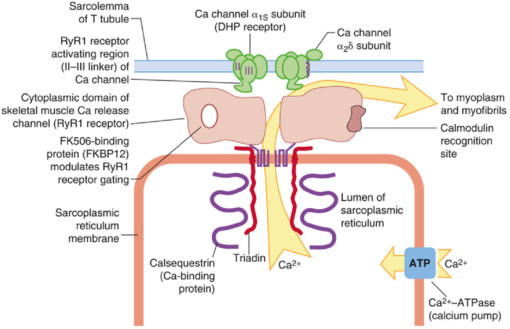
- Cav1.1 physically communicates - via conformational change, mediated by the adaptor protein STAC3 - with the Type 1 Ryanodine Receptor (RyR1), the Ca²⁺ release channel on the SR membrane.
- RyR1 opens, releasing a large bolus of Ca²⁺ from the SR lumen into the sarcoplasm.
- Ca²⁺ binds troponin C, displaces tropomyosin, and exposes actin's myosin-binding sites → muscle contraction.
- SERCA pumps (SR Ca²⁺-ATPases) then actively re-sequester Ca²⁺ back into the SR. When sarcoplasmic Ca²⁺ drops below 10⁻⁶ M, the muscle relaxes; the resting level is restored to ~10⁻⁷ M.
Step 2 - The Molecular Defect: RyR1 Mutations
RyR1 - Structure and Function
- Region 1 (N-terminal): amino acids 35-614
- Region 2 (Central/sarcoplasmic foot): amino acids 2129-2458
- Region 3 (C-terminal transmembrane/pore): amino acids 3916-4942
Step 3 - The Triggering Event and Ca²⁺ Cascade
- The mutant RyR1 channel opens uncontrollably - it cannot be adequately suppressed by normal inhibitory signals (Mg²⁺, calmodulin).
- Ca²⁺ floods out of the SR into the sarcoplasm in a sustained, unregulated manner.
- Sarcoplasmic Ca²⁺ rises far above the contractile threshold (10⁻⁶ M) and cannot be normalized.
- Excitation-coupled Ca²⁺ entry (ECCE): MH mutations enhance entry of extracellular Ca²⁺ into the cell, not just release from SR stores.
- Store-operated Ca²⁺ entry (SOCE): SR Ca²⁺ depletion activates additional sarcolemmal channels to import more extracellular Ca²⁺, further worsening the crisis.
Step 4 - The Hypermetabolic Cascade
| Pathological Process | Mechanism |
|---|---|
| Sustained muscle contracture / rigidity | Ca²⁺ remains above the contractile threshold; muscle cannot relax |
| Massively increased ATP consumption | SERCA pumps work maximally trying (and failing) to re-sequester Ca²⁺; sustained actin-myosin cycling also consumes ATP |
| Heat production (hyperthermia) | ATP hydrolysis and futile Ca²⁺ cycling generate enormous heat - temperatures can rise at 1°C per 5 minutes |
| Increased O₂ consumption and CO₂ production | Aerobic and anaerobic metabolism surge to regenerate ATP; early sign is rising PaCO₂ |
| Metabolic acidosis | Lactate accumulates from anaerobic glycolysis; CO₂ retention adds a respiratory component |
| Rhabdomyolysis | Sustained Ca²⁺ activates Ca²⁺-dependent proteases (calpains), phospholipases, and eventually leads to sarcomere destruction; CK peaks on day 2-3 |
| Hyperkalemia | Massive release of intracellular K⁺ from damaged myocytes; compounded by rhabdomyolysis |
| Hypocalcemia | Ca²⁺ is sequestered into damaged cells and precipitates with phosphate from lysed muscle |
Step 5 - Systemic Complications
- Acute kidney injury - from myoglobinuria (myoglobin precipitates in renal tubules, especially in an acid environment)
- Cardiac arrhythmias / arrest - from hyperkalemia, acidosis, and hyperthermia
- Disseminated intravascular coagulopathy (DIC) - released thromboplastins from lysed muscle activate the coagulation cascade
- Multi-organ failure - if not treated promptly
Genetics Summary
| Gene | Protein | % of MHS cases |
|---|---|---|
| RYR1 | Type 1 Ryanodine Receptor | 50-80% |
| CACNA1S | Cav1.1 (α1s subunit of DHPR) | ~1% |
| Unknown loci | - | ~20-50% |
Non-Anesthetic Triggers
- Vigorous exercise in a hot environment (exertional heat stroke variant)
- Neuroleptic agents - trigger a clinically similar syndrome (neuroleptic malignant syndrome) by raising myoplasmic Ca²⁺ via a different mechanism; occurs in ~0.2% of patients receiving neuroleptics
Why Dantrolene Works
- Miller's Anesthesia, 10e, pp. 4525-4536 - primary molecular and pathophysiological detail
- Katzung's Basic & Clinical Pharmacology, 16e, p. 704
- Goldman-Cecil Medicine
- DiMaio's Forensic Pathology, 3e, p. 416
Search for recent clinical guidelines on MH diagnosis and management
"malignant hyperthermia" AND (guidelines OR management OR diagnosis)
malignant hyperthermia clinical guidelines 2023 2024 2025 MHAUS EMHG diagnosis management
"malignant hyperthermia" AND ("European Malignant Hyperthermia Group" OR EMHG OR dantrolene OR "clinical grading scale")
https://www.nysora.com/education-news/updated-guidelines-for…
PMID: 39482150
PMID: 41478797
PMID: 39171998
https://pmc.ncbi.nlm.nih.gov/articles/PMC12860801
Recent Clinical Guidelines on MH Diagnosis and Management
Key Guideline Documents (2024-2026)
| Guideline | Body | Published | Focus |
|---|---|---|---|
| Recognition and management of an MH crisis | European MH Group (EMHG) | Jan 2025 (BJA) | Acute crisis management |
| Investigation of MH susceptibility | EMHG | Feb 2026 (BJA) | Diagnostic pathways, contracture testing, genetics |
| JSA Guideline for MH management 2025 | Japanese Society of Anesthesiologists | 2025 (PMC) | Comprehensive perioperative management |
| Expert Review: MH in the ICU | Pinyavat et al., Critical Care Medicine | Dec 2024 | ICU-focused management |
Part 1 - Diagnosis
Clinical Recognition: Pattern-Based, Not Single-Sign
- Sudden, inappropriate rise in end-tidal CO₂ (ETCO₂) - often the first sign
- Increased O₂ consumption
- Mixed metabolic and respiratory acidosis
- Tachycardia
- Profuse sweating and skin mottling
- Rapidly rising core temperature (can reach +1°C per 5 min)
- Severe hyperkalemia
- Muscle rigidity (may include masseter spasm after succinylcholine)
- Elevated CK and myoglobin
- Dark/cola-colored urine (myoglobinuria)
- Ventricular arrhythmias, cardiac arrest
- DIC
MH Clinical Grading Scale (CGS)
| Process | Finding | Points |
|---|---|---|
| I - Muscle Rigidity | Generalized rigidity | 15 |
| Masseter rigidity | 15 | |
| II - Myonecrosis | CK >20,000 (post-succinylcholine) | 15 |
| CK >10,000 (no succinylcholine) | 15 | |
| Cola-colored urine | 10 | |
| Myoglobin in urine >60 mg/L | 5 | |
| Serum K⁺ >6 mEq/L | 3 | |
| III - Respiratory Acidosis | PETCO₂ >55 mmHg (controlled ventilation) | 15 |
| PaCO₂ >60 mmHg (controlled ventilation) | 15 | |
| Inappropriate hypercarbia | 15 | |
| Inappropriate tachypnea | 10 | |
| IV - Temperature Increase | Rapid temperature rise | 15 |
| Perioperative temperature >38.8°C | 10 | |
| V - Cardiac Involvement | Inappropriate tachycardia | 3 |
| Ventricular tachycardia or fibrillation | 3 | |
| VI - Family History | Positive family history | 15 |
Differential Diagnosis
- Inadequate anesthesia / analgesia
- Sepsis / malignant hyperpyrexia from infection
- Anaphylaxis
- Thyroid storm
- Pheochromocytoma
- Neuroleptic malignant syndrome (similar mechanism, different trigger)
- Serotonin syndrome
- Recreational drug toxicity (MDMA)
- Equipment malfunction (faulty CO₂ analyzer, rebreathing)
Confirmatory Testing (Post-Crisis)
In Vitro Contracture Test (IVCT) / Caffeine-Halothane Contracture Test (CHCT)
- Gold standard for confirming susceptibility
- Requires fresh skeletal muscle biopsy from a specialized center
- Muscle is exposed to caffeine and halothane; an abnormal contracture response at sub-threshold concentrations indicates susceptibility
- Classified as MHS (susceptible), MHE (equivocal), or MHN (normal)
- Must be done at an EMHG-accredited center
- A negative result does not definitively rule out MH (sensitivity is not 100%)
Genetic Testing
- Focused on RYR1 variants (identified in ~50-70% of susceptible individuals) and CACNA1S variants (<1%)
- The 2025 EMHG guideline introduces an updated curation system for classifying genetic variants by their pathogenic relevance to MH
- Negative genetic testing cannot rule out susceptibility
- Positive for a known pathogenic variant = MH genotype designation (new 2025 category)
- Important for family screening after a clinical event
Elevated resting CK
- Persistently elevated CK (>3x upper limit of normal) without other explanation should raise suspicion, particularly in patients with a family history of anesthetic deaths
Part 2 - Acute Management
Immediate Steps (First Minutes) - 2024 EMHG Protocol
- Remove volatile anesthetics (turn off vaporizer, disconnect from machine)
- Discontinue succinylcholine if still infusing
- Notify the surgical team; complete or abort surgery as quickly and safely as possible
- Set flow rate ≥10 L/min
- Double to triple normal minute ventilation to reduce anesthetic circuit concentrations and treat CO₂ elevation
- Coordinated multidisciplinary team response required
- In the US: call MHAUS hotline (1-800-MH-HYPER) for real-time expert guidance
- Propofol-based anesthesia is safe; benzodiazepines, opioids, non-depolarizing NMBAs are also safe
- Place on both inspiratory and expiratory limbs of the anesthesia breathing circuit
- Reduces residual volatile agent concentrations rapidly; now recommended as routine during suspected MH
Dantrolene - The Specific Antidote
| Parameter | EMHG 2024 / International Standard | JSA 2025 |
|---|---|---|
| Initial dose | 2-2.5 mg/kg IV | 1-2 mg/kg IV (per Japanese package insert) |
| Repeat dosing | Repeat 2-2.5 mg/kg every 10 minutes if symptoms persist | Repeat every 10 min, evaluate each time |
| Maximum dose | Up to 10 mg/kg or more if still effective; no absolute ceiling | No upper limit set; 7 mg/kg per Japanese package insert but continue if effective |
| Stop criterion | PaCO₂ <6 kPa (45 mmHg), decreasing temperature, improving rigidity | Same |
| Formulation | Dantrolene 20 mg vials dissolved in 60 mL sterile water; Ryanodex (nanosuspension, faster preparation) available | 20 mg/60 mL sterile water |
| Stock requirement | 36 vials immediately accessible + 24 additional within 1 hour (EMHG) | Flexible, institution-determined |
| Prophylactic use | NOT recommended preoperatively | NOT recommended |
Key note: A new formulation, Ryanodex (dantrolene nanosuspension), has improved solubility and allows much faster reconstitution, critical when every minute counts.
Supportive Care
| Complication | Management |
|---|---|
| Hyperthermia | Chilled IV normal saline (up to 50-60 mL/kg); surface cooling with ice packs, cooling blankets; cool peritoneal/bladder lavage in extremis; stop cooling once temperature <38°C to avoid overshoot |
| Acidosis | Hyperventilation to normalize PaCO₂; IV sodium bicarbonate if pH <7.2 |
| Hyperkalemia | Glucose-insulin therapy; calcium gluconate (to stabilize myocardium); sodium bicarbonate; avoid calcium channel blockers with dantrolene (risk of cardiac arrest) |
| Arrhythmias | Amiodarone, magnesium; beta-blockers (esmolol) for persistent tachycardia; avoid calcium channel blockers |
| Rhabdomyolysis / AKI | Aggressive IV crystalloid hydration; forced diuresis with furosemide; target urine output 1-3 mL/kg/hr to flush myoglobin from renal tubules |
| Monitoring | Arterial line for blood gas monitoring; urinary catheter; temperature probe |
Part 3 - Perioperative Management of Known MH-Susceptible Patients
- Avoid all triggering agents: all volatile halogenated anesthetics (halothane, isoflurane, sevoflurane, desflurane) and succinylcholine
- Safe agents: propofol, benzodiazepines, opioids, non-depolarizing NMBAs (e.g., rocuronium, vecuronium), nitrous oxide, local anesthetics, regional/neuraxial anesthesia
- Machine preparation: remove or close vaporizers; replace all disposable circuit components; OR use activated charcoal filters on both circuit limbs (now the preferred, faster method)
- Dantrolene availability: must be on hand before starting anesthesia; do not give prophylactically (causes muscle weakness)
- Preoperative counseling: document MH susceptibility clearly; inform patient, family, and all future healthcare providers; wear MedAlert identification
Part 4 - Post-Crisis Follow-Up
- All suspected MH reactions must be reported to the North American MH Registry (NAMHR) or equivalent national registry
- Refer patient and first-degree relatives to an EMHG-accredited testing center for CHCT/IVCT and/or genetic testing
- Elevated baseline CK levels warrant workup for underlying myopathy
- The 2024 Critical Care Medicine review (PMID: 39171998) specifically highlights that increasing use of inhaled anesthetics in ICU sedation makes MH awareness critical outside the OR setting as well
Summary of What's New (2024-2026)
- Consensus definition of a clinical MH event (EMHG 2025 - first ever)
- MH genotype as a new formal diagnostic designation, separate from CHCT result
- Updated EMHG variant curation system for RYR1/CACNA1S genetic variants
- Activated charcoal filters now routinely recommended during suspected MH (not just for susceptible patients)
- Flexibility in treatment protocols based on local resources (EMHG 2024)
- Specific hyperkalemia management protocols added
- ICU MH awareness emphasized given increasing inhaled anesthetic use in ICU sedation
Explain bronchial blockers with mechanism and varieties type in details
bronchial blocker types Arndt Cohen EZ-Blocker Uniblocker mechanism one-lung ventilation anesthesia
Bronchial Blockers - Mechanism, Types, and Clinical Use
What is a Bronchial Blocker?
Mechanism of Action
How OLV Works with a Bronchial Blocker
- Bronchial occlusion - the inflated cuff creates an airtight seal within the target mainstem bronchus (or lobar bronchus for selective lobar blockade).
- Gas absorption - residual gas distal to the cuff (oxygen and N₂) is gradually absorbed by pulmonary capillaries. Pure oxygen filling causes faster collapse than nitrogen-containing gas mixtures.
- Lung collapse - with no incoming gas, the lung parenchyma collapses over minutes (~17-26 min with BBs; slightly faster with DLTs).
- Continued ventilation - the contralateral lung receives 100% of tidal volume via the standard single-lumen tube (SLT).
- Suction of secretions from the isolated lung (limited by small diameter)
- CPAP oxygen insufflation to the collapsed lung to treat hypoxemia during OLV, without reinflating it
Fiberoptic Bronchoscopy: Essential for All BBs
- Standard 9-Fr blockers: ETT ≥7.0 mm ID with a bronchoscope <4.0 mm diameter
- Larger bronchoscopes require ETT ≥7.5 mm ID
Types of Bronchial Blockers
Category 1 - Integrated Blocker
1. Univent Tube (Fuji Systems, Tokyo, Japan)
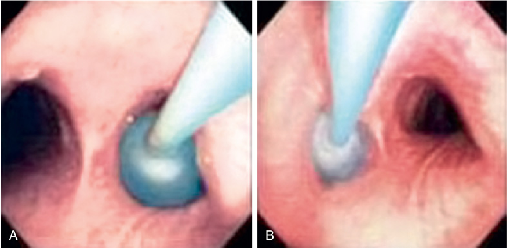
- A modified single-lumen silicone ETT with a small side channel bored into its wall housing a movable endobronchial blocker
- The blocker contains a high-volume, low-pressure balloon and is pre-angled to direct it into the desired bronchus
- The second-generation Torque Control Blocker Univent added a high-friction coefficient silicone tube body, greatly improving rotational control and directability
- Intubate the trachea with the Univent tube in the usual manner
- Under direct FOB vision, advance the blocker from its side channel into the target main bronchus
- Inflate the balloon under FOB visualization to confirm seal
- Single tube - no need to change to a standard SLT if postoperative ventilation is required
- Can apply CPAP or suction through the blocker lumen
- Available in a range of tube sizes
- The blocker channel increases the outer diameter of the tube, making it bulkier
- First generation was difficult to steer (tendency to torque); largely resolved in the second generation
- Particularly challenging to direct into the left main bronchus in some patients
Category 2 - Independent Blockers
2. Arndt Wire-Guided Endobronchial Blocker (Cook Critical Care, Bloomington, IN)
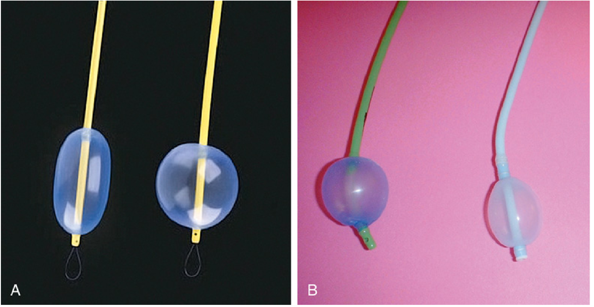
- A multi-lumen catheter with a retractable nylon wire loop at its distal end
- The loop acts as a snare to couple the blocker to the tip of the FOB
- Balloon available in spherical or elliptical shape
- Available in: 9 Fr (adult), 7 Fr (smaller adult), 5 Fr (pediatric)
- Requires ETT ≥8 mm for 9-Fr; ≥7 mm for 7-Fr; ≥4.5 mm for 5-Fr
- The FOB is threaded through the wire loop at the blocker's distal tip
- The FOB-blocker assembly is inserted through the multiport adapter into the ETT
- The FOB navigates to the target bronchus, dragging the blocker along
- Once the balloon is beyond the bronchial opening, the FOB is withdrawn
- The balloon is inflated under FOB visualization with 4-8 mL air
- The wire can then be removed, freeing the 1.6-mm central lumen for suction/CPAP
- The wire can now be re-inserted (current generation) to allow repositioning if the blocker migrates
- Includes a multiport adapter allowing uninterrupted ventilation during placement
- Spherical cuff: better for right mainstem bronchus; elliptical: suitable for either side
- The blocker is advanced somewhat blindly over the FOB - the tip can catch on the carina or ETT Murphy eye
- Requires an ETT of at least 8 mm ID (9-Fr version)
- Once wire is removed (original generation), repositioning requires re-threading
3. Cohen Flexitip Endobronchial Blocker (Cook Critical Care, Bloomington, IN)
- 9-Fr catheter with a rotating wheel at its proximal end that actively deflects the soft distal tip by >90 degrees
- Cuff is a distinctive blue-colored, pear-shaped, high-volume low-pressure balloon
- Cuff inflated via a 0.4-mm lumen inside the blocker wall
- Central lumen: 1.6 mm (suction/CPAP)
- Murphy eye: present
- Insert the blocker through the ETT past the tip, so the blocker tip extends beyond the ETT
- Pass the FOB alongside (not through the blocker) to visualize the carina
- Align the arrow marker on the distal shaft (visible bronchoscopically) toward the target bronchus
- Turn the proximal wheel to deflect the tip toward the target side
- Advance under direct FOB visualization; inflate cuff with 6-8 mL air
- The blocker and FOB do not need to pass through the ETT simultaneously - can be used with ETT as small as 7 mm
- Active tip deflection provides precise steering - particularly useful for left-sided placement
- Blue cuff is easily recognizable bronchoscopically
- More expensive than other blockers
- 9-Fr only (no pediatric option)
4. Fuji Uniblocker (Vitaid, Lewinston, NY)
- 9-Fr, silicone, balloon-tipped catheter with a fixed preformed "hockey-stick" (angled) distal tip
- No active steering mechanism - direction controlled by rotating the catheter body
- High-volume, low-pressure cuff
- Central lumen: 2 mm (the largest of all independent BBs)
- Includes a multiport adapter for passage through standard ETT
- Can also be placed outside/alongside the ETT through the glottis or tracheostomy
- Insert through the multiport adapter
- Rotate the blocker body (turning left or right) to aim the preformed angle toward the target bronchus
- Advance under FOB guidance; inflate cuff
- Largest inner lumen (2 mm) - best for secretion clearance
- Can be used extralaryngeally (alongside the ETT) - useful in pediatric patients where ETT space is limited
- Simple, no components to break
- No active steering - relies entirely on the fixed bend plus rotation, which can be challenging if anatomy is unusual
- Fixed angulation may not suit all patients
- Reported complication of posterior bronchial wall rupture (see below)
5. EZ-Blocker (Rusch / Teleflex, Dresden, Germany / Morrisville, NC)
- 7-Fr, 4-lumen, 75-cm Y-shaped bifurcated catheter
- Two distal limbs, each with an inflatable cuff and a central lumen
- Color-coded limbs: blue (left) and yellow (right) with matching colored pilot balloons
- The bifurcation is designed to mimic the tracheal carina - the Y-junction sits astride the carina
- Insert through a standard ETT (≥7.5 mm) using the included EZ-Multiport adaptor
- Advance until the Y-junction straddles the carina - both distal limbs enter the respective mainstem bronchi
- Inflate the balloon of the desired (operative) side under FOB guidance
- Only the selected lung collapses; the other lung continues to be ventilated normally
- No repositioning needed for bilateral procedures (both lumens are pre-positioned in each bronchus)
- Straightforward placement - the bifurcated design is self-positioning
- Color coding eliminates side confusion
- Central lumens are very small - inadequate for effective suctioning
- Reported complication of bronchial wall perforation, particularly in patients who received prior radiation therapy (fragile bronchial wall) - emphasizes the importance of FOB-guided advancement rather than blind insertion
- The Y-junction can shift off the carina
Comparison Table of All Independent Bronchial Blockers
| Feature | Arndt | Cohen | Uniblocker | EZ-Blocker |
|---|---|---|---|---|
| French size | 5, 7, 9 Fr | 9 Fr | 9 Fr | 7 Fr |
| Steering/guidance mechanism | Wire loop snares FOB | Rotating wheel deflects tip >90° | Fixed hockey-stick bend + rotation | Bifurcated Y-tip straddles carina |
| Minimum ETT size | 8 mm (9-Fr); 7 mm (7-Fr); 4.5 mm (5-Fr) | 7-8 mm | 8 mm | 7.5-8 mm |
| Central lumen | 1.6-1.8 mm | 1.6-1.8 mm | 2 mm | Too small for suction |
| Murphy eye | Yes (9-Fr) | Yes | No | No |
| Cuff shape | Spherical or elliptical | Pear-shaped | Elliptical | Elliptical |
| Key advantage | FOB-coupled precision guidance | Active tip deflection; fits 7-mm ETT | Largest lumen; extralaryngeal use | Pre-positioned for both bronchi; no repositioning |
| Key disadvantage | Blocker tip not seen during insertion; needs 8-mm ETT | Expensive | No steering; fixed bend | Lumen too small for suction; perforation risk if blind |
Preferred Clinical Indications for Bronchial Blockers Over DLT
| Clinical Situation | Reason BB is Preferred |
|---|---|
| Difficult/abnormal airway (prior neck/oral surgery, predicted difficult intubation) | Secure airway with SLT first (awake nasal/oral intubation or via tracheostomy); then add BB without changing tubes |
| Existing tracheostomy | BB passed through tracheostomy tube; DLT sizes don't fit |
| Need for postoperative mechanical ventilation | No tube exchange needed at end of surgery; avoids airway risks in an edematous airway |
| Prior contralateral pneumonectomy | Selective lobar blockade on remaining lung to improve oxygenation and surgical exposure |
| Pediatric patients | Smaller Arndt (5-Fr) or Fogarty catheter; DLTs are too large for small children |
| Critically ill patients requiring OLV in the ICU | BB placed through existing standard ETT without reintubation |
| Selective lobar collapse | BB can be advanced into a lobar bronchus (not possible with DLT) |
| Endobronchial lesion blocking mainstem bronchus | Contralateral DLT may be preferred but BB can sometimes be advanced past the lesion |
When DLT is Preferred Over Bronchial Blocker
- Both lungs must be ventilated independently (bilateral transplant, bilateral sympathectomy)
- Protection of dependent lung from contamination (massive hemoptysis, abscess) - BB cuff seal less reliable under high pressures
- Tumor occupying the mainstem bronchus / sleeve resection (bronchial anastomosis in operative field)
- Speed: DLT is faster to place and achieves faster lung collapse (~17 min DLT vs ~26 min BB)
Position Confirmation
- Fiberoptic bronchoscopy - primary method; visualize the cuff just at/below the bronchial orifice with the top of the cuff visible at the carina
- Clinical assessment: asymmetric chest movement, breath sounds
- Pressure/volume-flow profile changes
- Re-check in lateral decubitus - position must always be re-verified after repositioning the patient
Complications
| Complication | Details |
|---|---|
| Blocker migration / dislodgement | Most common issue; higher rate than DLT; blocker must be locked inside ETT |
| Cuff inflation in the trachea | Inflation above the carina causes bilateral airway obstruction, hypoxia, cardiac arrest - must be recognized and balloon deflated immediately |
| Failure to achieve lung collapse | Due to abnormal anatomy or inadequate seal; may require suction through lumen to hasten collapse |
| Balloon entrapment in staple line | During lobectomy - surgical team must be told of BB presence; withdraw BB several cm before stapling |
| Bronchial wall rupture | Reported with Fuji Uniblocker (rigid preformed tip) and EZ-Blocker (blind advancement) - use FOB guidance at all times |
| Balloon failure to deflate | Reported with Fuji blocker; test balloon inflation/deflation before use |
| Slow lung collapse | Inherent to all BBs due to small central lumen; suctioning through the lumen accelerates collapse |
| Postoperative sore throat / hoarseness | Less common with BBs than DLTs (meta-analysis of 39 RCTs) |
Fogarty Catheter - The Historical Option
- Miller's Anesthesia, 10e, pp. 7161-7168 - primary reference
- Barash Clinical Anesthesia, 9e, pp. 3194-3200 - comparison table and device-specific descriptions
- Morgan & Mikhail's Clinical Anesthesiology, 7e, pp. 1033-1034