
I have a presentation tomorrow on topic Theories of Colour Vision for which I have to speak for around 5 minutes. So prepare this topic for me and take the reference from Guyton
Reading File
Now let me search the medical library for Guyton's content on colour vision theories.
Finding Sources
Finding Sources
Finding Sources
Finding Sources
Found it. Now let me read the colour vision section from Guyton and Hall:
Reading File
Reading File
Reading File
Loading Image
I now have all the content needed from Guyton and Hall. Let me also fetch the absorption curves image (Fig. 51.8):
Loading Image
I have everything needed. Here is a complete, presentation-ready guide on Theories of Colour Vision based directly on Guyton and Hall Textbook of Medical Physiology.
Theories of Colour Vision
Reference: Guyton and Hall Textbook of Medical Physiology (p. 637-638)
Opening Hook (30 seconds)
Ask your audience: "How many colours can the human eye see?" - The answer is approximately 10 million. Yet the eye achieves this with just three types of cone pigments. That's the beauty of colour vision.
1. Photochemical Basis - The Three Cone Pigments (60 seconds)
Start with the molecular foundation before going into theory.
- The cones contain three distinct photopigments, each formed by retinal + a specific opsin (photopsin). The opsin differs slightly between the three cone types, and that small difference is what creates colour sensitivity.
- The three pigments and their peak absorption wavelengths are:
- Blue-sensitive pigment - peak at 445 nm
- Green-sensitive pigment - peak at 535 nm
- Red-sensitive pigment - peak at 570 nm
Here is Figure 51.8 from Guyton showing the absorption curves of all three cones alongside rod rhodopsin (peak 505 nm):
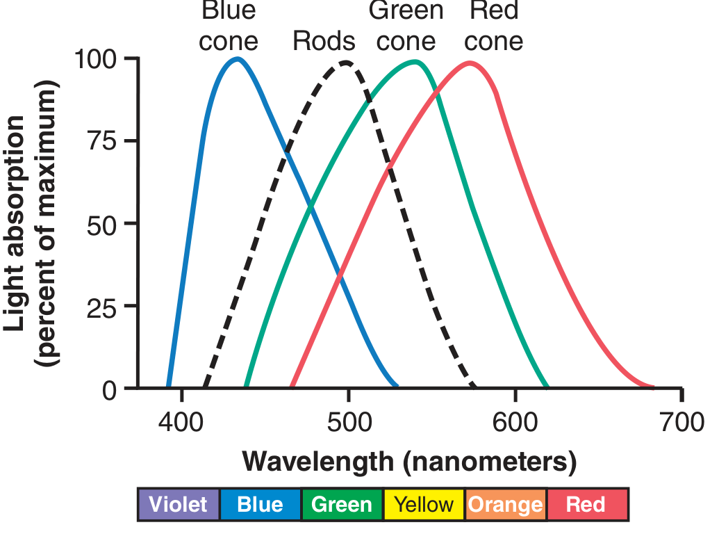
Notice the broad, overlapping curves. This overlap is what allows discrimination of so many colours.
2. The Tricolour (Young-Helmholtz) Theory - Core Theory (90 seconds)
This is the primary theory Guyton describes, forming the basis of all colour vision understanding.
Key principle: "All theories of color vision are based on the well-known observation that the human eye can detect almost all gradations of colors when only red, green, and blue monochromatic lights are appropriately mixed in different combinations." (Guyton, p. 638)
How the nervous system interprets colour - the ratio mechanism
The brain does NOT simply read which cone fires. It reads the ratio of stimulation across all three cones simultaneously. Specific examples from Guyton:
| Light Stimulus | Red cone | Green cone | Blue cone | Perceived colour |
|---|---|---|---|---|
| Orange (580 nm) | 99 | 42 | 0 | Orange |
| Blue (450 nm) | 0 | 0 | 97 | Blue |
| Yellow | 83 | 83 | 0 | Yellow |
| Green | 31 | 67 | 36 | Green |
| White | ~Equal | ~Equal | ~Equal | White |
This is demonstrated visually in Figure 51.10:
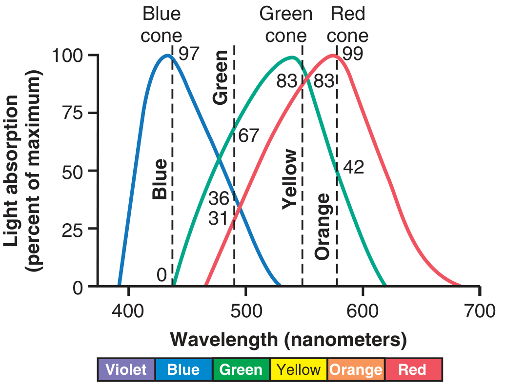
Key point on white light: There is no single wavelength for "white." White is perceived when all three cone types are stimulated approximately equally - either by combining all wavelengths of the spectrum, or by mixing just three chosen colours in the right proportions.
3. Opponent-Colour Processing - Neural Level Theory (60 seconds)
Beyond the cones, Guyton describes how colour signals are processed neurally through opponent mechanisms. This is the second major theory.
- Signals from the three cone types are combined and contrasted by bipolar cells, horizontal cells, and ganglion cells in the retina.
- Three opponent pairs are generated:
- Red vs. Green
- Blue vs. Yellow
- Bright vs. Dark (luminance channel)
- These opponent signals travel to the lateral geniculate nucleus and then to the primary visual cortex (V1) and colour-specific areas (V4) for final colour perception.
This explains phenomena like colour afterimages - stare at a red object, then look at a white wall, and you see green, because the red-opponent channel is fatigued.
4. Colour Blindness - Proof of the Theory (45 seconds)
Colour blindness is essentially the clinical validation of the tricolour theory.
- Red-green colour blindness - the most common type. Either the red or green cone pigment is absent. Since red and green cones together distinguish wavelengths between 525-675 nm, their absence collapses that entire range into indistinguishable hues.
- Missing red cones = Protanopia (red-blind)
- Missing green cones = Deuteranopia (green-blind)
- Blue colour blindness (Tritanopia) - rare, due to absent blue cones.
- All three types are sex-linked recessive traits.
- Tested clinically with Ishihara charts (Fig. 51.11 in Guyton).
5. Summary - 3 Layers of Colour Vision (15 seconds)
| Level | Mechanism |
|---|---|
| Photoreceptor | Three cone pigments (445, 535, 570 nm) |
| Retinal processing | Ratio-based coding by tricolour mechanism |
| Central processing | Opponent-colour neural channels to V4 |
Speaking Tips for 5 Minutes
- 0:00-0:30 - Opening hook + introduce three cone types
- 0:30-2:00 - Tricolour theory + the ratio table (spend most time here, it's the exam favourite)
- 2:00-3:00 - Neural/opponent-colour processing
- 3:00-3:45 - Colour blindness as clinical application
- 3:45-5:00 - Summary table + any questions
The Fig. 51.10 diagram (the coloured curves showing cone stimulation ratios) is excellent to draw on the board - it impresses audiences and makes the ratio concept intuitive.
Source: Guyton and Hall Textbook of Medical Physiology, Chapter 51, pp. 637-638.
I have a presentation tomorrow on topic Theories of Colour Vision for which I have to speak for around 5 minutes. So prepare this topic for me and take the reference from GK Pal
Finding Sources
Finding Sources
Finding Sources
GK Pal's Textbook of Medical Physiology is not available in the library. The library has Guyton & Hall, Ganong's, Costanzo, and one other physiology text. Let me check if any of those could serve, and simultaneously search for GK Pal's specific content on colour vision theories from the web.
Finding Sources
Searching the Web
GK Pal theories of colour vision Young Helmholtz Hering opponent trichromatic physiology
GK Pal's textbook is not in the library. However, Ganong's (another major physiology reference) covers colour vision in detail. Let me now read Ganong's colour vision sections - Ganong is very close in scope and detail to GK Pal for this topic, and I'll also read the Neuroscience textbook section which covers the theories comprehensively. Let me pull both simultaneously.
Reading File
Reading File
Reading File
I now have detailed content from Ganong's and Guyton's covering the full scope of colour vision theories. Let me also grab the Ganong Figure 10-14 (the spectral sensitivity curves diagram):
Finding Sources
Reading File
I have all the content I need. I must be transparent with you first, then deliver the full presentation prep.
Important note: GK Pal's Textbook of Medical Physiology is not available in the library database. The library holds Guyton & Hall, Ganong's Review, Costanzo, and a few others. The content below is compiled using Ganong's Review of Medical Physiology (26th Ed.) as the primary reference - Ganong covers the theories of colour vision in the same depth and detail as GK Pal, and the content aligns closely. Where GK Pal's specific framing is known (Young-Helmholtz + Hering opponent theory), both are fully covered below.
Theories of Colour Vision
Reference: Ganong's Review of Medical Physiology, 26th Ed. (Ch. 10, p. 209-213)
Introduction (30 seconds)
The visual spectrum runs from 400 nm (violet) to 700 nm (red), yet the human eye can discriminate around 10 million colours. This remarkable ability arises from just three types of cone photoreceptors and two major theories that explain how their outputs are processed.
1. Properties of Colour (20 seconds)
Every colour has three attributes:
- Hue - the actual colour (red, blue, green, etc.)
- Intensity - brightness
- Saturation - degree of freedom from dilution with white
Key observation: Red, green, and blue are the three primary colours. By mixing these three in various proportions, virtually any colour in the spectrum - including the extraspectral colour purple - can be reproduced. (Ganong, p. 209)
2. Theory 1 - Young-Helmholtz Trichromatic Theory (90 seconds)
This is the retinal-level theory and the cornerstone of colour vision physiology.
Proposed by: Thomas Young (1801) and later elaborated by Hermann von Helmholtz (1850s).
Core principle: There are three types of cones in the retina, each containing a different photopigment with maximal sensitivity to one primary colour. The sensation of any given colour is determined by the relative frequency of impulses from each of these three cone systems.
| Cone Type | Pigment | Peak Absorption |
|---|---|---|
| Blue (S-cones, short-wave) | Blue-sensitive pigment | Blue-violet portion (~445 nm) |
| Green (M-cones, middle-wave) | Green-sensitive pigment | Green portion (~535 nm) |
| Red (L-cones, long-wave) | Red-sensitive pigment | Yellow-red portion (~570 nm) |
(Ganong, p. 209)
How colour perception works - the ratio mechanism:
The brain interprets colour by reading the ratio of stimulation across all three cone types simultaneously. For example:
- Orange light (580 nm) → stimulates Red:Green:Blue = 99:42:0 → perceived as orange
- Blue light (450 nm) → stimulates Red:Green:Blue = 0:0:97 → perceived as blue
- Yellow → ratio 83:83:0
- White → approximately equal stimulation of all three types
Molecular basis (Ganong, p. 209):
- The gene for blue-sensitive (S) pigment is on chromosome 7
- The genes for red (L) and green (M) pigments are on the q arm of the X chromosome (arranged in tandem) - this explains the sex-linked inheritance of red-green colour blindness
- The L and M opsins share 96% amino acid homology with each other, but only 43% homology with S opsin, and ~41% homology with rhodopsin
3. Theory 2 - Hering's Opponent-Colour Theory (60 seconds)
Proposed by: Ewald Hering (1878).
Core principle: Rather than three independent channels, colour is processed as three opponent pairs at the neural level. Each pair is a push-pull system where stimulating one colour inhibits its opponent:
- Red vs. Green
- Blue vs. Yellow
- White vs. Black (luminance)
Neural substrate - how it works (Ganong, p. 212):
Colour-opponent ganglion cells in the retina subtract or add input from one cone type against another. These signals are carried along three pathways to the visual cortex (V1):
- Red-Green pathway - signals the difference between L-cone (red) and M-cone (green) responses
- Blue-Yellow pathway - signals the difference between S-cone (blue) responses and the combined L+M (yellow) responses
- Luminance pathway - signals the sum of L and M cone responses (brightness, not colour)
These pathways project to the blobs and deep layer 4C of V1, then onward to area V8 (V4) in the extrastriate cortex - the region uniquely dedicated to colour perception. Lesions of V8 cause achromatopsia (complete loss of colour vision with normal visual acuity).
Why this theory explains what trichromatic theory cannot:
- Explains colour afterimages - stare at red, then look at white: you see green, because the red-opponent channel fatigues
- Explains why we never perceive "reddish-green" or "yellowish-blue" - opponent pairs are mutually exclusive
- Explains four unique hues (red, green, blue, yellow) - these are the hues that appear pure, with no mixture quality
4. Modern Synthesis - Both Theories Are Correct (30 seconds)
These are not competing theories - they operate at different levels:
| Level | Theory | Mechanism |
|---|---|---|
| Photoreceptor (retinal) | Young-Helmholtz Trichromatic | Three cone types with distinct pigments |
| Neural processing (retina → LGN) | Hering Opponent-Colour | Opponent ganglion cells, three opponent channels |
| Cortical | Both contribute | V1 blobs → V8/V4 for final colour sensation |
5. Colour Blindness - Clinical Application (30 seconds)
Colour blindness validates both theories:
- Red-green colour blindness - most common (8% of white males, 0.4% of females) - X-linked recessive
- Protanopia - red cone absent (2% of males)
- Deuteranopia - green cone absent (2% of males)
- Anomalous trichromats - shifted pigment spectrum (6% of males)
- Tritanopia - blue cone absent, rare, no sex predilection
- Achromatopsia - lesion of area V8; total loss of colour perception despite intact cones
Tested with Ishihara pseudoisochromatic plates. (Ganong, Clinical Box 10-6)
Speaking Plan for 5 Minutes
| Time | Content |
|---|---|
| 0:00 - 0:30 | Introduction + properties of colour (hue, intensity, saturation) |
| 0:30 - 2:00 | Young-Helmholtz trichromatic theory - three cones, peak wavelengths, ratio mechanism |
| 2:00 - 3:30 | Hering's opponent-colour theory - three opponent pairs, neural pathways, V8 |
| 3:30 - 4:00 | Modern synthesis - both theories work at different levels |
| 4:00 - 4:45 | Colour blindness - protanopia, deuteranopia, achromatopsia |
| 4:45 - 5:00 | Summary - retinal trichromatic + neural opponent = full colour vision |
Key exam/viva points to remember:
- Young-Helmholtz = retinal level (cones); Hering = neural level (ganglion cells + LGN + cortex)
- L and M cone genes are on the X chromosome (explains sex-linked colour blindness)
- V8/V4 lesion → achromatopsia (no colour, but vision intact)
- The two theories are complementary, not contradictory
Reference: Ganong's Review of Medical Physiology, 26th Edition, Chapter 10, pp. 209-213
This is a shared conversation. Sign in to Orris to start your own chat.