
Give a short note like flow chart to explain the japanese encephalitis
Reading File
Japanese Encephalitis (JE) is a viral encephalitis caused by a flavivirus, transmitted by Culex mosquitoes, with pigs and wading birds as amplifying hosts. Let me pull detailed content from the medical library.
Searching sources...
Finding Sources
Finding Sources
Good hits. Let me read the most comprehensive sources in parallel - Park's PSM (epidemiology/public health focus), Goldman-Cecil (clinical), and Bradley-Daroff (neurology).
Reading File
Reading File
Reading File
Excellent - I now have rich, detailed information from Goldman-Cecil Medicine and Bradley-Daroff's Neurology. Let me now compile a clear flowchart-style note.
Japanese Encephalitis - Flowchart Note
1. AGENT & TRANSMISSION
Causative Agent
└── Japanese Encephalitis Virus (JEV)
    ├── Family: Flaviviridae, Genus: Flavivirus
    └── RNA virus (same family as Dengue, Zika, West Nile)

Vector
└── Culex tritaeniorhynchus mosquito
    ├── Ground-dwelling; breeds in flooded rice fields
    └── Peak biting: dusk to dawn (outdoors)

Transmission Cycle
Amplifying Hosts ──────────────────────────────────────────────┐
(Pigs + Wading Birds/Ducks)                                    │
        ↑          ↓                                            │
   Culex mosquito ← bites ← Pig/Bird (high viremia)            │
        │                                                       │
        ↓ bites human (DEAD-END HOST)                           │
       Human (low viremia - cannot amplify)
2. PATHOGENESIS
Infected Mosquito Bite
        │
        ▼
Virus replicates in SKIN
        │
        ▼
Spreads to Draining Lymph Nodes + Spleen
        │
        ▼
PRIMARY VIREMIA
(usually sub-clinical - most infections stop here → asymptomatic)
        │
        ▼ (in ~0.5% of infections)
SECONDARY VIREMIA
        │
        ▼
End-Organ Invasion
├── Brain (thalamus, basal ganglia, midbrain, cerebellum)
└── Spinal Cord
        │
        ▼
Apoptotic cell death + Inflammatory immune response
        │
        ▼
ENCEPHALITIS / MENINGOENCEPHALITIS
Only 1 in 200 infections results in severe disease. The majority are asymptomatic.
3. CLINICAL SPECTRUM
Incubation Period: 6-16 days
        │
        ▼
┌──────────────────────────────────────────────────────────────┐
│                     CLINICAL SPECTRUM                        │
├──────────────────────────────────────────────────────────────┤
│ ASYMPTOMATIC ──── Most common (>99% of infections)           │
│                                                              │
│ MILD FEBRILE ILLNESS                                         │
│  └── Fever, headache, nausea, vomiting, abdominal pain       │
│                                                              │
│ ASEPTIC MENINGITIS                                           │
│  └── Meningismus without brain parenchymal involvement       │
│                                                              │
│ ENCEPHALITIS (Severe, ~0.5%)                                 │
│  ├── Prodrome (2-4 days): Fever, headache, drowsiness        │
│  ├── Excitability / Delirium                                 │
│  ├── Seizures (common, especially in children)               │
│  ├── Hyperthermia                                            │
│  ├── Expressionless facies + Axial rigidity                  │
│  ├── Limb tremors + Myoclonus                                │
│  ├── Erratic eye movements + Cranial nerve palsies           │
│  ├── Ataxia (cerebellar)                                     │
│  ├── Limb paresis (LMN-type weakness)                        │
│  └── Coma → Death                                            │
└──────────────────────────────────────────────────────────────┘
4. DIAGNOSIS
Clinical Suspicion (endemic area + encephalitis)
        │
        ▼
┌─────────────────────────────────────────┐
│          INVESTIGATIONS                 │
├─────────────────────────────────────────┤
│ Blood:                                  │
│  └── JEV-specific IgM ELISA (serum)     │
│       Detectable by Day 7 of illness    │
│                                         │
│ Lumbar Puncture (CSF):                  │
│  ├── Pleocytosis: 10-500 WBC/μL         │
│  │    (early: PMN; late: lymphocytes)   │
│  ├── Protein: 50-100 mg/dL (elevated)   │
│  ├── Glucose: NORMAL                    │
│  └── JEV IgM ELISA in CSF              │
│       Sensitivity 65-70%, Spec 90-100%  │
│                                         │
│ MRI Brain:                              │
│  └── T2 hyperintensity in THALAMUS      │
│       (also: basal ganglia, midbrain,   │
│        cerebellum)                      │
│                                         │
│ Gold Standard:                          │
│  └── Plaque Reduction Neutralization   │
│       Test (PRNT)                       │
│  OR 4-fold rise in IgG titer            │
│                                         │
│ Early disease:                          │
│  └── RT-PCR (blood/CSF - detects        │
│       virus before antibody appears)    │
└─────────────────────────────────────────┘
5. TREATMENT
NO SPECIFIC ANTIVIRAL THERAPY AVAILABLE

(Interferon alfa-2a failed in RCT - no benefit on mortality or sequelae)

SUPPORTIVE CARE ONLY:
├── Control seizures (anticonvulsants)
├── Manage raised intracranial pressure
├── Airway and respiratory support (if comatose)
└── Prevention of secondary infections
6. PROGNOSIS
Case Fatality Rate: 20-30%
        │
        ├── Poor prognosis: prolonged fever, seizures, coma,
        │    high CNS viral load
        │
        └── Survivors (30-50% have long-term neurological sequelae):
             ├── Parkinsonism
             ├── Seizure disorders
             ├── Paresis / Paralysis
             ├── Cognitive impairment / Mental retardation
             └── Neuropsychiatric disorders
7. PREVENTION & CONTROL
PRIMARY PREVENTION
├── VACCINATION (most effective)
│    ├── Live-attenuated vaccine
│    ├── Recommended: Children 1-15 years (endemic areas)
│    ├── Travelers: 2 doses at Day 0 and Day 28
│    └── Highly efficacious even after first dose
│
├── VECTOR CONTROL
│    ├── Larvivorous fish in breeding sites
│    ├── Outdoor fogging with malathion (outbreaks)
│    └── NOT indoor residual spray (vectors are outdoor resters)
│
└── PERSONAL PROTECTION
     ├── Bed nets (dusk to dawn)
     ├── Protective clothing (full body cover)
     └── Keep pigs away from human dwellings

SECONDARY PREVENTION
├── Early diagnosis and case management
├── Sentinel surveillance at tertiary hospitals
└── Health education / community awareness
QUICK SUMMARY TABLE
| Feature | Detail |
|---|---|
| Virus | Flavivirus (RNA) |
| Vector | Culex tritaeniorhynchus |
| Amplifying hosts | Pigs + wading birds |
| Incubation | 6-16 days |
| % progressing to encephalitis | ~0.5% (1 in 200) |
| CSF glucose | Normal |
| Key MRI finding | T2 hyperintensity in thalamus |
| Diagnosis (first-line) | JEV-specific IgM ELISA (serum/CSF) |
| Treatment | Supportive only (no antiviral) |
| CFR | 20-30% |
| Neurological sequelae | 30-50% of survivors |
| Vaccine | Live-attenuated, 2-dose regimen |
Sources: Goldman-Cecil Medicine; Bradley and Daroff's Neurology in Clinical Practice; Park's Textbook of Preventive and Social Medicine
Give a note on rabies
Rabies is a fatal viral encephalitis caused by a Rhabdovirus, transmitted through the bite of an infected animal. Once symptoms appear, it is virtually 100% fatal - making prevention and post-exposure prophylaxis the cornerstone of management.
Searching sources...
Finding Sources
Finding Sources
Good sources found. Let me read the most comprehensive ones in parallel - Bradley-Daroff (neurology detail), Goldman-Cecil (clinical), Harrison's (pathogenesis), and Park's PSM (epidemiology/prevention).
Reading File
Reading File
Reading File
Finding Sources
Good detail so far. Let me read more on clinical phases, diagnosis, and PEP from Harrison's and Goldman-Cecil.
Reading File
Reading File
Now let me get the PEP details and fetch the pathogenesis diagram.
Reading File
Loading Image
Excellent - I have all the information needed plus the pathogenesis diagram. Now let me compile the full comprehensive note.
RABIES - A Comprehensive Note
1. AGENT & EPIDEMIOLOGY
| Feature | Detail |
|---|---|
| Causative Agent | Rabies virus |
| Family | Rhabdoviridae |
| Genus | Lyssavirus |
| Morphology | Bullet-shaped, single-stranded RNA virus |
| Annual Cases | ~55,000 deaths/year worldwide |
| Distribution | Endemic in Africa, Asia, Latin America, Eastern Europe |
| Main reservoir (developing world) | Domestic dog (most human cases) |
| Main reservoir (USA/developed) | Bats (most human cases) |
| Other reservoirs | Skunks, foxes, raccoons, coyotes, wolves |
2. TRANSMISSION
ANIMAL (infected) ──bite──► HUMAN
                             OR
                  ──scratch──► HUMAN (if saliva contacts wound)
                             OR
           ──mucous membrane exposure──► HUMAN
                             OR
           ──organ/tissue transplant──► HUMAN (rare)
                             OR
           ──aerosol (lab/cave exposure)──► HUMAN (very rare)

❌ Human-to-human transmission is NOT documented (except organ transplant)
⚠️ Bat bites may be unrecognized (tiny puncture wounds)
3. PATHOGENESIS
The pathogenesis diagram below (from Harrison's, 22nd Ed.) summarizes the viral journey:
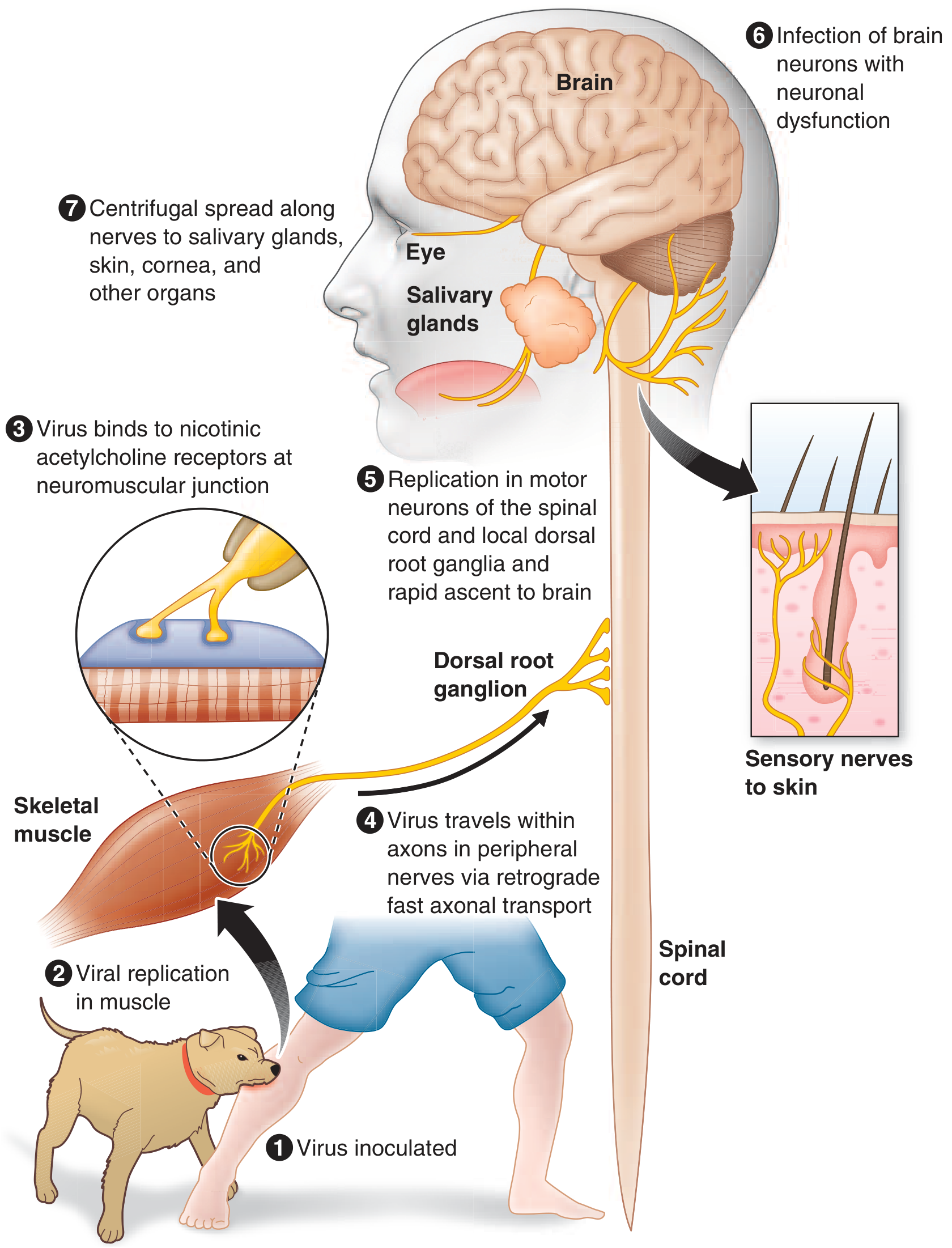
① Virus inoculated at bite site (skin/subcutaneous tissue)
        │
        ▼
② Viral replication in MUSCLE (days to weeks, near the bite site)
        │
        ▼
③ Virus binds nicotinic acetylcholine receptors
   at neuromuscular junctions
        │
        ▼
④ Retrograde fast axonal transport along peripheral nerves
   (centripetal spread, ~250 mm/day; 12-hr delay at each synapse)
        │
        ▼
⑤ Replication in spinal cord motor neurons +
   Dorsal root ganglia → rapid ascent to brain
        │
        ▼
⑥ Brain infection → Neuronal dysfunction
   (brainstem, limbic system maximally affected)
        │
        ▼
⑦ Centrifugal spread along autonomic/sensory nerves
   → Salivary glands, skin, cornea, heart, adrenal glands
   → Virus shed in SALIVA (enables transmission)
Key concept: The long incubation period is because the virus stays near the bite site for most of the incubation period - this is the window for effective PEP.
Pathological findings:
- Negri bodies - eosinophilic cytoplasmic inclusions in neurons (especially Purkinje cells of cerebellum and pyramidal cells of hippocampus) - pathognomonic
- Babes nodules - microglial nodules in CNS parenchyma
- Surprisingly mild degenerative neuronal changes despite fatal outcome
- Neuronal dysfunction (not death) drives clinical disease
4. CLINICAL STAGES
┌────────────────────────────────────────────────────────────────┐
│ STAGE 1: INCUBATION PERIOD                                     │
│ Duration: Usually 20-90 days (range: days to >1 year)          │
│ Symptoms: NONE                                                  │
│ Shorter with: multiple bites, facial bites, deep wounds        │
└─────────────────────────┬──────────────────────────────────────┘
                          │
                          ▼
┌────────────────────────────────────────────────────────────────┐
│ STAGE 2: PRODROME                                              │
│ Duration: 2-10 days                                            │
│ ├── Fever, malaise, headache, anorexia, nausea, vomiting       │
│ ├── Anxiety / agitation                                        │
│ └── PATHOGNOMONIC: Paresthesias, pain, or PRURITUS             │
│      at/near the BITE SITE (50-80% of patients)                │
│      (due to infection of dorsal root/cranial sensory ganglia) │
└─────────────────────────┬──────────────────────────────────────┘
                          │
                          ▼
┌────────────────────────────────────────────────────────────────┐
│ STAGE 3: ACUTE NEUROLOGICAL PHASE                              │
│                                                                │
│ TWO FORMS:                                                     │
│                                                                │
│ A. ENCEPHALITIC / FURIOUS RABIES (~80%)                        │
│    ├── Agitation, hyperexcitability, combativeness             │
│    ├── Hallucinations, confusion, seizures                     │
│    ├── Fever (may reach 105-107°F)                             │
│    ├── HYDROPHOBIA - involuntary painful contraction of        │
│    │    pharynx/larynx/diaphragm on swallowing water           │
│    │    (due to brainstem dysfunction)                         │
│    ├── AEROPHOBIA - same spasms triggered by a draft of air    │
│    ├── "Foaming at the mouth" (hypersalivation + pharyngeal    │
│    │    dysfunction)                                           │
│    ├── Autonomic hyperactivity:                                │
│    │    ├── Hypersalivation, gooseflesh                        │
│    │    ├── Cardiac arrhythmias, priapism                      │
│    │    └── SIADH or Diabetes insipidus                        │
│    └── Episodes of lucidity between hyperexcitability          │
│         (become shorter as disease progresses)                 │
│                                                                │
│ B. PARALYTIC / DUMB RABIES (~20%)                              │
│    ├── Flaccid muscle weakness (LMN type)                      │
│    ├── Begins in bitten limb → spreads to quadriparesis        │
│    ├── Facial weakness common                                  │
│    ├── Sphincter involvement                                   │
│    ├── NO hydrophobia / aerophobia                             │
│    ├── Mimics Guillain-Barré syndrome                          │
│    └── Survives a few days longer than encephalitic form       │
└─────────────────────────┬──────────────────────────────────────┘
                          │
                          ▼
┌────────────────────────────────────────────────────────────────┐
│ STAGE 4: COMA                                                  │
│ ├── Cardiopulmonary failure                                    │
│ ├── Noncardiogenic pulmonary edema                             │
│ ├── GI hemorrhage                                              │
│ └── DEATH (virtually 100% fatal once symptoms appear)          │
└────────────────────────────────────────────────────────────────┘
5. DIAGNOSIS
CLINICAL SUSPICION
(encephalitis + animal bite history + hydrophobia/aerophobia)
        │
        ▼
┌────────────────────────────────────────────────────────────────┐
│                      INVESTIGATIONS                            │
├────────────────────────────────────────────────────────────────┤
│ CSF:                                                           │
│  ├── Mild mononuclear pleocytosis (<100 WBC/μL) in >50%       │
│  ├── Mildly elevated protein                                   │
│  └── Glucose: NORMAL                                           │
│      (Severe pleocytosis >1000 → look for alternate diagnosis) │
│                                                                │
│ Imaging (CT/MRI):                                              │
│  ├── CT head: Usually NORMAL                                   │
│  └── MRI: T2/FLAIR signal in brainstem, basal ganglia,        │
│       thalamus, midbrain (variable; nonspecific)               │
│                                                                │
│ Specific Tests:                                                │
│  ├── RT-PCR: saliva, CSF, skin biopsy, brain tissue           │
│  ├── Skin biopsy (nape of neck): Rabies antigen in            │
│  │    sensory nerve endings (immunofluorescence/               │
│  │    immunohistochemistry)                                    │
│  ├── Serum/CSF IgM/IgG antibodies                             │
│  │    (not detectable until 2nd week; patient may die first)   │
│  └── Brain biopsy/autopsy: Negri bodies (H&E stain);          │
│       immunofluorescence (more sensitive)                      │
└────────────────────────────────────────────────────────────────┘
6. POST-EXPOSURE PROPHYLAXIS (PEP)
PEP is effective when started promptly BEFORE symptoms develop.
EXPOSURE OCCURS
        │
        ▼
STEP 1: WOUND CARE (IMMEDIATE - most important first step)
        ├── Wash wound thoroughly with soap and water (≥15 min)
        ├── Apply povidone-iodine or alcohol
        └── Do NOT suture primarily (if possible)
        │
        ▼
STEP 2: ASSESS RISK (Was the animal rabid? Vaccine status?)
        │
        ▼
STEP 3: PASSIVE IMMUNIZATION
        └── Rabies Immunoglobulin (RIG)
             ├── Human RIG (HRIG): 20 IU/kg
             └── Equine RIG (ERIG): 40 IU/kg
             Inject as much as possible INTO and AROUND wound
             Remainder given IM at distant site
             Given ONLY on Day 0 (with 1st vaccine dose)
        │
        ▼
STEP 4: ACTIVE IMMUNIZATION (Rabies Vaccine)
        └── 4-dose regimen (post-2010):
             Day 0 → Day 3 → Day 7 → Day 14
             (Previously 5-dose; 4-dose is now standard)
             IM into deltoid (NOT gluteal region)

        ⚠️ Previously vaccinated individuals:
             → Give vaccine on Day 0 and Day 3 ONLY
             → NO RIG needed
7. PRE-EXPOSURE PROPHYLAXIS (PrEP)
For: Veterinarians, animal handlers, travelers to endemic areas, lab workers
3 doses: Day 0 → Day 7 → Day 21 or Day 28

Even after PrEP, if exposed: still need 2 booster doses (Day 0 + Day 3)
NO RIG required after pre-exposure vaccination
8. TREATMENT (Once Symptomatic)
NO PROVEN SPECIFIC TREATMENT

Antiviral attempts (ribavirin, interferon-alpha): FAILED
Milwaukee Protocol (therapeutic coma): 1 survivor reported;
    subsequent attempts → fatal outcome

Management = PALLIATIVE / SUPPORTIVE CARE only:
├── Seizure control
├── Sedation for agitation
├── Airway management
├── Cardiovascular support
└── Minimize patient suffering
9. PREVENTION (Public Health)
├── DOG VACCINATION (mass campaigns) - single most effective measure
├── Animal bite surveillance and stray dog control
├── Pre-exposure vaccination of high-risk groups
├── Post-exposure prophylaxis for ALL exposures
├── Public health education: seek care after animal bites
└── Avoid handling wild animals / bats
QUICK REFERENCE SUMMARY
| Feature | Key Point |
|---|---|
| Virus | Lyssavirus (Rhabdoviridae), RNA, bullet-shaped |
| Transmission | Bite/scratch/mucous membrane (mainly dog, bat) |
| Incubation | 20-90 days (up to 1 year+) |
| Pathognomonic prodrome sign | Pain/pruritus at bite site |
| Pathognomonic brain finding | Negri bodies |
| Clinical forms | Furious (80%) vs Paralytic (20%) |
| Hallmark clinical signs | Hydrophobia, aerophobia |
| Paralytic rabies mimics | Guillain-Barré syndrome |
| CSF glucose | Normal |
| MRI | T2 signal in brainstem / basal ganglia |
| Best diagnostic test | RT-PCR (saliva/CSF) + skin biopsy (nape) |
| Treatment | Supportive only - virtually 100% fatal |
| PEP vaccine doses | 4 doses: Day 0, 3, 7, 14 |
| RIG dose (human) | 20 IU/kg |
| Mortality once symptomatic | ~100% |
Sources: Harrison's Principles of Internal Medicine 22E; Goldman-Cecil Medicine; Bradley and Daroff's Neurology in Clinical Practice
This is a shared conversation. Sign in to Orris to start your own chat.