
Lap chole
Laparoscopic cholecystectomy is a minimally invasive surgical procedure to remove the gallbladder, most commonly for symptomatic gallstones or cholecystitis. Key steps include trocar placement, pneumoperitoneum creation, dissection of Calot's triangle, clipping and dividing the cystic duct and artery, and extraction of the gallbladder.
Searching sources...
Reading File
The query is broad - "lap chole" could mean many things. Let me clarify before diving deep.
Asking for Preferences
What specifically about laparoscopic cholecystectomy do you need?
Calors triangle
Calot's triangle (hepatocystic triangle) is bounded by the cystic duct inferiorly, the common hepatic duct medially, and the inferior surface of the liver superiorly. The cystic artery (usually a branch of the right hepatic artery) runs within it. Safe dissection of this triangle is the key step in lap chole to achieve the "critical view of safety."
Searching sources...
Finding Sources
Searching Images
Calot's triangle anatomy cystic duct cystic artery hepatocystic triangle
Finding Sources
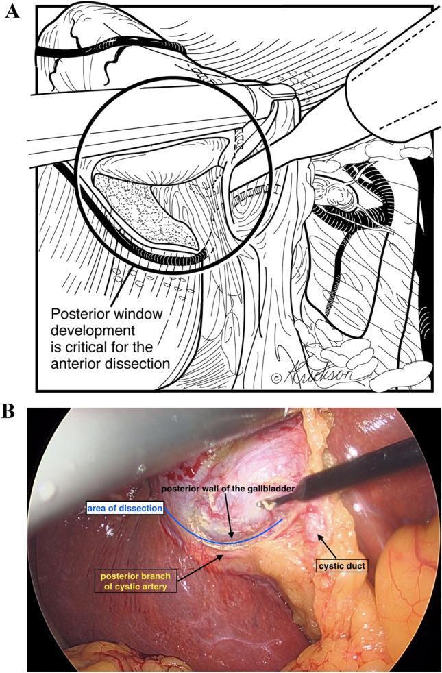
Reading File
Reading File
Reading File
Loading Image
Loading Image
Calot's Triangle (Hepatocystic Triangle)
Definition & Boundaries
Original description (Calot, 1891):
| Border | Structure |
|---|---|
| Inferior | Cystic duct |
| Medial | Common hepatic duct |
| Superior | Cystic artery |
Modern/contemporary definition (hepatocystic triangle) - now the preferred term:
| Border | Structure |
|---|---|
| Inferior/lateral | Cystic duct + medial wall of gallbladder |
| Medial | Common hepatic duct |
| Superior | Inferior surface of the liver |
The cystic artery now lies within this larger triangle rather than forming its roof. This modern definition is what surgeons use during lap chole.
(- Bailey & Love's Surgery 28e, p. 1254; Mulholland & Greenfield Surgery 7e, p. 2781)
Contents
- Cystic artery - the main structure to identify and clip
- Cystic lymph node (Lund's node / node of Mascagni) - lies superficial to the cystic artery; useful landmark when anatomy is difficult
- Connective/areolar tissue
- Occasionally: accessory bile ducts, aberrant right hepatic duct
Cystic Artery - Origin & Variations
The diagram above (Mulholland & Greenfield) shows 6 key variants:
| Variant | Detail |
|---|---|
| A - Normal | Arises from right hepatic artery, behind common hepatic duct |
| B | Off gastroduodenal artery (GDA) |
| C | Double cystic artery - one off GDA |
| D | Off proper hepatic artery |
| E | Anterior to common bile duct |
| F | Off replaced right hepatic artery |
In ~15% of cases, the right hepatic artery and/or cystic artery cross in front of the common hepatic duct and cystic duct - a dangerous variant.
Surgical Significance: Critical View of Safety (CVS)
Before clipping anything in Calot's triangle, the surgeon must achieve the Critical View of Safety:
- The hepatocystic triangle is cleared of all fat and fibrous tissue
- Only two structures are seen entering the gallbladder (cystic duct + cystic artery)
- The lower one-third of the gallbladder is separated from the liver bed
This prevents the most feared complication - bile duct injury - which occurs when the common bile duct is mistaken for the cystic duct.
Anatomical Hazards
- Aberrant right hepatic duct - most common anomaly causing injury during lap chole. Most dangerous variant: cystic duct joining a low-lying aberrant right sectoral duct.
- Classic anatomy (textbook normal) is present in only ~30% of individuals - anomalies are the rule.
- A posterior cystic artery can be easily injured if not recognized.
- The right hepatic artery may "caterpillar loop" onto the gallbladder surface with a very short cystic artery arising from it.
(- Maingot's Abdominal Operations, p. 1017-1018; Schwartz's Principles of Surgery 11e)
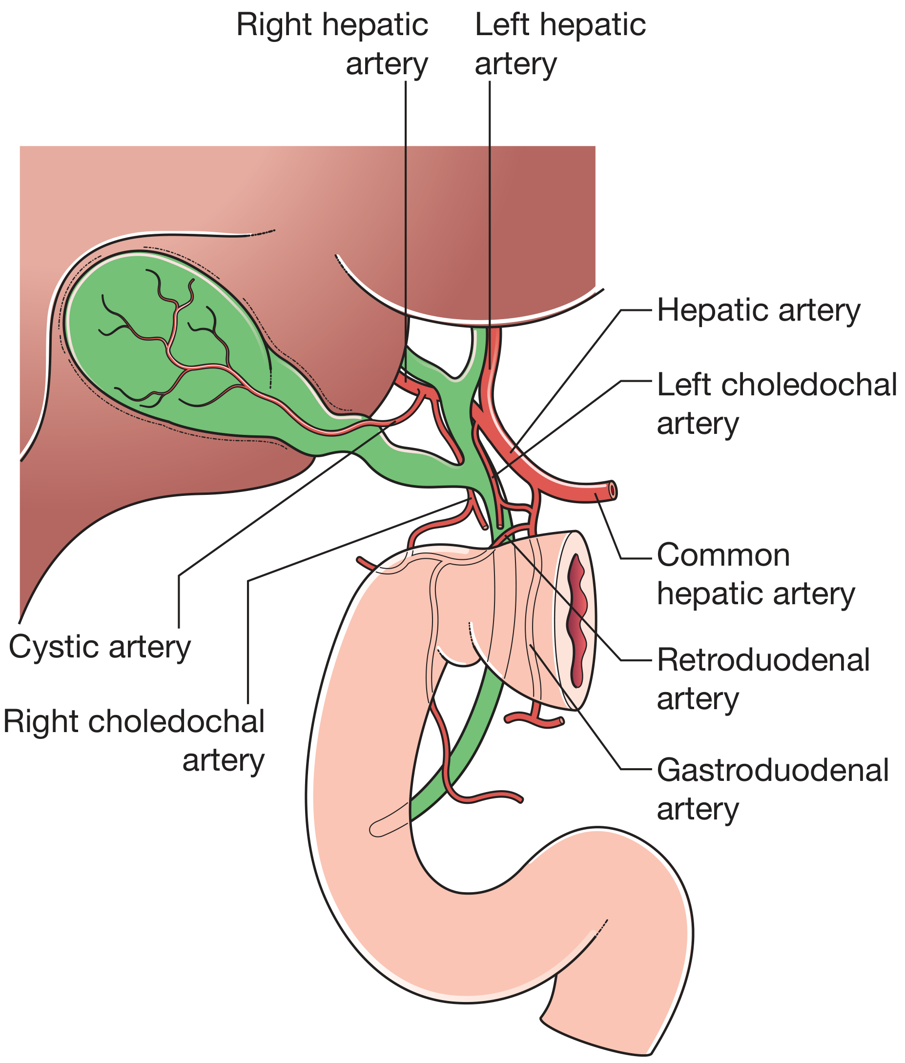
Arterial anatomy of the gallbladder region - Bailey & Love's 28e
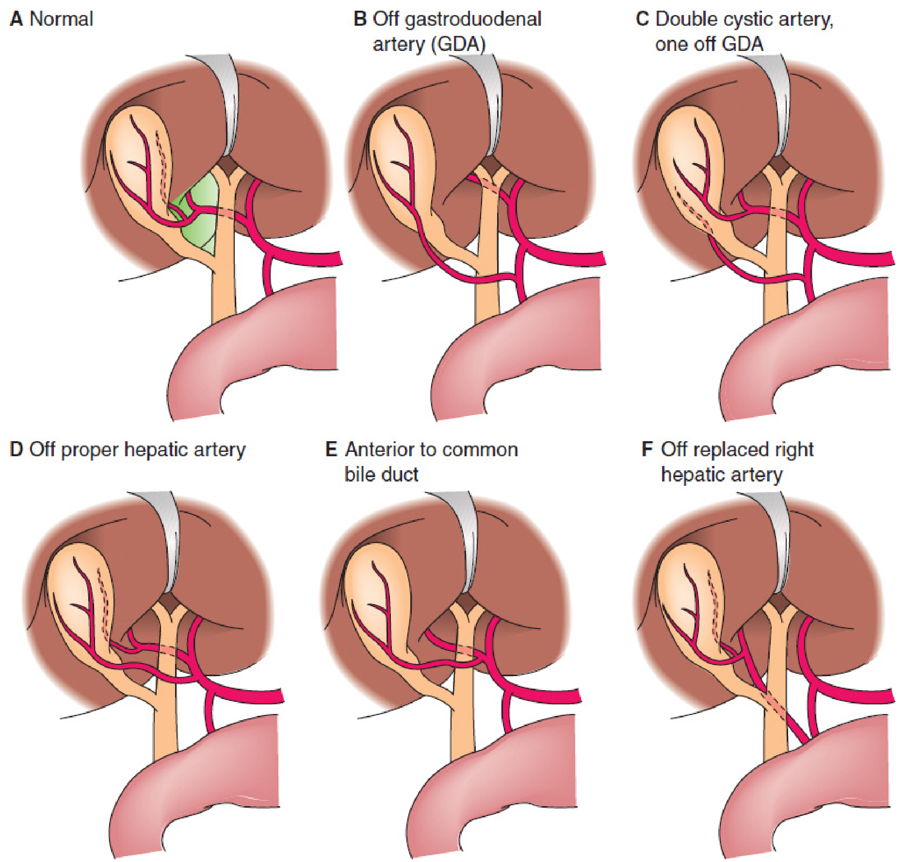
Variations of the cystic artery. Modern Calot's/hepatocystic triangle marked in green (A) - Mulholland & Greenfield Surgery 7e
Lap chole poss open
Conversion from laparoscopic to open cholecystectomy (lap to open) is indicated when safe dissection cannot be completed laparoscopically. Common reasons include dense adhesions, bleeding, bile duct injury, unclear anatomy, and Mirizzi syndrome. The conversion rate is approximately 2-15% and is not considered a failure.
Searching sources...
Finding Sources
Reading File
Reading File
Laparoscopic to Open Conversion (Lap to Open)
Key Principle
Conversion is not a failure - it is sound surgical judgment. It should always be discussed with the patient pre-operatively. (- Schwartz's Principles of Surgery 11e)
Absolute Contraindications to Lap Chole (Primary Open)
These patients should go straight to open:
| Condition | Reason |
|---|---|
| Haemodynamic instability | Cannot tolerate pneumoperitoneum |
| Uncontrolled coagulopathy | Bleeding risk |
| Frank peritonitis | Contamination, visibility |
| Severe COPD | Cannot tolerate raised intraabdominal pressure from CO₂ |
| Severe CCF (EF <20%) | Cannot tolerate pneumoperitoneum |
(- Schwartz's Principles of Surgery 11e, p. 1439)
Indications for Intraoperative Conversion
| Indication | Detail |
|---|---|
| Unclear anatomy / cannot achieve CVS | Most common reason - inability to identify cystic duct/artery safely |
| Dense adhesions | Previous upper abdominal surgery, chronic cholecystitis |
| Uncontrolled bleeding | Cystic artery, port-site, or hepatic bleeding |
| Bile duct injury | Recognised injury requiring open repair |
| No progress over a set time | "If you're not moving forward, convert" |
| Acute cholecystitis >72 hours | Conversion rate rises significantly after this window |
| Mirizzi syndrome | Stone impacted in cystic duct compressing CHD |
| Gangrene / empyema of gallbladder | Friable tissue, poor planes |
| Cholecystoenteric fistula | Complex anatomy |
| Cirrhosis / portal hypertension | Bleeding from collaterals |
Conversion Rates (approximate)
| Setting | Rate |
|---|---|
| Elective (uncomplicated) | ~5% |
| Acute cholecystitis | ~9.5-35% (higher after 72 h) |
| Emergent / complicated gallstone disease | 10-30% |
(- Schwartz's 11e; Maingot's Abdominal Operations)
Relative Contraindications (increased risk, not absolute - attempt lap first)
Previously considered absolute contraindications, now treated as risk factors for difficult lap chole:
- Acute cholecystitis
- Gangrene / empyema
- Biliary-enteric fistula
- Obesity
- Pregnancy (second trimester safest if needed)
- Ventriculoperitoneal shunt
- Cirrhosis
- Previous upper abdominal surgery
Situations Where Cholecystectomy (Lap or Open) Should be Avoided Entirely
Use percutaneous cholecystostomy instead when:
- Absolute contraindication to general anaesthesia (e.g. respiratory failure)
- Recent MI or acute exacerbation of severe comorbidity
In acute cholecystitis with significant comorbidities: antibiotics + cholecystostomy tube, then elective lap chole 6-8 weeks later.
(- Maingot's Abdominal Operations)
Practical Rule
"One should not hesitate to convert to open if significant adhesions or inflammation are identified during laparoscopy."
- Maingot's Abdominal Operations
The threshold to convert should be low, early, and proactive - converting early is safer than persisting and causing a bile duct injury.
Calor triangle
I covered Calot's triangle in detail two messages ago! Here's a quick summary, and the full answer is above for reference.
Calot's Triangle - Quick Summary
Boundaries
| Original (1891) | Modern (Hepatocystic Triangle) | |
|---|---|---|
| Inferior | Cystic duct | Cystic duct |
| Medial | Common hepatic duct | Common hepatic duct |
| Superior | Cystic artery | Inferior surface of liver |
Contents
- Cystic artery (target structure to clip)
- Lund's node (cystic lymph node) - landmark for the cystic artery in difficult dissections
- Connective tissue, occasionally aberrant ducts
Surgical Significance
- Must achieve the Critical View of Safety (CVS) before clipping:
- Triangle cleared of all fat/fibrous tissue
- Only two structures seen entering gallbladder (cystic duct + cystic artery)
- Lower 1/3 of gallbladder separated from liver bed
- Classic anatomy present in only ~30% of individuals - always expect variants
- Most feared complication: bile duct injury from mistaking CBD for cystic duct
Key Cystic Artery Variants
- Normally off right hepatic artery (behind CHD)
- In 15%: right hepatic artery/cystic artery cross in front of CHD - dangerous!
- Can arise from GDA, proper hepatic, or replaced right hepatic artery
Scroll up for the full detailed answer with diagrams.
Lap chole possible open
This was covered two messages ago! Here's the full summary for quick reference:
Laparoscopic Cholecystectomy, Possible Open - Quick Summary
Core Principle
Conversion is NOT a failure - it is sound surgical judgment. Always discuss with patient pre-operatively.
Absolute Contraindications to Lap Chole (go straight to open)
| Condition |
|---|
| Haemodynamic instability |
| Uncontrolled coagulopathy |
| Frank peritonitis |
| Severe COPD |
| Severe CCF (EF <20%) |
Intraoperative Indications to Convert
| Indication |
|---|
| Cannot achieve Critical View of Safety (CVS) |
| Dense adhesions |
| Uncontrolled bleeding |
| Recognised bile duct injury |
| No progress over a set time period |
| Acute cholecystitis >72 hours |
| Mirizzi syndrome |
| Gangrene / empyema |
| Cholecystoenteric fistula |
| Cirrhosis / portal hypertension |
Conversion Rates
| Setting | Rate |
|---|---|
| Elective | ~5% |
| Acute cholecystitis | ~9.5-35% |
| Emergent/complicated | 10-30% |
Golden Rule
Convert early - persisting and causing a bile duct injury is far worse than converting.
Scroll up for the full detailed answer with all relative contraindications, pregnancy management, and cholecystostomy indications.
What is lap chole
Your credits are exhausted. You can purchase a top-up or subscribe to a plan to continue using Orris. If you're already subscribed, your daily credits will reset tomorrow.
This is a shared conversation. Sign in to Orris to start your own chat.