
Differentiate between adult hemoglobin and fetal hemoglobin
The key differences between adult hemoglobin (HbA) and fetal hemoglobin (HbF) lie in their subunit composition, oxygen affinity, and physiological roles. HbA has two alpha and two beta chains (α2β2), while HbF has two alpha and two gamma chains (α2γ2). The gamma chains in HbF bind 2,3-BPG less avidly than beta chains, giving HbF a higher oxygen affinity - this allows the fetus to extract oxygen from maternal blood across the placenta.
Searching sources...
Reading File
Finding Sources
Finding Sources
Reading File
Reading File
Reading File
Finding Sources
Reading File
Reading File
Reading File
Now I have comprehensive information from multiple authoritative textbooks. Let me also fetch the oxygen-hemoglobin dissociation curve image referenced in Lippincott.
Reading File
I now have all the information needed for a thorough, well-sourced answer. Let me read the key developmental changes figure text from Lippincott and the 2,3-BPG section for a complete picture.
Reading File
I now have everything needed for a thorough, textbook-cited answer. Let me fetch the developmental changes in globin production figure.
Loading Image
Here is a comprehensive, textbook-sourced comparison of adult hemoglobin (HbA) and fetal hemoglobin (HbF):
Adult Hemoglobin (HbA) vs. Fetal Hemoglobin (HbF)
1. Structural Composition
| Feature | HbA (Adult) | HbF (Fetal) |
|---|---|---|
| Subunit structure | α₂β₂ | α₂γ₂ |
| Alpha chains | Present (identical to HbF) | Present (identical to HbA) |
| Non-alpha chains | β (beta) chains | γ (gamma) chains |
| Gene family | β-globin gene family (chromosome 11) | β-globin gene family (chromosome 11) |
The only structural difference is the substitution of the two β chains with two γ chains. The α chains are identical in both. - Costanzo Physiology, 7th Ed.
2. Proportion in the Body
| Stage | HbA | HbF |
|---|---|---|
| Fetus (last months of gestation) | Minimal/absent | ~60% of total hemoglobin |
| At birth | Starting to rise | Still dominant |
| By ~6 months after birth | Dominant (>90%) | Declining rapidly |
| Normal adult | ~90–97% (including HbA1c) | <2% (concentrated in F cells) |
HbA synthesis starts in bone marrow around the 8th month of pregnancy and gradually replaces HbF during the first year of life. - Lippincott Illustrated Reviews: Biochemistry, 8th Ed.
3. Oxygen Affinity - The Key Functional Difference
HbF has a higher affinity for O₂ than HbA. This is the most important physiological distinction, and it exists because of how each hemoglobin interacts with 2,3-bisphosphoglycerate (2,3-BPG).
Mechanism:
- In HbA, 2,3-BPG binds to positively charged residues (including His H21) on the β chains, stabilizing the low-affinity T (deoxy) state and causing O₂ to be released more readily.
- In HbF, the γ chains have Ser at position H21 instead of His. Serine cannot form the same salt bridge, so 2,3-BPG binds much more weakly to HbF.
- With less 2,3-BPG stabilization of the T state, HbF stays more in the R (oxy) state → higher O₂ affinity.
"In the fetal hemoglobin, residue H21 of the γ subunit is Ser rather than His. Since Ser cannot form a salt bridge, BPG binds more weakly to HbF than to HbA. The lower stabilization afforded to the T state by BPG helps account for HbF having a higher affinity for O₂ than HbA."
- Harper's Illustrated Biochemistry, 32nd Ed.
"If both HbA and HbF are stripped of their 2,3-BPG, they then have a similar oxygen affinity."
- Lippincott Illustrated Reviews: Biochemistry, 8th Ed.
4. The O₂ Dissociation Curve
HbF's O₂ dissociation curve is shifted to the left compared to HbA, reflecting its higher O₂ affinity (lower P50). This means:
- HbF loads O₂ more readily at any given PO₂
- HbF releases O₂ less readily to tissues
This left shift is the physiological basis for placental O₂ transfer - HbF in fetal blood can "steal" O₂ from maternal HbA at the placenta.
5. Physiological Role
| Feature | HbA | HbF |
|---|---|---|
| Primary role | O₂ transport in adults | O₂ extraction from mother via placenta |
| Why designed this way | Release O₂ to peripheral tissues | Must extract O₂ from maternal circulation where PO₂ is relatively low |
| 2,3-BPG sensitivity | High - strongly decreases O₂ affinity | Low - weakly decreases O₂ affinity |
"Hemoglobin F is therefore critical to facilitate movement of O₂ from the maternal to the fetal circulation, particularly at later stages of gestation where oxygen demand increases."
- Ganong's Review of Medical Physiology, 26th Ed.
6. Developmental Switching (Hemoglobin Switching)
The figure below (from Lippincott's) shows the developmental timeline of globin chain production:
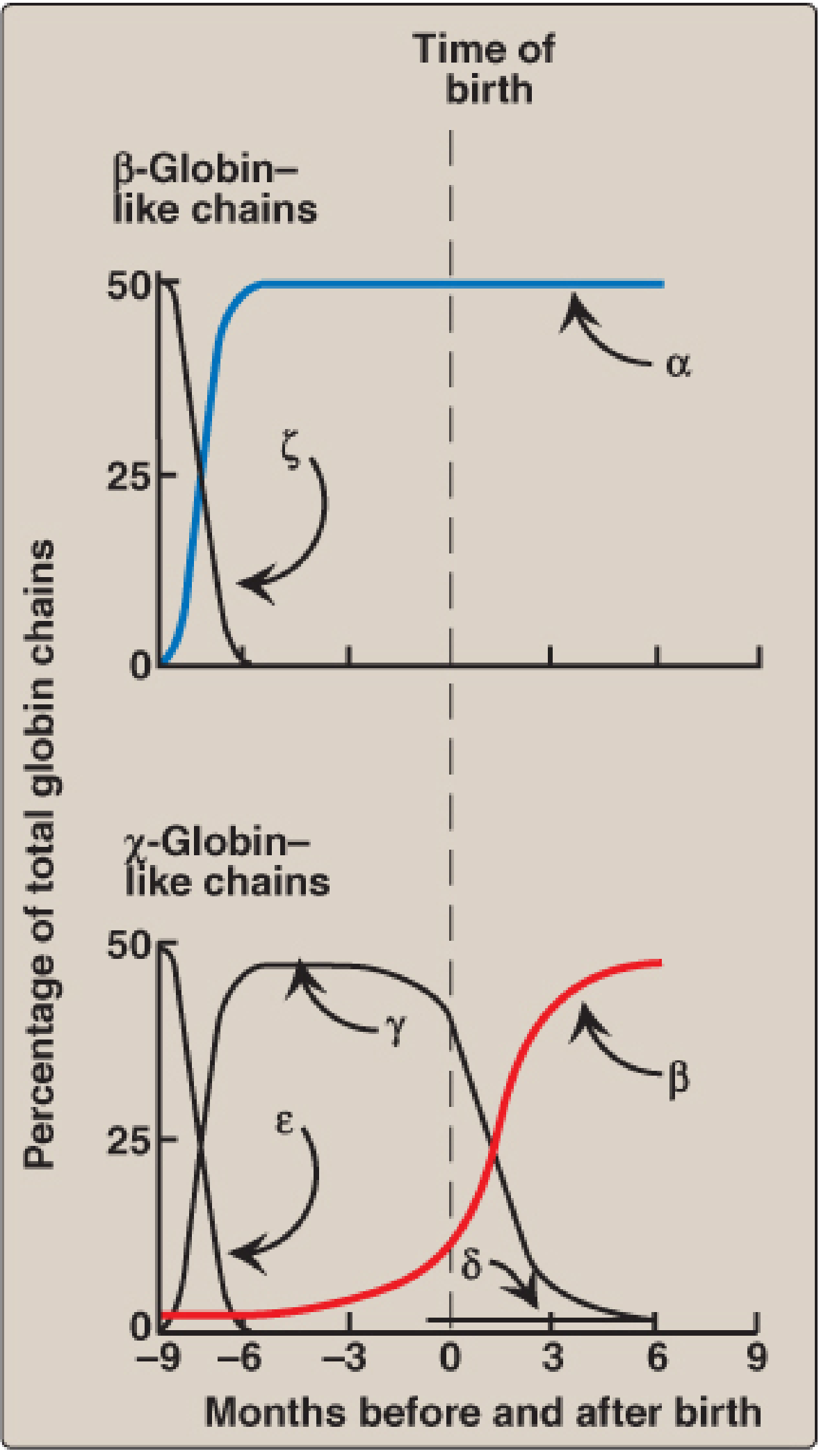
Three stages of hemoglobin exist across development:
- Embryonic (~weeks 1-8): Gower 1 (ζ₂ε₂) and Gower 2 (α₂ε₂) - produced in yolk sac
- Fetal (~week 8 to birth): HbF (α₂γ₂) - produced in fetal liver/spleen, then bone marrow
- Adult (after birth): HbA (α₂β₂) - produced in bone marrow; replaces HbF within ~6 months
The switch from γ to β chain production is regulated by transcription factors and oxygen availability. Relative hypoxia favors HbF production. - Basic Medical Biochemistry, 6th Ed.
7. Summary Comparison Table
| Parameter | HbA | HbF |
|---|---|---|
| Chains | α₂β₂ | α₂γ₂ |
| O₂ affinity | Lower | Higher (left-shifted curve) |
| P50 (approx.) | ~26-27 mmHg | ~20 mmHg |
| 2,3-BPG binding | Strong (β-chain His H21) | Weak (γ-chain Ser H21) |
| Normal adult % | ~90-97% | <2% |
| Period of dominance | Post-birth/adult life | Fetal life |
| Site of synthesis | Bone marrow | Fetal liver → bone marrow |
| Clinical relevance | Glycated → HbA1c (diabetes monitoring) | Elevated in β-thalassemia, sickle cell disease (protective) |
8. Clinical Relevance of HbF in Adults
In disease states such as sickle cell anemia and β-thalassemia, higher residual levels of HbF are protective because:
- HbF cannot polymerize like HbS under deoxygenation
- HbF can partially compensate for absent/reduced β-chain production in β-thalassemia
Hydroxyurea is used clinically to induce HbF production (by stimulating γ-globin gene expression), reducing the severity of sickle cell crises. - Goodman & Gilman's Pharmacological Basis of Therapeutics
Individuals who persistently express HbF past infancy (>2% in adults) have Hereditary Persistence of Fetal Hemoglobin (HPFH), which is clinically benign. - Basic Medical Biochemistry, 6th Ed.
This is a shared conversation. Sign in to Orris to start your own chat.