
Titanium mesh for bone grafting !! Explain in simple terms
Reading File
Now let me search the medical library and web simultaneously.
Finding Sources
Searching the Web
titanium mesh bone grafting explained simple terms dental oral surgery
Searching PubMed
titanium mesh bone regeneration grafting
Now let me read the most relevant textbook sections and extract the Nature article for clinical details.
Reading File
Reading Web Page
https://www.nature.com/articles/s41368-020-00107-z
Let me also fetch the surgical image to include visually.
Loading Image
Here is a clear, simple explanation of titanium mesh for bone grafting, drawing from authoritative surgical textbooks and current research.
Titanium Mesh for Bone Grafting - Explained Simply
The Basic Idea
Think of titanium mesh as a tiny, flexible cage or basket made of metal. When a patient is missing bone - whether from an injury, surgery (like removing a tumor), tooth loss, or infection - the body cannot always fill that gap on its own. The mesh acts as a scaffold or tent that:
- Holds the space open where new bone needs to grow
- Contains bone graft material (crushed bone chips) inside it
- Protects the graft while new bone slowly forms around it
Why Titanium Specifically?
Titanium has a unique set of properties that make it ideal for the body:
| Property | What it means in practice |
|---|---|
| High stiffness | Keeps the shape, doesn't collapse under pressure |
| Low density | Lightweight - doesn't burden the patient |
| Corrosion resistant | Doesn't rust or degrade inside the body |
| Biocompatible | The body tolerates it well - no rejection |
| Malleable | Surgeons can bend/cut it to fit any shape of defect |
Where Is It Used?
Titanium mesh for bone grafting shows up in many surgical specialties:
1. Dental/Jaw Surgery (Most common use)
When someone loses teeth, the jawbone shrinks over time. To place a dental implant, you need enough bone. The mesh is placed over the defect, packed with bone graft chips, and covered with gum tissue. Over 6-9 months, new bone fills the space. This technique is called Guided Bone Regeneration (GBR).
2. Spine Surgery
After removing a damaged vertebra (corpectomy), the gap needs support. A titanium mesh cage is packed with bone chips and wedged in to restore the column's height and stability, as described in Rockwood and Green's Fractures in Adults (10th Ed, 2025).
3. Jaw Reconstruction (Cancer / Trauma)
After removing a jaw tumor or repairing a traumatic fracture, a titanium mesh "crib" is filled with crushed bone graft and bone morphogenetic protein (BMP) to rebuild the missing mandible - Cummings Otolaryngology, Head and Neck Surgery.
4. Skull / Chest Wall Repair
Large defects in the skull or chest wall can be patched using titanium mesh alone or combined with bone grafts, as noted in Sabiston Textbook of Surgery.
The Surgical Image
Below is an actual surgical photo from Cummings Otolaryngology showing a titanium mesh plate with a graft crib placed in a jaw (mandible) reconstruction:
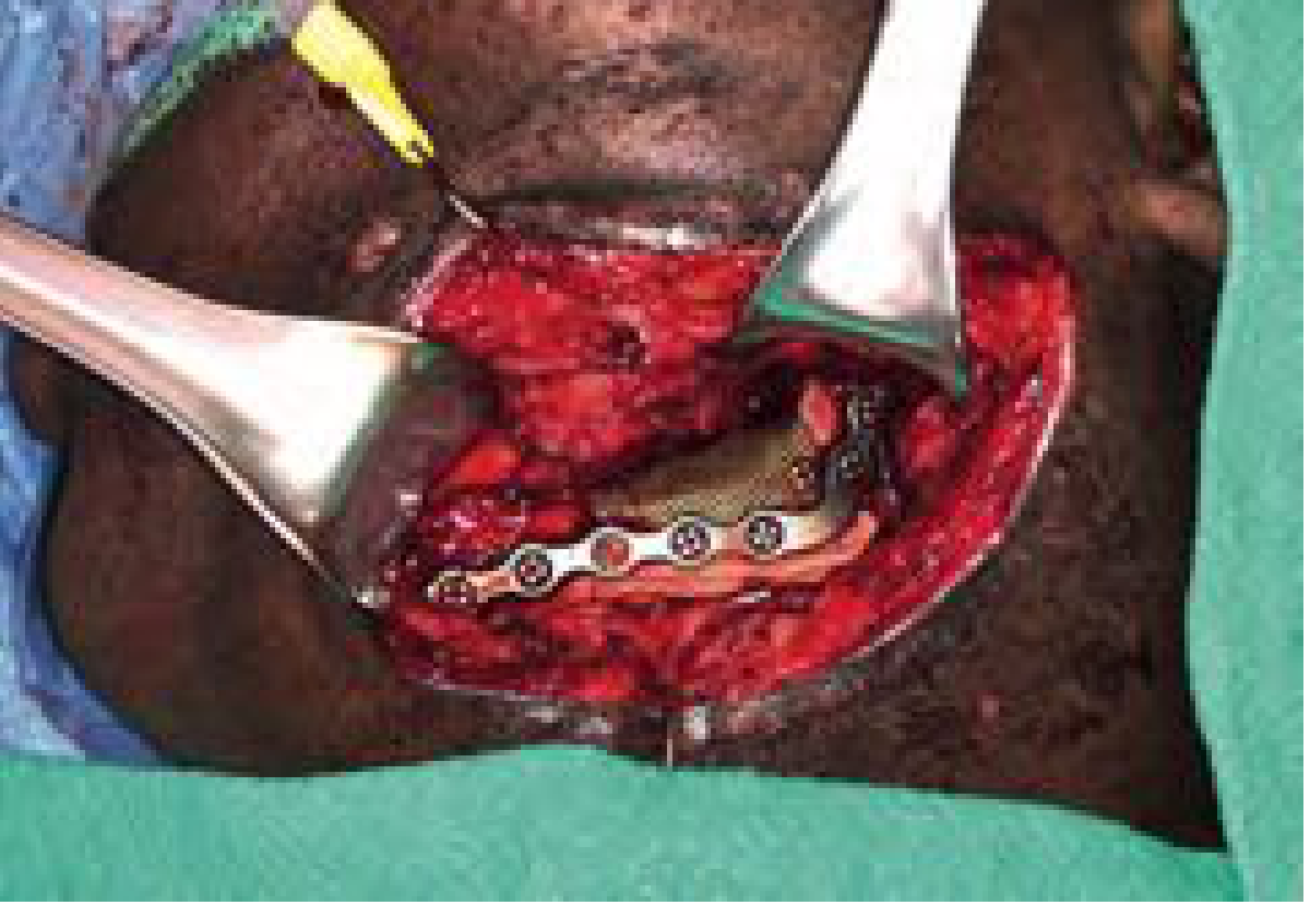
The golden-colored mesh and the silver reconstruction plate working together to hold the shape of the lower jaw.
Step-by-Step: What Happens During the Procedure?
- Surgeon opens the surgical site and exposes the bony defect
- Bone graft material is prepared - this can be:
- Your own bone (autograft, e.g., from the hip or skull)
- Donor/processed bone (allograft)
- Synthetic bone substitutes
- Mesh is shaped - cut and bent to perfectly fit the defect (or now often custom-made using CAD/CAM 3D technology)
- Mesh is packed with bone chips and placed over the defect
- Screws or sutures hold the mesh in place against existing bone
- Soft tissue (gum or skin) is closed over it to protect the site
- Over months, new bone grows into and around the mesh
- Mesh may be removed in a second surgery, or left in place permanently (depending on location and purpose)
CAD/CAM: The Modern Upgrade
Traditional titanium mesh was cut and bent by hand in the operating room - imprecise and time-consuming. Modern computer-designed (CAD/CAM) custom titanium mesh is:
- 3D-printed to exactly match the patient's anatomy (from CT scans)
- More precise fit = less surgery time
- Better bone volume outcomes
A recent 2025 meta-analysis (Ragucci et al., PMID 40844656) confirmed CAD/CAM mesh achieves better bone regeneration results compared to hand-bent conventional mesh.
Main Complication: Mesh Exposure
The #1 problem with titanium mesh is that the overlying gum or skin can split open, exposing the mesh - this happens in roughly 20-30% of cases (and up to 66% in some reports), according to the Nature review on titanium mesh in oral implantology.
- Early exposure (within 4 weeks): often from sharp mesh edges cutting through the tissue
- Late exposure: from pressure or thin overlying tissue
- The good news: exposed mesh doesn't always mean failure - often the area can be managed with trimming and antibiotics without losing the graft
Summary in One Sentence
Titanium mesh is a strong, body-friendly metal basket that holds bone graft material in place, keeps the space open, and gives the body the protected environment it needs to grow new bone where it's missing.
Sources:
- Cummings Otolaryngology Head and Neck Surgery - Reconstructive Options
- Rockwood and Green's Fractures in Adults, 10th Ed (2025) - Reconstruction
- Campbell's Operative Orthopaedics, 15th Ed (2026)
- Sabiston Textbook of Surgery - Trunk and Chest Wall
- Titanium mesh bone augmentation - Nature/IJOS Review (2020)
- Systematic review: Sabri et al., 2024, PMID 39283219
- Meta-analysis: Ragucci et al., 2025, PMID 40844656
This is a shared conversation. Sign in to Orris to start your own chat.