
Xanthomas eruption
Reading File
Eruptive xanthomas are yellow-orange papules that appear suddenly due to severe hypertriglyceridemia. They are a cutaneous sign of a lipid metabolism disorder, most commonly seen when triglycerides exceed 1000-2000 mg/dL. The lesions result from dermal macrophages engulfing triglyceride-laden chylomicrons.
Searching sources...
Finding Sources
Searching PubMed
eruptive xanthoma hypertriglyceridemia
Finding Sources
Searching the Web
eruptive xanthomas clinical features causes treatment 2024
Reading File
Finding Sources
Loading Image
Reading File
Here is a thorough overview of eruptive xanthomas, drawn from Andrews' Diseases of the Skin and supported by current literature.
Eruptive Xanthomas
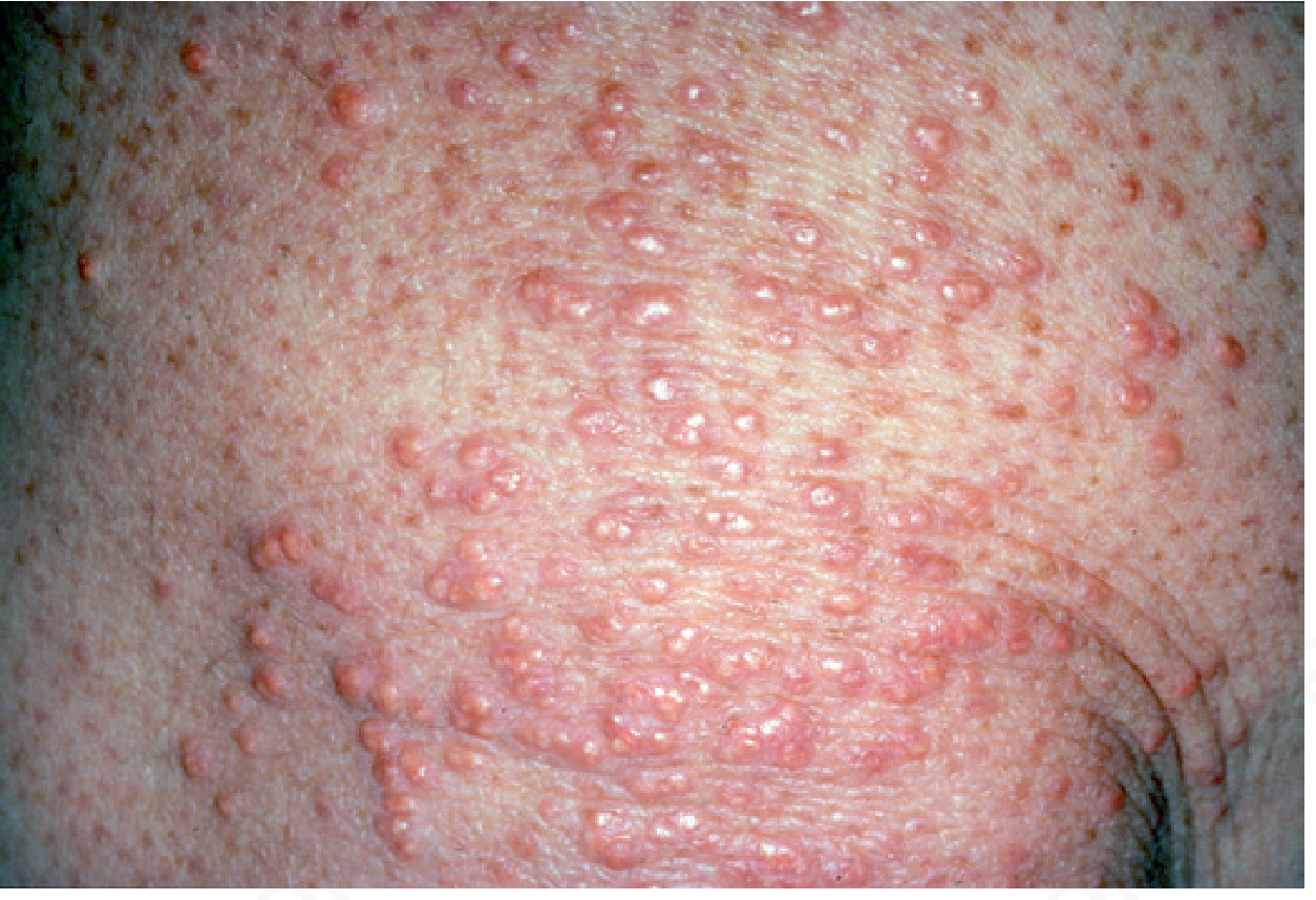
Definition
Eruptive xanthomas (xanthoma eruptivum) are small yellowish-orange to reddish-brown papules that appear suddenly in crops over the body. They result from the deposition of lipid-laden macrophages (foam cells) in the superficial dermis, driven by severe hypertriglyceridemia.
Clinical Features
| Feature | Detail |
|---|---|
| Morphology | 2-5 mm yellow-orange or red-brown papules, often with an erythematous halo |
| Distribution | Buttocks, extensor surfaces of arms and thighs, knees, inguinal and axillary folds, oral mucosa |
| Onset | Sudden, in "crops" |
| Symptoms | Variable pruritus; may be tender |
| Koebner phenomenon | Can occur (lesions appear at sites of trauma) |
Pathogenesis
When plasma triglycerides exceed ~1,000 mg/dL (11.2 mmol/L), circulating chylomicrons overwhelm the reticuloendothelial system. Macrophages in the dermis phagocytose excess lipid, becoming "foam cells," which accumulate visibly as papules. At triglyceride levels above 2,000 mg/dL, the risk of acute pancreatitis rises to 10-20%.
Histology: Foamy macrophages in the superficial dermis with a lymphocytic infiltrate.
Causes / Associated Conditions
Primary (Genetic) Disorders
| Fredrickson Type | Lipoprotein Abnormality | Notes |
|---|---|---|
| Type I (HLP I) - Chylomicronemia | Excess chylomicrons | Lipoprotein lipase (LPL) deficiency - presents in childhood with eruptive xanthomas, pancreatitis, lipemia retinalis |
| Type III - Familial dysbetalipoproteinemia | Elevated IDL + chylomicron remnants | Eruptive or tuberous xanthomas; palmar crease xanthomas are pathognomonic |
| Type IV - Familial hypertriglyceridemia | Elevated VLDL | Eruptive xanthomas if triglycerides markedly elevated |
| Type V | Elevated VLDL + chylomicrons | Common cause of adult chylomicronemia syndrome |
Secondary (Acquired) Causes
- Poorly controlled type 2 diabetes mellitus (most common)
- Chronic renal failure
- Hypothyroidism
- Drug-induced: estrogens, corticosteroids, systemic retinoids
- Alcohol excess
- Metabolic syndrome / obesity
Key clinical pearl: Eruptive xanthomas are seen in ~8.5% of patients presenting with severe hypertriglyceridemia (TG >1,772 mg/dL). They should always prompt urgent lipid workup.
Associated Systemic Findings
- Lipemia retinalis (retinal vessels appear creamy-white)
- Hepatosplenomegaly (triglyceride deposition in liver/spleen)
- Acute pancreatitis (5% risk at TG >1,000 mg/dL; 10-20% at TG >2,000 mg/dL)
- Lactescent (milky) serum on centrifugation
Differential Diagnosis
- Granuloma annulare (generalized)
- Molluscum contagiosum
- Subepidermal calcified nodules
- Xanthoma disseminatum (normolipemic)
- Sarcoidosis
- Eruptive syringomas
Diagnosis
- Fasting lipid panel - triglycerides typically >1,000 mg/dL
- Skin biopsy - foamy macrophages (foam cells) in superficial dermis with lymphocytic infiltrate
- Serum appearance - lactescent or milky
- Workup for secondary causes: fasting glucose/HbA1c, TSH, renal function
Treatment
Treatment is directed at the underlying lipid disorder. Eruptive xanthomas resolve spontaneously within weeks to months once triglycerides are controlled.
Lipid-Lowering Therapy
- Fibrates (e.g., fenofibrate) - first-line for severe hypertriglyceridemia
- Omega-3 fatty acids (high-dose) - reduce VLDL synthesis
- Statins - useful if combined hyperlipidemia
- Nicotinic acid - reduces VLDL; limited by side effects
Dietary / Lifestyle
- Strict low-fat diet (critical in chylomicronemia syndrome)
- Alcohol cessation
- Tight glycemic control in diabetics
Specific/Advanced
- Alipogene tiparvovec (gene therapy) for monogenic LPL deficiency
- Volanesorsen (antisense oligonucleotide targeting ApoC-III) for familial chylomicronemia syndrome - a newer option
Skin lesions
The papules do not require specific dermatologic treatment - they resolve with systemic triglyceride reduction.
Prognosis
- Lesions resolve within weeks of initiating effective lipid-lowering treatment
- Main risks are from the underlying disorder: pancreatitis and cardiovascular disease
- Recurrence occurs if the metabolic trigger (e.g., poor diabetes control) returns
Sources: Andrews' Diseases of the Skin, p. 617; Cleveland Clinic Journal of Medicine - Eruptive xanthoma as a warning sign; DermNet NZ - Xanthoma; Hegele RA (2026), Approach to the Adult Patient with Chylomicronemia [PMID: 41472374]
Is associated with poorly controlled diabetes mellitus
Yes, eruptive xanthomas are most commonly associated with poorly controlled type 2 diabetes mellitus. Here is why:
Mechanism: Diabetes → Hypertriglyceridemia → Eruptive Xanthomas
In poorly controlled diabetes, insulin deficiency or resistance leads to:
-
Reduced lipoprotein lipase (LPL) activity - LPL normally breaks down triglycerides in chylomicrons and VLDL. Insulin is required to activate LPL. Without adequate insulin action, LPL activity falls sharply.
-
Increased VLDL production - The liver produces excess VLDL due to elevated free fatty acid flux (from uninhibited lipolysis in adipose tissue).
-
Result: Severe hypertriglyceridemia - Triglycerides accumulate in the bloodstream (often >1,000 mg/dL).
-
Dermal macrophage overload - Circulating chylomicrons and VLDL particles are phagocytosed by dermal macrophages, forming foam cells → visible as eruptive xanthoma papules.
Key Points
- Eruptive xanthomas can be the first presenting sign of previously undiagnosed diabetes mellitus
- They are seen most often in type 2 DM, but can occur in type 1 DM with poor control
- The hypertriglyceridemia in DM is reversible - once glycemic control is restored, LPL activity recovers, triglycerides fall, and lesions resolve within weeks
- Poorly controlled DM is an example of a secondary (acquired) cause of hypertriglyceridemia, unmasking a polygenic predisposition in susceptible individuals
Clinical Implication
Any patient presenting with eruptive xanthomas should have:
- Fasting glucose / HbA1c checked immediately
- A full fasting lipid panel
- Assessment for other secondary causes (hypothyroidism, renal failure, drugs)
As stated in Andrews' Diseases of the Skin: "Eruptive xanthomas are seen most often in poorly controlled type 2 diabetes mellitus."
Acute pancreatitis treatment
Acute pancreatitis treatment centers on fluid resuscitation, analgesia, nutritional support, and managing complications. Mild cases resolve with supportive care; severe cases need ICU-level management. The underlying cause (gallstones, hypertriglyceridemia, alcohol) must also be addressed.
Searching sources...
Finding Sources
Searching PubMed
"acute pancreatitis"[MeSH Terms] AND management
Finding Sources
Searching PubMed
"acute pancreatitis" treatment guidelines
Reading File
Reading File
Reading File
Reading File
Excellent - the Rosen's Emergency Medicine textbook has comprehensive treatment content in that same block. Let me compile everything now.
Acute Pancreatitis - Treatment
Diagnosis First (Quick Review)
Diagnosis requires ≥2 of 3 criteria (Revised Atlanta Classification):
- Abdominal pain characteristic of acute pancreatitis (epigastric, radiating to back)
- Serum lipase (preferred) or amylase >3x upper limit of normal
- Characteristic findings on CT/MRI
Note: Lipase is more sensitive and specific than amylase. The degree of elevation does not correlate with severity.
Severity Classification
| Grade | Features |
|---|---|
| Mild | No organ failure, no local/systemic complications; resolves in 1 week |
| Moderately Severe | Transient organ failure (<48 hrs) OR local complications (e.g., pseudocyst, necrosis) |
| Severe | Persistent organ failure (>48 hrs), single or multi-organ; mortality up to 30% |
Scoring tools: APACHE II, Ranson criteria, BISAP score, CT Severity Index (CTSI)
Treatment
1. Fluid Resuscitation (Cornerstone)
- Aggressive IV hydration is the most important early intervention
- Lactated Ringer's (LR) is the preferred fluid - reduces SIRS and progression compared to normal saline
- Rate: 250-500 mL/hour in the first 12-24 hours (or 1.5-2x maintenance)
- Goal: urine output >0.5 mL/kg/hr, HR <100 bpm, MAP >65 mmHg
- Reassess frequently - over-resuscitation causes abdominal compartment syndrome
2. Analgesia
- Opioids (e.g., IV morphine or hydromorphone) - effective and safe; the old concern that morphine worsens pancreatitis via sphincter of Oddi spasm is not clinically significant
- NSAIDs (e.g., ketorolac) - useful adjunct
- Adequate pain control improves patient comfort and reduces stress response
3. Nutrition
- Early oral/enteral feeding (within 24-48 hours) is now strongly recommended - even in severe cases
- Nil per os (NPO) is no longer standard for mild-moderate disease
- Start with a low-fat, soft diet as soon as tolerated; not necessary to wait for pain/enzyme resolution
- In severe cases: nasojejunal (NJ) tube feeding is preferred over parenteral nutrition
- Total parenteral nutrition (TPN) only if enteral route is not tolerated or contraindicated
- Enteral nutrition preserves gut barrier, reduces bacterial translocation, lowers infectious complications
4. Antibiotics
- NOT indicated prophylactically in acute pancreatitis, even in necrotizing disease
- Use antibiotics only if there is confirmed infected necrosis or another concurrent infection (cholangitis, bacteremia)
- Empiric broad-spectrum antibiotics (e.g., imipenem, meropenem) if infected pancreatic necrosis is suspected (fever + deterioration + gas in necrosis on CT)
5. Imaging
- CT with IV contrast is NOT routine at admission
- Indicated if: diagnostic uncertainty, failure to improve after 48-72 hours, or suspected complication
- Best performed ≥72 hours after onset - early CT underestimates necrosis
- CT Severity Index (CTSI) helps grade severity and predict outcomes
6. Cause-Specific Treatment
| Cause | Specific Management |
|---|---|
| Gallstone pancreatitis | ERCP + sphincterotomy if cholangitis or persistent biliary obstruction; laparoscopic cholecystectomy before discharge (mild) or after recovery (severe) |
| Hypertriglyceridemia | Insulin infusion (activates LPL), IV heparin (transiently), fibrates; strict low-fat diet; plasmapheresis in refractory severe cases |
| Alcohol | Cessation counseling; thiamine supplementation |
| Drug-induced | Stop offending agent |
| Hypercalcemia | Treat underlying cause (e.g., hyperparathyroidism) |
7. Management of Complications
| Complication | Management |
|---|---|
| Pancreatic pseudocyst | Observe if asymptomatic; endoscopic/surgical drainage if symptomatic or infected |
| Infected necrosis (abscess) | Step-up approach: percutaneous drainage → endoscopic necrosectomy → surgical necrosectomy |
| ARDS | ICU, mechanical ventilation, lung-protective strategy |
| AKI / renal failure | IV fluids, dialysis if needed |
| DIC | FFP, platelets, cryoprecipitate |
| Hypocalcemia | IV calcium gluconate |
| Hyperglycemia | Insulin infusion |
8. What NOT to Do
- Do not give prophylactic antibiotics routinely
- Do not keep the patient NPO unnecessarily
- Do not do early CT routinely
- Do not delay cholecystectomy in gallstone pancreatitis if mild
Updated Guidelines (2025)
The IAP/APA/EPC/IPC/JPS Revised Guidelines on Acute Pancreatitis 2025 (PMID: 40651900) - the most current international consensus - reaffirms: goal-directed fluid resuscitation with LR, early enteral nutrition, restrictive antibiotic use, and step-up approach for infected necrosis.
Sources: Rosen's Emergency Medicine, Chapter 77; Sleisenger & Fordtran's GI and Liver Disease; IAP/APA 2025 Guidelines [PMID: 40651900]; Management of Gallstone Pancreatitis (JAMA Surg, 2024) [PMID: 38691369]


84452.jpg
+2 more
It occurs in genital area
analyse_media
analyse_media
analyse_media
Finding Sources
Finding Sources
Finding Sources
Reading File
Reading File
Finding Sources
Reading File
Reading File
The images show lesions highly consistent with Condyloma Acuminata (Genital Warts) - caused by Human Papillomavirus (HPV). This is quite different from xanthomas. Here is a full analysis:
Image Analysis
Image 1 (84452): Two discrete, well-circumscribed plaques with a honeycombed/cobblestone internal architecture - pinkish-red, moist surface. Classic lobulated condyloma morphology.
Image 2 (84449): A single cauliflower-like, exophytic, moist papule with lobulated projections on a mucosal surface - highly characteristic of condyloma acuminata.
Image 3 (84446): A cluster of small vesicles/papules on an erythematous base in a herpetiform grouping. This image raises the differential of Herpes Simplex Virus (HSV) genitalis in addition to condyloma.
Condyloma Acuminata (Genital Warts)
Etiology
- Caused by Human Papillomavirus (HPV), most commonly types 6 and 11 (low-risk, rarely associated with malignancy)
- HPV types 16 and 18 are high-risk and associated with dysplasia and squamous cell carcinoma
- Transmitted via sexual contact (vaginal, anal, oral) or skin-to-skin contact; rarely by perinatal transmission
Sites of Occurrence
- Perianal skin and anal canal
- Genital region (penis, vulva, vagina, cervix)
- Inguinal folds, oral mucosa
Clinical Features
- Lesions range from small, flesh-colored papules to large cauliflower-like exophytic masses
- May be asymptomatic, or cause pruritus, burning, bleeding, discharge, or pain
- Application of 5% acetic acid turns lesions white (acetowhitening) - aids identification
Diagnosis
- Clinical diagnosis in most cases
- Biopsy with histopathology to confirm - especially important in immunocompromised patients to rule out high-grade squamous intraepithelial neoplasia (HSIL) or malignancy
- Colposcopy / anoscopy / sigmoidoscopy to assess extent
Differential Diagnosis
| Condition | Distinguishing Feature |
|---|---|
| Condylomata lata (syphilis) | Flat, broad-based; RPR/VDRL positive |
| Molluscum contagiosum | Umbilicated papules; molluscum bodies on histology |
| Herpes genitalis (HSV) | Painful vesicles/ulcers; Tzanck smear/PCR positive |
| Seborrheic keratoses | Dry, stuck-on appearance; non-mucosal |
| Skin tags | Soft, pedunculated; no viral etiology |
| Anal/squamous cell carcinoma | Ulcerated, indurated; biopsy confirms |
Treatment
Treatment is individualized based on lesion size, number, location, and whether first episode or recurrent.
Patient-Applied (Self-administered)
| Agent | Mechanism | Use |
|---|---|---|
| Imiquimod 5% cream | Immune response modifier (Toll-like receptor 7 agonist) | Apply 3x/week for up to 16 weeks; most effective for keratinized skin |
| Podophyllotoxin (Podofilox) 0.5% | Antimitotic - arrests cell division | Apply twice daily for 3 days, rest 4 days; repeat up to 4 cycles |
| Sinecatechins 15% ointment | Green tea catechins - antiviral/immunomodulatory | Apply 3x/day for up to 16 weeks |
Provider-Applied (Office/Clinic)
| Agent/Procedure | Notes |
|---|---|
| Cryotherapy (liquid nitrogen) | Freeze-thaw cycles; effective for limited lesions; combination with podophyllin improves outcomes |
| Trichloroacetic acid (TCA) 80-90% | Chemical cauterization; useful for mucosal/vaginal lesions |
| Podophyllin resin 25% | Applied by provider, washed off after 1-4 hrs; NOT for mucosal use in pregnancy |
| Surgical excision / shave excision | For large or bulky disease |
| CO2 laser ablation | Effective for extensive or vestibular involvement; ~70% response in perigenital disease |
| Electrofulguration/cautery | Tissue destruction; outpatient |
Operating Room (Severe/Extensive Disease)
- Surgical excision under anesthesia
- CO2 laser under anesthesia
- Clearance rate: 60-90% after surgery; recurrence rate: 20-30% within first few months
Special Situations
- Pregnancy: Podophyllin and podophyllotoxin are contraindicated; imiquimod use requires caution. TCA and cryotherapy preferred.
- Immunosuppressed (HIV, transplant): Higher recurrence rates; biopsy all lesions to exclude HSIL; may need more aggressive treatment
- Pediatric condyloma: Perinatal transmission possible; CO2 laser and imiquimod used; consider sexual abuse evaluation
No Single Superior Treatment
As noted in Current Surgical Therapy: "There is no evidence to suggest that one treatment is significantly superior to another" - choice depends on lesion characteristics and clinical context.
Prevention
- HPV vaccination (Gardasil 9) - protects against HPV types 6, 11, 16, 18 and five additional high-risk types
- Recommended for ages 9-26 years (can be given up to age 45)
- Condom use reduces but does not eliminate transmission (skin beyond condom coverage can still transmit)
Note on Image 3: The herpetiform clustered vesicles with erythematous base in that image raise the possibility of co-existing Herpes Simplex Virus (HSV) infection - HSV and HPV can co-exist. Viral swab / PCR of the vesicular lesions would help differentiate. HSV treatment requires acyclovir/valacyclovir, not the treatments listed above.
Sources: Current Surgical Therapy 14e, p. 370; Sabiston Textbook of Surgery; Campbell Walsh Wein Urology
Treatment for male patients
Treatment for genital warts (condyloma acuminata) in male patients follows the same general principles but has some male-specific considerations regarding site (penile shaft, glans, foreskin, scrotum, perianal). The treatment is chosen based on lesion location, size, and number.
Searching sources...
Finding Sources
Finding Sources
Reading File
Finding Sources
Finding Sources
Searching the Web
genital warts condyloma acuminata treatment male CDC guidelines 2024
Reading File
Reading Web Page
https://www.bashh.org/_userfiles/pages/files/resources/agw_2…
Here is a focused guide on the treatment of genital warts (condyloma acuminata) in male patients, organized by lesion site and treatment modality.
Treatment of Condyloma Acuminata in Male Patients
Site-Specific Considerations in Males
| Site | Common in Males | Special Considerations |
|---|---|---|
| Penile shaft | Yes | Well-keratinized skin - most treatments work well |
| Glans penis / Corona | Yes | Thin mucosa - avoid podophyllin resin; prefer TCA, imiquimod |
| Foreskin (prepuce) | Yes | Often hiding lesions; retract fully to examine |
| Urethral meatus | Yes | Special management needed (see below) |
| Scrotum | Yes | Sensitive skin - cryotherapy preferred |
| Perianal / anal canal | Yes | Very common site; may need anoscopy |
Treatment Options
A. Patient-Applied (Self-administered at Home)
1. Podophyllotoxin (Podofilox) 0.5% solution or gel
- How: Apply to warts twice daily for 3 consecutive days, then 4 days rest - repeat up to 4 cycles
- Best for: External genital and perianal warts on keratinized skin
- Not for: Urethral meatus, intra-anal, or mucosal surfaces; avoid >10 cm² area
- Side effects: Local irritation, erosion, burning
- Note: Solution (0.5%) is preferred over 0.15% cream where easily applied (BASHH 2024 guideline)
2. Imiquimod 5% cream
- How: Apply 3 times per week at bedtime, wash off after 6-10 hours - up to 16 weeks
- Mechanism: Immune response modifier (Toll-like receptor 7 agonist) - stimulates local interferon and cytokine response
- Best for: Keratinized skin; external penile, perianal lesions; also useful as adjunct after ablation to reduce recurrence
- Side effects: Local erythema, erosion, itching, flu-like symptoms
- Lower recurrence rates than ablative methods alone
3. Sinecatechins 15% ointment (green tea polyphenols)
- Apply 3 times daily for up to 16 weeks
- Alternative for external genital warts
B. Provider-Applied (Clinic/Office)
1. Cryotherapy (Liquid Nitrogen)
- Freeze-thaw technique applied directly to the lesion; repeat every 1-2 weeks
- Effective for penile shaft, scrotal, perianal lesions
- For urethral meatus warts: cryotherapy is an option if the wart base is clearly visible on meatal eversion (examine with otoscope/meatoscope)
- Combination with podophyllotoxin has improved outcomes in men
- No systemic effects; fume extraction needed for anal lesions
2. Trichloroacetic Acid (TCA) 80-90%
- Applied precisely with a cotton-tipped swab by the clinician
- Safe on mucosal surfaces (glans, urethral meatus, intra-anal)
- Useful for recurrent lesions after initial clearance
- Repeat weekly as needed
3. Electrocautery / Fulguration
- Highly effective for perianal and multiple lesions
- Needle-tip cautery allows precise targeting with minimal surrounding tissue damage
- For large perianal disease: electrocautery fulguration is the mainstay - lesions turned white, then eschar formed and curetted
- Risk of smoke plume containing viable virus - appropriate PPE required
4. Surgical Excision (Shave/Tangential excision)
- For large, bulky, or resistant lesions
- Allows tissue for histopathology (important in immunosuppressed patients)
- Suture closure is contraindicated for penile lesions (distorts anatomy, impairs healing)
5. CO2 Laser Ablation
- Most effective for extensive, confluent, or multi-site disease
- ~70% response rate for perigenital lesions
- Requires appropriate ventilation/fume extraction (viable HPV in laser plume)
6. Photodynamic Therapy (PDT - 5-aminolevulinic acid)
- Emerging evidence specifically for urethral condyloma in male patients
- Effective and well-tolerated for intraurethral warts
C. Urethral Meatus / Intraurethral Warts (Male-Specific)
This is a unique challenge in male patients:
- Examine the urethral orifice with a good light source and meatoscope/otoscope
- Cryotherapy - if wart base is clearly visible
- PDT (photodynamic therapy) - increasing evidence for effectiveness
- CO2 laser or electrosurgery - preferred for deeper intraurethral involvement
- Podophyllotoxin and TCA can be used at the visible meatal surface only
- Urology referral recommended for intraurethral disease
D. Perianal / Intra-anal Warts (Management Strategy)
Per Current Surgical Therapy 14e:
Initial presentation (larger burden):
- Operating room under sedation - allows thorough examination, acetic acid counterstaining, representative biopsy for histology, then electrocautery fulguration
- Islands of normal skin can be preserved even in confluent disease
- For circumferential disease: stage treatment (treat half at a time, allow healing, return in 3-4 weeks)
Recurrence after initial clearance:
- Office-applied TCA for small recurrent lesions
- Imiquimod or podofilox self-applied at home
- Best approach: combination of ablative therapy + imiquimod adjunct - reduces recurrence rate significantly
Summary Table
| Treatment | Route | Best Site in Males | Recurrence Rate |
|---|---|---|---|
| Podophyllotoxin 0.5% | Patient-applied | Penile shaft, perianal | Moderate |
| Imiquimod 5% | Patient-applied | Penile shaft, perianal | Lower (immune-based) |
| Cryotherapy | Provider | Penile, scrotal, perianal, meatus | Moderate |
| TCA 80-90% | Provider | Glans, meatus, intra-anal | Moderate |
| Electrocautery | Provider | Perianal, extensive disease | 20-30% |
| CO2 Laser | Provider | Extensive/multi-site | 20-30% |
| Surgical excision | Provider | Large/resistant lesions | 20-30% |
| PDT | Provider | Urethral/intraurethral | Good evidence |
Important Points for Male Patients
- No single treatment is superior - choice depends on site, burden, and recurrence history
- Recurrence is common (20-30%) within the first few months - counsel the patient
- Biopsy all lesions in immunosuppressed males (HIV, transplant) - high co-incidence of HSIL
- Test for other STIs: syphilis (RPR), HIV, gonorrhea, chlamydia
- Partner notification and treatment - sexual partners should be examined
- HPV vaccination (Gardasil 9): Recommended up to age 45 - even in those already infected, reduces reinfection with other HPV types
- Spontaneous resolution occurs in up to 75% within 2 years without treatment, but treatment is recommended due to risk of spread, transmission to partners, and rare malignant transformation
Sources: Current Surgical Therapy 14e, p. 370; Pfenninger & Fowler's Procedures for Primary Care; BASHH National Guideline for Anogenital Warts 2024; CDC STI Treatment Guidelines
This is a shared conversation. Sign in to Orris to start your own chat.