
Reading File
Now let me search the medical library for all relevant topics simultaneously.
Finding Sources
Finding Sources
Finding Sources
Finding Sources
Reading File
Reading File
Reading File
Finding Sources
Now let me get the Renshaw cell and reciprocal innervation content from Ganong and Kandel in parallel.
Reading File
Reading File
Reading File
Reading File
I now have excellent content from multiple authoritative textbooks. Let me also fetch the key diagram images.
Loading Image
Loading Image
Loading Image
I now have all the material needed for a comprehensive answer. Here is the full response:
Stretch Reflex, Muscle Spindle Receptor, Reciprocal Innervation & Renshaw Cell Inhibition
1. Stretch Reflex (Myotatic Reflex) [2 marks]
The stretch reflex (also called the myotatic reflex) is the simplest spinal reflex, defined as: a reflex contraction of a muscle in response to its sudden passive stretching. Whenever a muscle is stretched suddenly, excitation of the spindle receptors causes reflex contraction of the large skeletal (extrafusal) muscle fibers of that same stretched muscle, and of closely allied synergistic muscles. Its purpose is to resist sudden changes in muscle length, to damp oscillatory movements, and to maintain muscle tone.
Key features:
- Monosynaptic - the afferent (Ia fiber) synapses directly on the anterior horn motor neuron (shortest possible reflex arc latency)
- Ipsilateral - acts on the same muscle that was stretched
- Homonymous - acts on the same muscle from which the signal originated
Classic clinical example: Knee jerk reflex - striking the patellar tendon stretches the quadriceps, triggering a dynamic stretch reflex that causes the lower leg to kick forward.
2. Receptor Involved: The Muscle Spindle [6 marks]
The receptor for the stretch reflex is the muscle spindle (a proprioceptor).
Structure
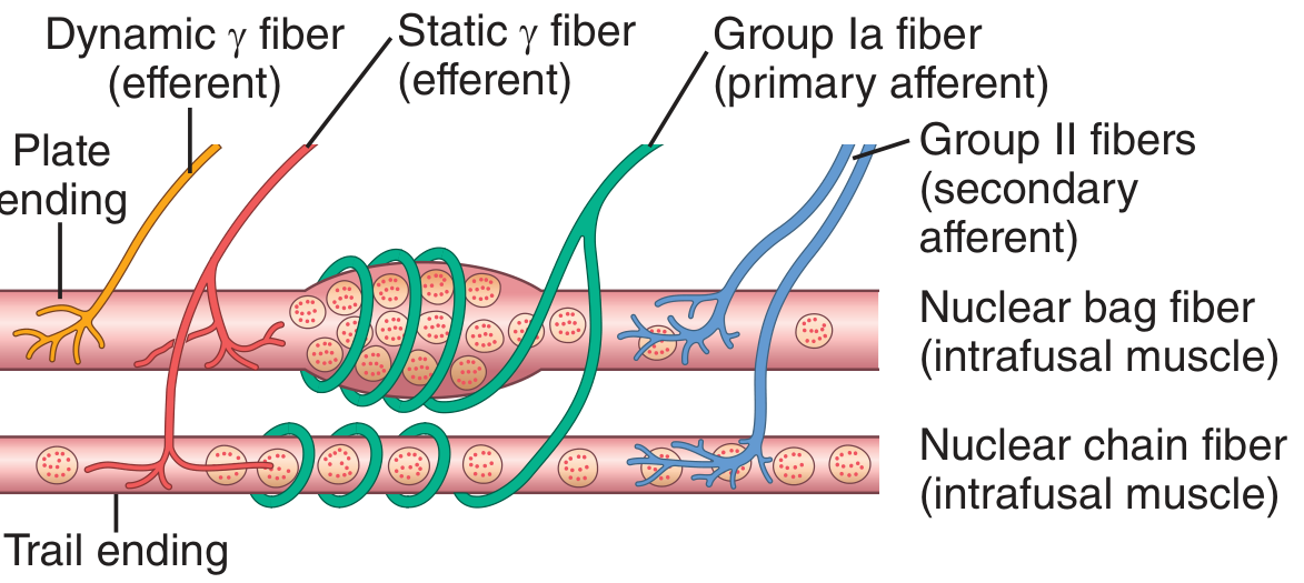
Each muscle spindle is 3-10 mm long and is built around 3-12 intrafusal muscle fibers attached to the surrounding large extrafusal skeletal muscle fibers. There are two types of intrafusal fibers:
| Feature | Nuclear Bag Fiber | Nuclear Chain Fiber |
|---|---|---|
| Number per spindle | 1-3 | 3-9 |
| Nuclei arrangement | Clustered in a bag | In a chain |
| Response | Dynamic (detects rate of stretch) | Static (detects sustained length) |
The central (equatorial) region has few actin/myosin filaments - it acts as a sensory receptor. The polar ends contract when stimulated by gamma (γ) motor neurons.
Sensory (Afferent) Innervation
Primary afferent ending (annulospiral ending):
- Formed by a Type Ia fiber (17 µm diameter)
- Wraps around the equatorial region of both nuclear bag and nuclear chain fibers
- Conduction velocity: 70-120 m/sec (fastest in the body)
- Responds to both dynamic (rate of stretch) and static length changes
Secondary afferent ending (flower-spray ending):
- Formed by Type II fiber (~8 µm diameter)
- Located on one or both sides of the primary ending, mainly on nuclear chain fibers
- Responds primarily to sustained (static) stretch
- Terminates on interneurons in the cord, providing delayed polysynaptic signals
Motor (Efferent) Innervation
- Alpha (α) motor neurons - innervate the extrafusal muscle fibers (the main muscle body)
- Gamma (γ) motor neurons - innervate the polar contractile ends of the intrafusal fibers
- Dynamic γ fibers - innervate nuclear bag fibers (increase spindle sensitivity to rate of stretch)
- Static γ fibers - innervate nuclear chain fibers (increase spindle sensitivity to length)
- Alpha-gamma coactivation: During voluntary movement, both α and γ neurons are activated together, preventing the spindle from going "silent" during muscle shortening, thus maintaining spindle sensitivity throughout contraction.
3. Reflex Arc of the Stretch Reflex (Diagram)
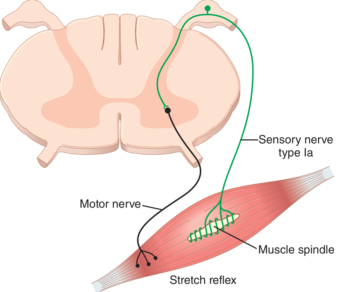
The reflex arc consists of:
- Receptor - Muscle spindle (in stretched muscle)
- Afferent limb - Type Ia fiber → enters via dorsal root
- Integration center - Anterior horn of spinal cord (monosynaptic connection)
- Efferent limb - Alpha motor neuron → ventral root
- Effector - Extrafusal skeletal muscle fibers (contraction of stretched muscle)
The main arc is monosynaptic (one synapse, shortest latency). Type II fibers additionally activate interneurons for polysynaptic pathways.
4. Reciprocal Innervation [2 marks]
Definition: When the stretch reflex excites (contracts) one muscle (the agonist), it simultaneously inhibits the motor neurons supplying the antagonist muscles. This phenomenon is called reciprocal inhibition, and the neuronal circuit that mediates this is called reciprocal innervation.
Mechanism (Ganong's Physiology):
- Ia afferent fibers from the stretched agonist muscle spindle enter the spinal cord
- A collateral branch of each Ia fiber synapses on an inhibitory interneuron (the Ia inhibitory interneuron) in the cord gray matter
- This inhibitory interneuron then synapses on the alpha motor neurons supplying the antagonist muscles
- This is an example of postsynaptic inhibition (IPSP in the antagonist motor neuron)
Functional significance: When a flexor muscle contracts (e.g., biceps during bicep curl), the extensor (e.g., triceps) must simultaneously relax - this reciprocal inhibition allows smooth, coordinated movement without the two muscle groups opposing each other. Descending corticospinal pathways can also modulate the Ia inhibitory interneuron to allow co-contraction at joints when needed.
5. Renshaw Cell Inhibition [2 marks]
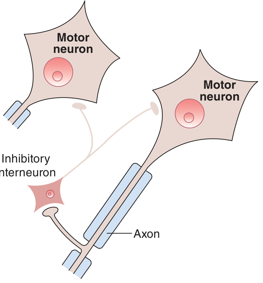
Definition: Renshaw cell inhibition (also called recurrent collateral inhibition or negative feedback inhibition) is a form of postsynaptic inhibition in which a motor neuron's own axon collateral feeds back to inhibit that same motor neuron via a specialized inhibitory interneuron called the Renshaw cell.
Circuit (Ganong's & Kandel's Neuroscience):
- The alpha motor neuron axon, as it exits the anterior horn, gives off a recurrent collateral branch
- This collateral synapses on a Renshaw cell (an inhibitory interneuron in the anterior horn)
- The Renshaw cell releases glycine (inhibitory neurotransmitter)
- Glycine causes IPSPs on the same motor neuron (and on neighboring motor neurons of synergist muscles, and on Ia inhibitory interneurons)
Functional significance:
- Acts as a negative feedback system to regulate motor neuron excitability
- Stabilizes firing rates - prevents motor neurons from firing at excessively high frequencies
- When motor neuron activity increases, Renshaw cell inhibition increases proportionally, capping output
- Also inhibits synergist motor neurons and Ia inhibitory interneurons, helping to coordinate overall joint motor output
- Clinical relevance: Tetanus toxin preferentially blocks glycine release from Renshaw cells → loss of recurrent inhibition → sustained tonic spasm of muscles
Sources: Guyton & Hall Textbook of Medical Physiology, Ch. 55; Ganong's Review of Medical Physiology, 26th Ed., Ch. 12; Kandel's Principles of Neural Science, 6th Ed., Ch. 32.
This is a shared conversation. Sign in to Orris to start your own chat.