
What is junctional rhythm
"junctional rhythm" OR "junctional ectopic tachycardia"
junctional rhythm ECG inverted P waves
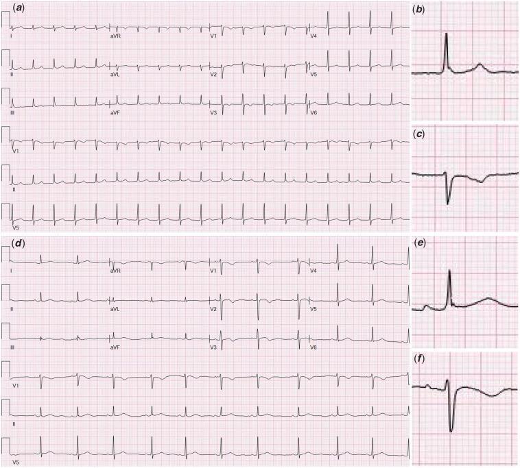
A multi-panel comparative electrocardiogram (ECG) demonstrating the diagnostic features of junctional tachycardia versus normal sinus rhythm. Panel (a) shows a 12-lead ECG of junctional tachycardia characterized by a rapid, narrow-complex rhythm with a rate exceeding 100 bpm and an absence of normal preceding P waves. Magnified inserts (b) and (c) highlight Lead II and V1 respectively, revealing retrograde (inverted) P waves occurring after the QRS complex, indicating atrial depolarization originating from the atrioventricular (AV) junction. Panel (d) displays a standard 12-lead ECG of normal sinus rhythm for comparison. Magnified inserts (e) and (f) for Lead II and V1 demonstrate normal morphology with upright P waves consistently preceding each QRS complex at a regular rate. This diagnostic image is intended for cardiology education to illustrate electrophysiological changes in junctional arrhythmias and the importance of P-wave morphology and timing in rhythm identification. This case is clinically relevant in the context of MIS-C and pediatric myocarditis where AV nodal inflammation may occur.
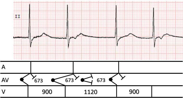
This diagnostic image contains a single-lead electrocardiogram (ECG) rhythm strip from lead II, paired with a corresponding ladder diagram to illustrate cardiac conduction. The ECG demonstrates group beating with variable R-R intervals (900ms and 1120ms), absence of normal sinus P waves, and the presence of retrograde P waves (inverted deflections following the QRS complexes). The visual evidence points to a junctional rhythm with retrograde atrial conduction and occasional reciprocal (echo) beats. The ladder diagram below the tracing provides a schematic representation of conduction across three levels: Atria (A), Atrioventricular node (AV), and Ventricles (V). It specifically denotes timing intervals (673ms, 900ms, 1120ms) and uses standard symbols to indicate points of origin (dots), antegrade and retrograde conduction pathways (angled lines), and conduction block (perpendicular bars). This material is designed for intermediate to advanced cardiovascular education, specifically focusing on complex arrhythmia interpretation and the electrophysiological mechanism of re-entrant junctional rhythms.
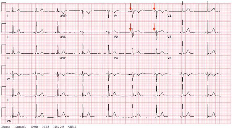
A standard 12-lead electrocardiogram (ECG) recorded at 25 mm/s and 10 mm/mV, demonstrating sinus bradycardia at approximately 53 beats per minute. The tracing shows consistent P waves (highlighted with red arrows in leads V1, V2, V4, and V5) preceding each QRS complex, indicating a transition from a previous junctional rhythm to a normal sinus rhythm. The QRS complexes are narrow and exhibit normal morphology, with positive deflections (dominant R waves) in leads I, II, aVF, and V4-V6. Lead aVR shows a characteristic negative deflection. T waves are upright in the lateral and inferior leads and appropriately inverted in aVR. The image serves as a clinical example of sinus rhythm resolution following a transient arrhythmia. The transition to sinus rhythm is evidenced by the clear P-wave morphology and consistent PR intervals across the limb and precordial leads.
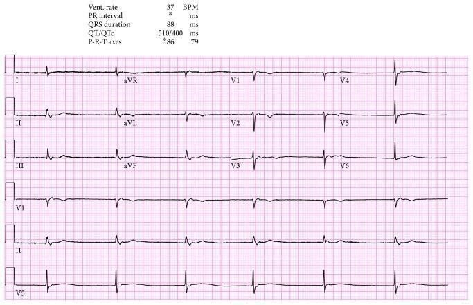
A standard 12-lead electrocardiogram (ECG) displayed on a pink grid, illustrating a severe bradycardia with a heart rate of 37 beats per minute. The rhythm is characterized by a junctional escape rhythm, evidenced by the absence of visible P waves preceding the QRS complexes and a narrow QRS duration of 88 ms. The clinical data printed at the top indicates a QT/QTc interval of 510/400 ms. Morphology analysis shows relatively low QRS amplitude in limb leads with higher amplitudes in precordial leads V4-V6. T waves are upright in lateral leads (I, aVL, V5, V6) and inverted in aVR, with no significant ST-segment elevation or depression noted across the leads. This visual demonstrates common findings associated with sick sinus syndrome or high-grade atrioventricular block where a junctional pacemaker has taken over at its intrinsic slow rate. The image is a primary diagnostic tool for evaluating cardiac rhythm disturbances and conduction system disease in a clinical setting.
- Normally the SA node fires faster than the AV node, which suppresses the AV node and all other slower pacemakers.
- If the SA node discharges slowly or its impulses fail to reach the AV node, junctional escape beats appear. A run of these beats is a junctional rhythm. - Tintinalli's Emergency Medicine, p. 147
- Enhanced AV nodal automaticity can also override the sinus node. This gives an accelerated junctional rhythm or junctional tachycardia. - Tintinalli's Emergency Medicine, p. 147
- There are no normal sinus P waves with a normal PR interval.
- The QRS complex is narrow.
- A retrograde P wave is uncommon. It is an inverted P wave right next to the QRS, either just before or just after it.
| Type | Ventricular rate |
|---|---|
| Junctional (escape) rhythm | 40-60 beats/min |
| Accelerated junctional rhythm | 60-100 beats/min |
| Junctional tachycardia | >100 beats/min |
- Slow or sustained escape rhythms: sinus bradycardia, pauses after premature beats, heart failure, myocarditis, hypokalemia, digitalis toxicity, and sick sinus syndrome or high-grade AV block.
- Accelerated rhythm or tachycardia: medication toxicity (especially digitalis), acute rheumatic fever, and inferior myocardial ischemia.
- Other triggers: digitalis toxicity commonly causes AV junctional rhythm (Katzung's Basic and Clinical Pharmacology). Radiofrequency slow pathway ablation can produce a transient accelerated junctional rhythm (Braunwald's Heart Disease). Volatile anesthetic agents can also cause it (Morgan and Mikhail's Clinical Anesthesiology).
- Isolated, infrequent escape beats usually need no treatment.
- For symptomatic sustained escape rhythms, treat the underlying cause. Atropine can speed up the SA node and improve AV nodal conduction.
- Accelerated junctional rhythm and junctional tachycardia are managed by treating the cause. Examples are stopping digoxin and correcting potassium.