
Brachial artery and anastomosis around elbow
Reading File
The brachial artery is the main artery of the arm, running from the lower border of teres major to the cubital fossa where it bifurcates. The anastomosis around the elbow is a network formed by branches above and below the joint that protects perfusion if the brachial artery is ligated.
Searching sources...
Finding Sources
Finding Sources
Finding Sources
Reading File
Reading File
Finding Sources
Reading File
Reading File
Reading File
Now let me fetch the key anatomical images and also get the cubital fossa content:
Loading Image
Loading Image
Finding Sources
Reading File
Finding Sources
Reading File
Reading File
Loading Image
Loading Image
I now have comprehensive information from multiple authoritative textbooks. Here is a full, well-organized answer:
Brachial Artery and Anastomosis Around the Elbow
Brachial Artery - Overview
The brachial artery is the major arterial supply to the arm. It is the direct continuation of the axillary artery, beginning at the inferior border of teres major (or lower border of pectoralis major, depending on the reference). It runs down the arm and terminates in the cubital fossa (antecubital fossa), where it bifurcates into the radial and ulnar arteries, usually at the apex of the fossa. - Gray's Anatomy for Students
Course in the Arm
- Proximal arm: Lies on the medial side of the humerus, in the medial bicipital groove, where its pulse is palpable and it can be compressed against the humerus in an emergency.
- Distal arm: Moves laterally to a position midway between the medial and lateral epicondyles.
- Cubital fossa: Crosses anteriorly to the elbow joint, lying just medial to the biceps brachii tendon, covered by the bicipital aponeurosis (which protects it).
- The median nerve runs immediately medial to it throughout its course; in the arm it crosses the brachial artery from lateral to medial. - Imaging Anatomy: Bones, Joints, Vessels and Nerves
Relationship to Median Nerve
The median nerve is formed lateral to the axillary artery, crosses superficial to the brachial artery from lateral to medial in the arm, and enters the cubital fossa medial to both the biceps tendon and brachial artery. It then passes between the heads of pronator teres. - Rockwood & Green's Fractures in Adults
Branches of the Brachial Artery
| Branch | Notes |
|---|---|
| Profunda brachii (deep brachial artery) | Largest branch; arises at lower border of teres major. Passes with radial nerve through triangular interval and along radial groove. Divides into radial collateral and middle collateral arteries |
| Superior ulnar collateral artery | Arises distal to profunda brachii; runs alongside the ulnar nerve; anastomoses posteriorly |
| Inferior ulnar collateral artery | Arises more distally, just above the medial epicondyle near the olecranon fossa |
| Nutrient arteries to humerus | Pass through foramen in anteromedial humeral shaft |
| Muscular branches | Numerous, to arm muscles |
- General Anatomy and Musculoskeletal System (THIEME Atlas) | Color Atlas of Human Anatomy Vol. 2
The profunda brachii arises as a single trunk in 55-70% of cases. It may also arise from the axillary artery (22%), as a common trunk with the superior ulnar collateral artery (22%), or rarely from the circumflex humeral arteries.
Cubital Fossa
The cubital fossa is a triangular depression anterior to the elbow, bounded by:
- Laterally: brachioradialis
- Medially: pronator teres
- Base: imaginary line between medial and lateral epicondyles
- Floor: brachialis muscle
- Roof: skin and superficial fascia (+ bicipital aponeurosis anteriorly)
Contents (lateral to medial): biceps tendon → brachial artery → median nerve
The radial nerve runs just under the brachioradialis lip (lateral margin) and divides into its superficial and deep branches here. The ulnar nerve does NOT pass through the cubital fossa - it passes posterior to the medial epicondyle. - Gray's Anatomy for Students
Anastomosis Around the Elbow (Cubital Anastomosis)
This is a periarticular vascular network encircling the elbow joint, formed by descending branches from above (brachial artery and profunda brachii) meeting ascending recurrent branches from below (radial and ulnar arteries).
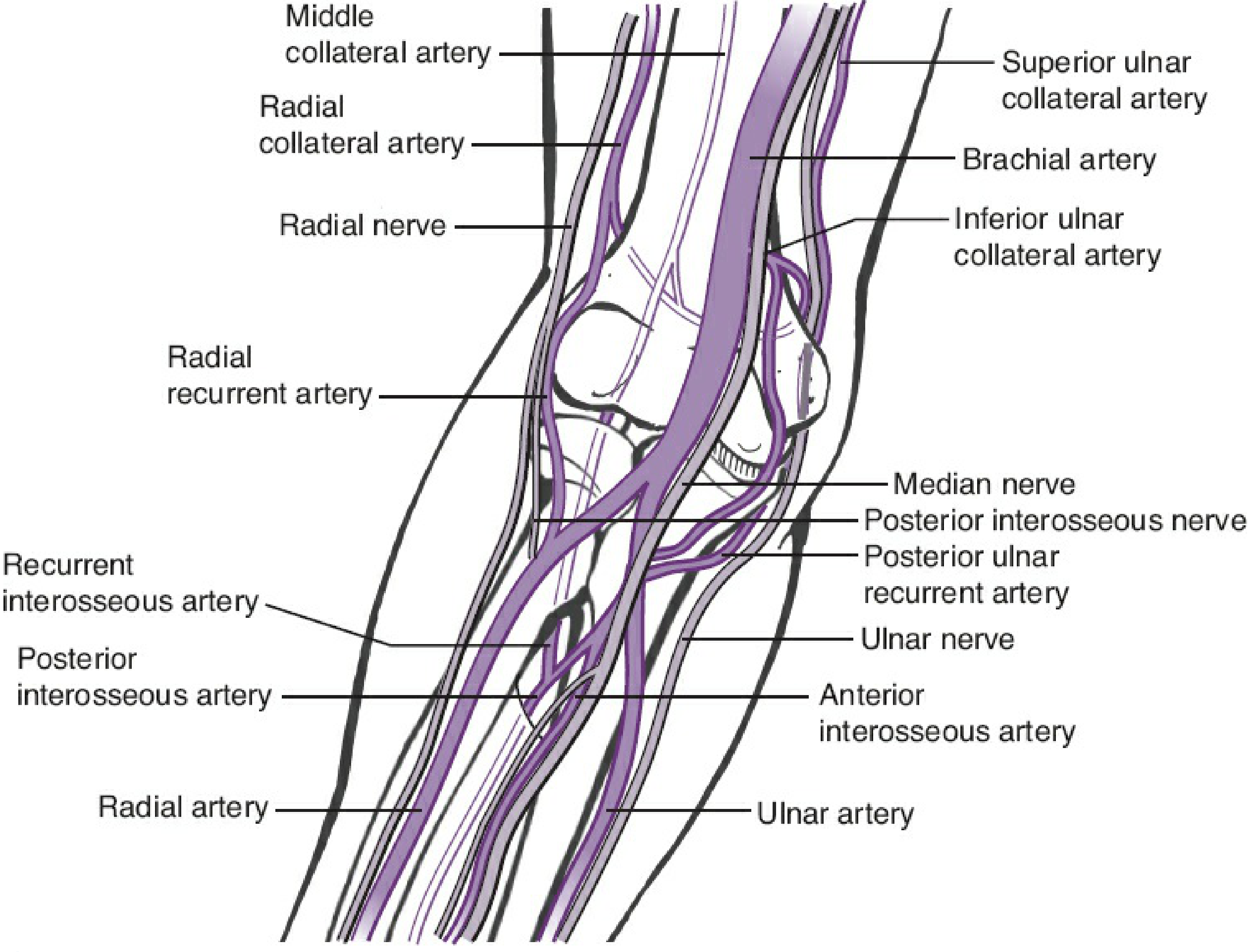
Anastomosis around the elbow showing collateral and recurrent vessels (Rockwood & Green's Fractures in Adults)
Three Vascular Arcades (from Rockwood & Green's)
| Arcade | Formed by | Supplies |
|---|---|---|
| Lateral | Interosseous recurrent + radial recurrent + radial collateral arteries | Capitellum, radial head, lateral epicondyle, lateral trochlea |
| Medial | Superior ulnar collateral + inferior ulnar collateral + anterior & posterior ulnar recurrent arteries | Medial epicondyle, medial trochlea |
| Posterior | Medial collateral artery + contributions from medial & lateral arcades | Olecranon fossa, supracondylar area |
Detailed Components (THIEME Atlas)
Descending (from above):
- Radial collateral artery (from profunda brachii) - communicates via radial recurrent artery
- Middle collateral artery (from profunda brachii) - communicates via interosseous recurrent artery
- Superior ulnar collateral artery (from brachial artery) - communicates via posterior ulnar recurrent artery
- Inferior ulnar collateral artery (from brachial artery) - communicates via anterior ulnar recurrent artery
Ascending recurrent (from below):
- Radial recurrent artery (1st branch of radial artery)
- Anterior ulnar recurrent artery (from ulnar artery)
- Posterior ulnar recurrent artery (from ulnar artery)
- Recurrent interosseous artery (from posterior interosseous artery)
Clinical Significance
-
Ligation of brachial artery: Because of the cubital anastomosis, the brachial artery can be safely ligated distal to the origin of the profunda brachii without compromising blood supply to the elbow region - collateral flow is maintained through the anastomotic network. - THIEME Atlas; Color Atlas of Human Anatomy Vol. 2
-
Radial artery harvest for grafts: The patent cubital anastomosis permits removal of the radial artery as a graft (e.g., for coronary bypass), as collateral circulation is provided by recurrent vessels via the ulnar artery. - Color Atlas Vol. 2
-
Supracondylar process and Struthers' ligament: In ~0.7% of people, a bony outgrowth (supracondylar process) above the medial epicondyle connects to the medial epicondyle via Struthers' ligament, forming a fibro-osseous supracondylar canal that can trap and compress both the brachial artery and the median nerve. - THIEME Atlas
-
Superficial brachial artery variant (4-12%): Courses superficial to the median nerve instead of deep to it - more vulnerable to injury. It is an important fetal vessel and normally regresses postnatally. - Imaging Anatomy Vol. 3
-
Supracondylar fractures in children: The brachial artery is at risk of injury (kinking, entrapment, or laceration) from the proximal fragment, particularly in extension-type supracondylar fractures. The robust anastomotic network may maintain some distal perfusion even with brachial artery injury, which can give a falsely reassuring pink hand.
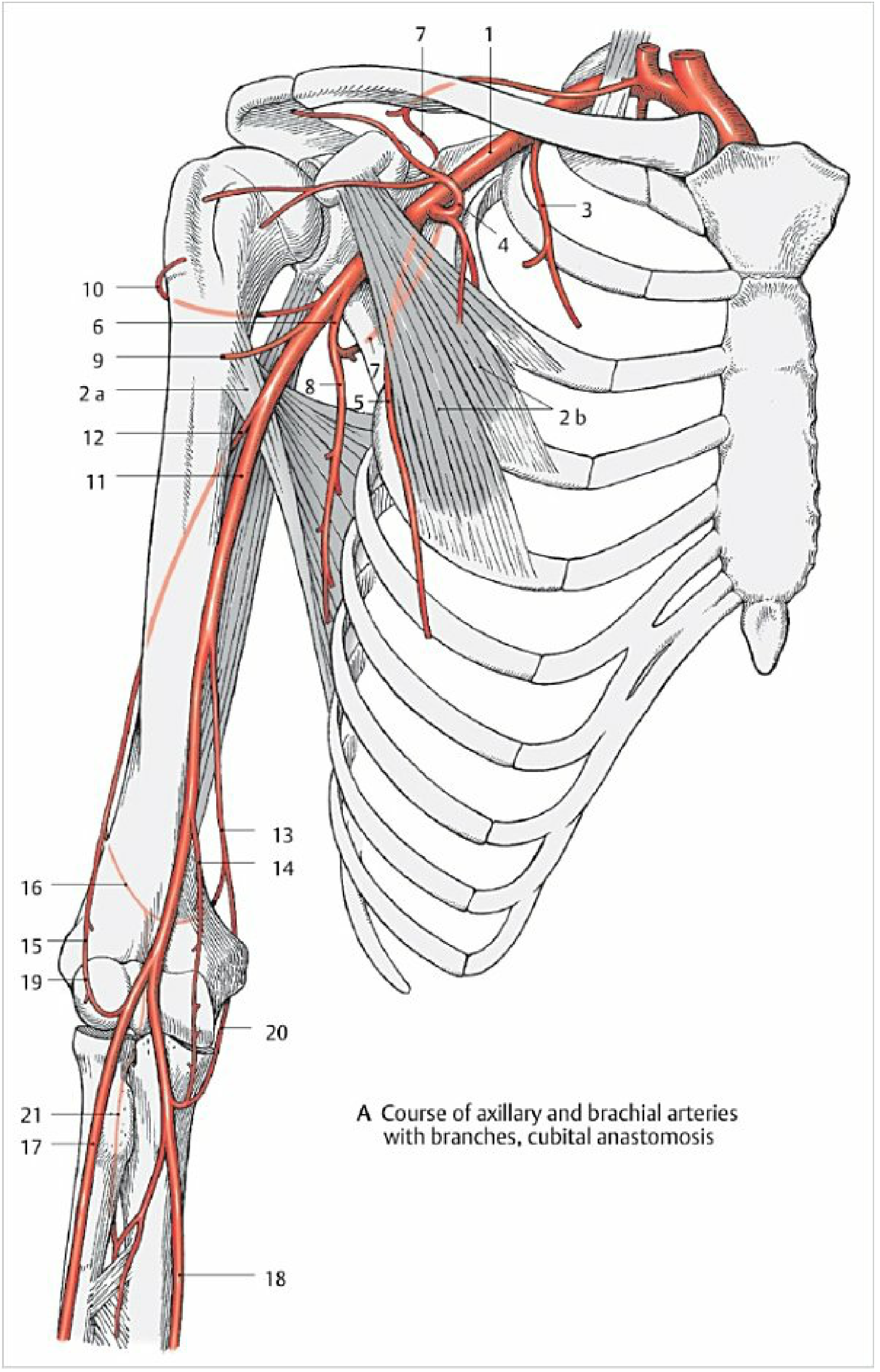
Course of the axillary and brachial arteries with cubital anastomosis - Color Atlas of Human Anatomy Vol. 2
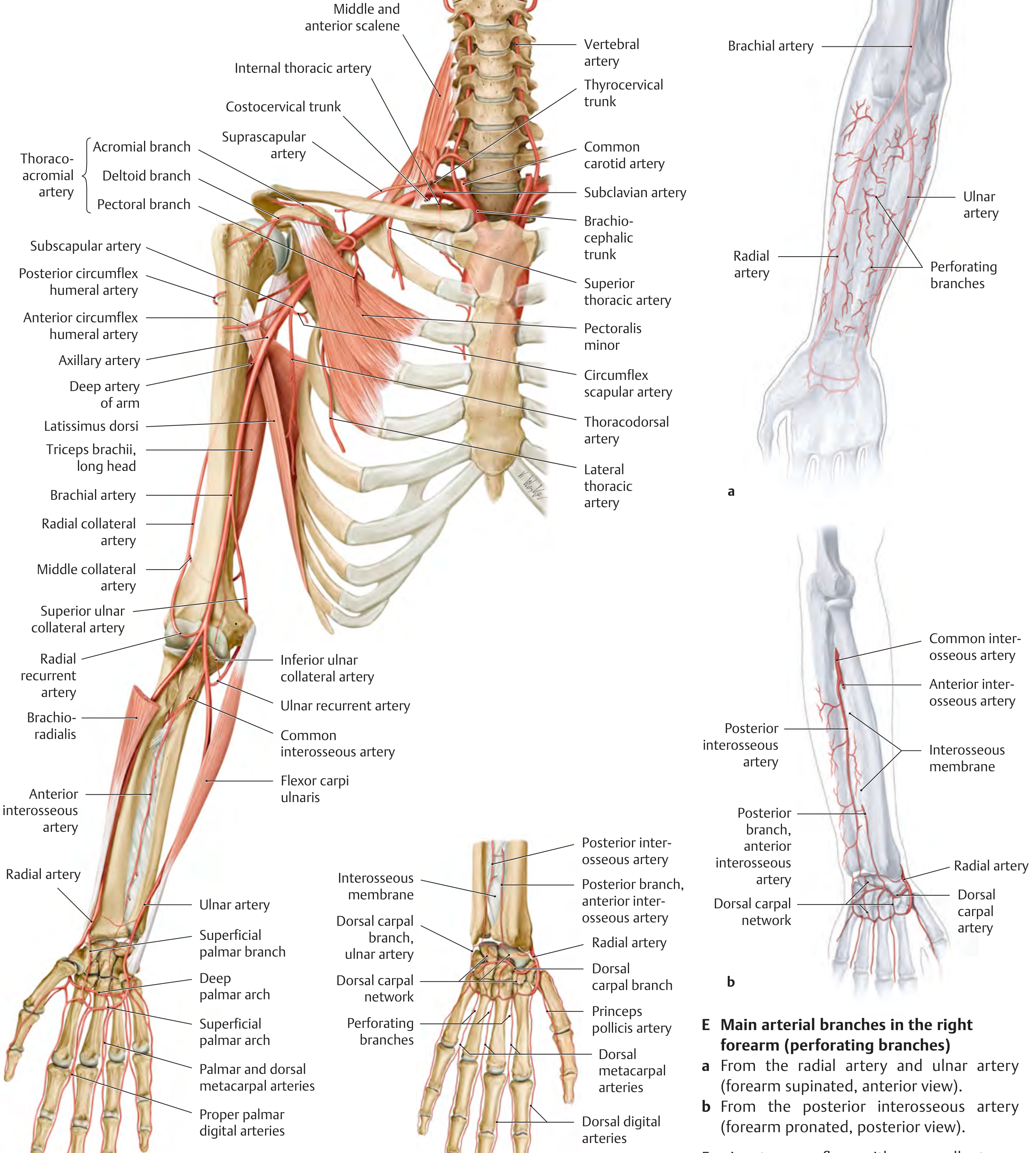
Upper limb arterial overview showing collateral and recurrent branches - THIEME Atlas of General Anatomy
Sources:
- Gray's Anatomy for Students, 4th Ed
- General Anatomy and Musculoskeletal System (THIEME Atlas)
- Color Atlas of Human Anatomy Vol. 2 - Internal Organs
- Imaging Anatomy: Bones, Joints, Vessels and Nerves Vol. 3
- Rockwood and Green's Fractures in Adults, 10th Ed (2025)
This is a shared conversation. Sign in to Orris to start your own chat.