
Role of club cells
Club cells (formerly called Clara cells) are non-ciliated, dome-shaped secretory cells found in the bronchiolar epithelium. Their main roles are: secreting Club Cell Secretory Protein (CCSP/CC16), which has anti-inflammatory properties; producing surfactant-like lipoproteins to protect airway surfaces; detoxifying inhaled toxins via cytochrome P450 enzymes; and serving as progenitor cells that can regenerate bronchiolar epithelium after injury.
Searching sources...
Reading File
Finding Sources
Searching Images
club cells Clara cells bronchiolar epithelium histology
Searching PubMed
"club cells" bronchiolar function secretory
Reading File
Reading File
Reading File
Loading Image
Reading File
Here is a comprehensive overview of club cells, drawn from multiple authoritative pulmonary and histology textbooks:
Club Cells (Formerly Clara Cells)
Location and Morphology
Club cells are non-ciliated secretory cells found in the bronchiolar epithelium, predominantly in the terminal and respiratory bronchioles. They constitute roughly 22% of the total epithelial cell number in these regions. Their defining feature is a characteristic dome-shaped or "club-like" apical surface that bulges into the airway lumen - particularly prominent in rodents.
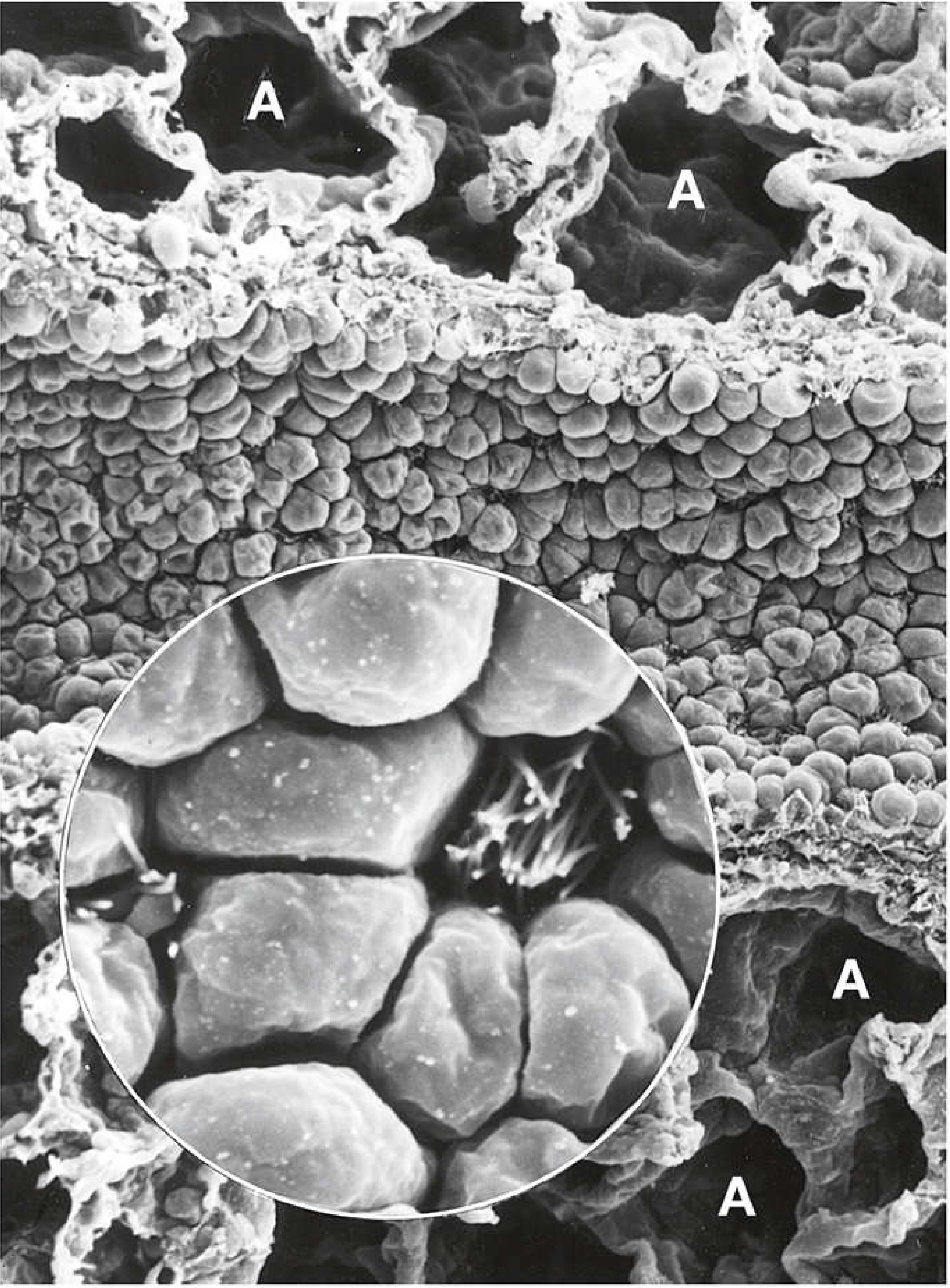
SEM of a terminal bronchiole: the rounded, dome-shaped apical surfaces of club cells are clearly visible, surrounding alveoli (A). The inset shows individual club cells at higher magnification.
Ultrastructurally, club cells display features of protein-secreting cells:
- Well-developed basal rough ER (rER)
- Lateral or supranuclear Golgi apparatus
- Membrane-bound electron-dense secretory granules (~500-600 nm diameter)
- Abundant smooth ER cisternae in the apical cytoplasm (especially in rodents)
Functions of Club Cells
1. Secretion of Club Cell Secretory Protein (CCSP / CC16 / CC10)
Club cells are defined by the expression of secretoglobin 1A1 (SCGB1A1), also called:
- Club cell secretory protein (CCSP)
- CC16 (16-kDa protein)
- CC10 or uteroglobin
This protein is an abundant component of airway secretions. It has immunomodulatory and anti-inflammatory properties (demonstrated in animal studies). CCSP is a clinically useful pulmonary biomarker:
- CCSP levels in BAL fluid decrease in smokers, COPD, and interstitial lung disease - due to club cell damage
- Serum CCSP levels may increase during lung injury because of leakage across the air-blood barrier
2. Surface-Active Lipoprotein Secretion
Club cells secrete a surface-active lipoprotein that prevents luminal adhesion if airway walls collapse during expiration. This is functionally analogous to the surfactant role of type II alveolar cells in the alveoli.
3. Host Defense
Club cells produce several important defensive factors:
- Surfactant proteins A, B, and D - key to innate immunity and alveolar stability
- Complement factor C3 - involved in opsonization and immune activation
- The function of these secretions contributes to the protective lining layer of the distal lung
4. Xenobiotic Detoxification (Cytochrome P450 Enzymes)
Within the lung, club cells are the primary site of cytochrome P450 monooxygenase activity. They are heavily involved in the detoxification of inhaled xenobiotics (foreign chemical compounds, environmental toxins, drugs). This is why club cells are also particularly vulnerable to certain inhaled toxins (e.g., naphthalene, which selectively destroys them in animal models).
5. Progenitor / Stem Cell Function
Club cells play a major role in bronchiolar epithelial regeneration and homeostasis:
- Under normal conditions, proliferation of club cells maintains bronchiolar epithelial homeostasis
- A specialized subset called "variant club cells" (previously "variant Clara cells") - located at neuroepithelial body niches and bronchioalveolar duct junctions - acts as progenitor cells under pathologic conditions
- At the bronchioalveolar duct junction, bronchioalveolar stem cells (BASCs) co-express CCSP (club cell marker) AND surfactant protein C (AT2 cell marker), allowing them to give rise to both airway and alveolar epithelial cells after injury
- Club cells show high plasticity - they can differentiate into multiple cell types, including ciliated cells
Clinical Significance
| Condition | Club Cell Change |
|---|---|
| COPD | Decreased CCSP in airways; club cells undergo metaplasia |
| Asthma | Reduced club cell numbers; altered CC16 levels |
| Acute Lung Injury | Club cell depletion; serum CCSP rises (air-blood barrier leak) |
| Naphthalene toxicity | Selective destruction of club cells (used experimentally) |
| Lung adenocarcinoma | Tumor cells often show ultrastructural features of both club cells and type II alveolar cells |
Summary Table
| Role | Mechanism/Product |
|---|---|
| Secretory | CCSP (CC16), surface-active lipoprotein |
| Host defense | Surfactant proteins A/B/D, complement C3 |
| Anti-inflammatory | CCSP (immunomodulatory) |
| Detoxification | Cytochrome P450 enzymes (xenobiotics) |
| Progenitor/repair | Variant club cells, BASCs at bronchioalveolar junctions |
Sources: Histology: A Text and Atlas (Junqueira), p. 1795 | Fishman's Pulmonary Diseases and Disorders, p. 58 | Murray & Nadel's Textbook of Respiratory Medicine, pp. 117, 192
Role of surfactant
Pulmonary surfactant is a lipoprotein complex secreted by type II pneumocytes that lines the alveolar surface. Its primary role is to reduce alveolar surface tension, preventing alveolar collapse (atelectasis) at end-expiration. It also has immune defense functions via surfactant proteins A and D, and stabilizes alveoli of different sizes by varying surface tension with alveolar radius.
Searching sources...
Finding Sources
Searching PubMed
"pulmonary surfactant" function alveolar
Reading File
Reading File
Reading File
Reading File
Searching Images
pulmonary surfactant alveoli surface tension type II pneumocyte lamellar bodies
Reading File
Loading Image
Finding Sources
Reading File
Reading File
Here is a detailed account of pulmonary surfactant, drawn from leading physiology and respiratory medicine textbooks:
Role of Pulmonary Surfactant
What Is Surfactant?
Pulmonary surfactant is a complex mixture of lipids (~90%) and proteins (~10%) secreted into the alveolar space by type II alveolar (AT2) cells. The most important lipid component is dipalmitoylphosphatidylcholine (DPPC), which accounts for roughly 50-60% of surfactant lipid. Other components include unsaturated phosphatidylcholine and phosphatidylglycerol.
DPPC is an amphipathic molecule - hydrophobic on one end, hydrophilic on the other. DPPC molecules align themselves at the air-liquid alveolar interface, with their hydrophobic tails attracted to each other and their hydrophilic heads repelled. This disrupts the attracting forces between liquid molecules that would otherwise create high surface tension.
Core Physical Role: Reducing Surface Tension
By the Law of Laplace, the collapsing pressure within a sphere is:
P = 2T / r
Where T = surface tension, r = radius. This means small alveoli (small r) face a higher collapsing pressure than large ones - and would collapse without surfactant.
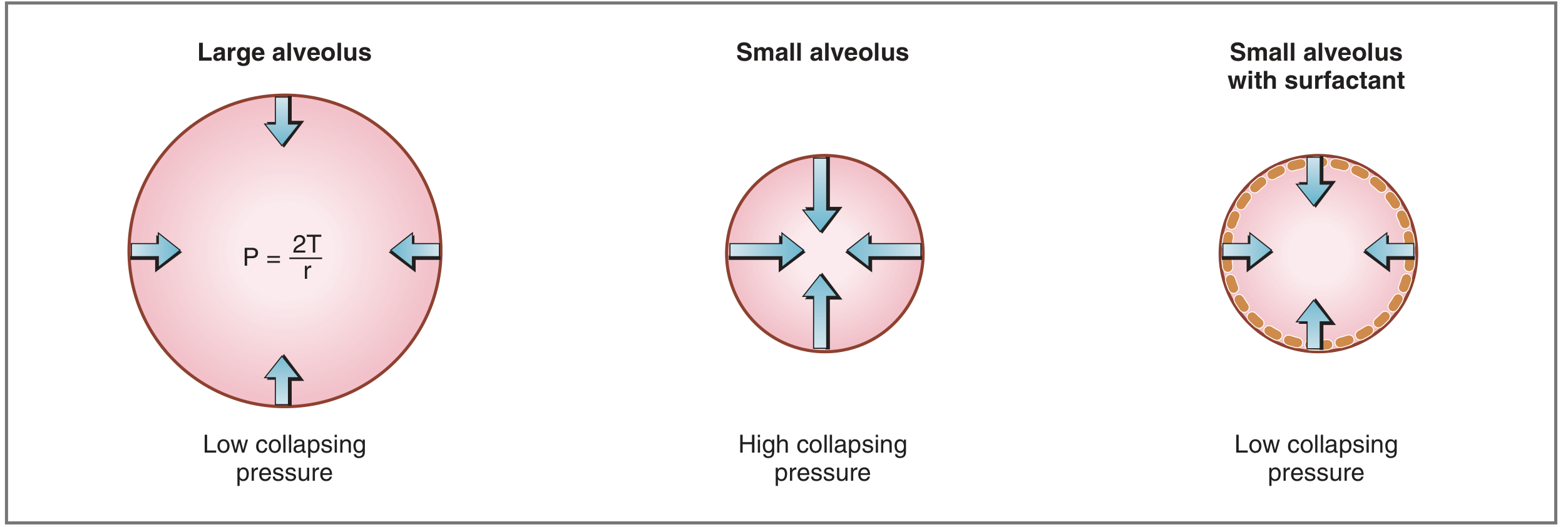
Left: large alveolus - low collapsing pressure. Middle: small alveolus without surfactant - high collapsing pressure. Right: small alveolus with surfactant - collapsing pressure restored to low.
Key consequences of reducing surface tension:
- Prevents alveolar collapse (atelectasis) - especially at end-expiration and in small alveoli
- Stabilizes alveoli of different sizes - by reducing surface tension more in smaller alveoli (where DPPC molecules are more tightly packed), surfactant equalizes pressures across alveoli and prevents air flowing from small to large alveoli
- Increases lung compliance - reduces the elastic recoil force at any given volume, decreasing the work of breathing during inspiration
Surfactant Secretion and Turnover
- 10-20% of the surfactant pool is secreted hourly from AT2 cells via regulated exocytosis of lamellar bodies (LBs)
- After exocytosis, lamellar body contents reorganize into tubular myelin, which then spreads as a monolayer film at the air-liquid interface
- Secretion is stimulated by: hyperventilation, deep breaths/sighs, mechanical stretch (via calcium propagation through gap junctions between AT1 and AT2 cells), beta-agonists, and ATP
- After forming the surface film, surfactant is recycled back into AT2 cells or degraded by alveolar macrophages
Surfactant Proteins
Four proteins are associated with surfactant, each with distinct roles:
| Protein | Type | Key Role |
|---|---|---|
| SP-A | Hydrophilic (collectin) | Regulates surfactant synthesis/secretion (homeostasis); innate immune defense against viruses, bacteria, fungi |
| SP-B | Hydrophobic | Essential for adsorption/spreading of surfactant film; LB formation; transformation to tubular myelin; absence is uniformly fatal |
| SP-C | Hydrophobic | Aids DPPC orientation; maintains the thin film layer; 1% of total protein mass |
| SP-D | Hydrophilic (collectin) | Primarily innate immune defense; binds gram-negative bacteria and microorganisms; regulates inflammation |
SP-A in detail
SP-A is the most abundant surfactant protein. Its C-type lectin carbohydrate recognition domain (CRD) binds to DPPC, microorganisms, and macrophages. It controls feedback regulation of surfactant secretion by AT2 cells and has immunomodulatory properties. In humans, two genes (SFTPA1 and SFTPA2) encode SP-A on chromosome 10.
SP-B in detail
SP-B is a homodimer with five amphipathic helices that interact with the surfactant monolayer surface. It facilitates phospholipid adsorption and spreading onto the alveolar surface. SP-B deficiency causes complete loss of all hydrophobic surfactant function (because it is also needed for SP-C processing via lamellar bodies), resulting in uniformly fatal respiratory failure unless lung transplantation is performed.
Immune Defense Functions
Beyond biophysics, surfactant proteins participate actively in innate lung immunity:
- SP-A and SP-D bind pathogens (viruses, bacteria, fungi) and promote phagocytosis by alveolar macrophages via opsonization
- SP-D also has dual pro- and anti-inflammatory signaling depending on its conformation (intact multimer = anti-inflammatory via CRD; disrupted to monomers = pro-inflammatory via collagenous tail)
- SP-D is upregulated in asthma, hyperoxic injury, bleomycin injury, and Pneumocystis pneumonia; elevated serum SP-D is a biomarker for IPF, COPD, and post-transplant lung dysfunction
- Complement factor C3 is also produced by club cells in conjunction with surfactant proteins
Surfactant Development and Hormonal Regulation
- Surfactant synthesis in the fetus begins around gestational week 24 and is reliably present by week 35
- Synthesis is modulated by: cortisol (stimulates), insulin, prolactin, thyroxine
- Antenatal corticosteroids given to mothers at risk of preterm delivery accelerate surfactant production and reduce neonatal mortality
Clinical Significance
| Condition | Surfactant Abnormality | Consequence |
|---|---|---|
| Neonatal RDS (IRDS / hyaline membrane disease) | Absent surfactant (premature birth) | Atelectasis, hypoxemia, stiff lungs |
| ARDS | Surfactant inactivation (epithelial injury, inflammation) | Hyaline membranes, respiratory failure |
| SP-B deficiency | Absent SP-B and SP-C | Uniformly fatal neonatal respiratory failure |
| ABCA3 mutations | Absent lamellar bodies | Neonatal respiratory failure |
| Pulmonary alveolar proteinosis (PAP) | Surfactant overproduction/impaired clearance | Alveolar filling with surfactant material; "crazy-paving" on CT |
| COPD / smoking | Reduced CCSP (club cell marker); altered surfactant | Impaired airway protection and repair |
Sources: Costanzo Physiology 7th Edition, p. 210 | Murray & Nadel's Textbook of Respiratory Medicine, pp. 84-87 | Histology: A Text and Atlas (Junqueira), pp. 1794-1808 | Ganong's Review of Medical Physiology, 26th Ed.
How does surfactant reduce surface tension
Surfactant reduces surface tension by inserting DPPC molecules at the air-liquid interface of alveoli. DPPC is amphipathic - its hydrophobic tails cluster together while its hydrophilic heads face the aqueous lining. This intermolecular arrangement physically disrupts the cohesive forces between water molecules that generate surface tension, lowering it from ~70 mN/m (pure water) to near zero at end-expiration.
Diving deeper...
Finding Sources
Finding Sources
Reading File
Reading File
Reading File
Reading File
Reading File
Loading Image
Loading Image
Here is a focused, mechanistic explanation drawn from multiple physiology and pulmonary medicine textbooks:
How Surfactant Reduces Surface Tension
1. The Problem: Surface Tension at the Air-Liquid Interface
The alveoli are lined with a thin film of fluid. At any air-water interface, water molecules are strongly attracted to each other by intermolecular cohesive forces. At the surface, molecules are only pulled inward and sideways (not outward into air), creating a net inward force - this is surface tension. It causes the liquid surface to contract, which for a sphere (like an alveolus) generates a collapsing pressure (Law of Laplace: P = 2T/r).
Surface tension values:
- Pure water: ~72 dynes/cm
- Alveolar fluid without surfactant: ~50 dynes/cm
- Alveolar fluid with normal surfactant: 5-30 dynes/cm (up to 12x lower than pure water)
Without surfactant, the collapsing pressure in an average alveolus (radius ~100 μm) would be ~18 cm H₂O - 4.5 times higher than normal. This would cause widespread alveolar collapse and pulmonary edema.
2. The Mechanism: DPPC at the Interface
The key agent is dipalmitoylphosphatidylcholine (DPPC), which makes up ~50-60% of surfactant lipid. DPPC is unique because both its fatty acid side chains are saturated (palmitoyl = C16 saturated), allowing them to pack very densely.
The molecular mechanism works as follows:
DPPC is amphipathic - it has:
- A hydrophilic phosphocholine head (attracted to water)
- Two hydrophobic palmitoyl tails (repelled by water)
When DPPC reaches the alveolar surface, it does not dissolve uniformly in the fluid. Instead, it inserts itself at the air-liquid interface, oriented with its hydrophilic heads in the water and its hydrophobic tails pointing into the air. This forms a monomolecular film (monolayer).
This monolayer physically interposes DPPC molecules between water molecules at the surface. The intermolecular forces between adjacent DPPC molecules replace and break up the strong water-water cohesive forces that generate surface tension. The result: surface tension drops dramatically.
3. Why Saturated Tails Are Critical - and the Problem at 37°C
Saturated C16 acyl chains (palmitoyl) pack densely at the air-liquid interface, which is what makes surface tension reduction so effective. However, this dense packing (semicrystalline "gel phase") normally only occurs at a phase transition of 41°C - above physiologic temperature. At 37°C, pure DPPC alone forms a rigid gel phase that cannot spread or move rapidly with the expansion and compression of breathing.
This is where SP-B and SP-C become essential. These hydrophobic surfactant proteins:
- Enable DPPC to adsorb rapidly to the air-liquid interface at 37°C despite the gel-phase limitation
- Facilitate spreading and compression of the DPPC monolayer during the respiratory cycle
- Allow surfactant to maintain low surface tension during the dynamic compression of expiration
As Murray & Nadel states: "DPPC-enriched phospholipid films reduce surface tension at the air-liquid interface, but these lipids alone demonstrate slow film formation and cannot provide optimal surfactant function. Surfactant proteins play a [critical] role."
4. Dynamic Variation of Surface Tension with Alveolar Size
A critical and elegant property of surfactant is that its surface tension varies with alveolar volume - and this is what stabilizes alveoli:
-
During expiration (alveolus shrinks): The same amount of surfactant is compressed into a smaller surface area, so DPPC molecules pack more tightly. Surface tension falls toward zero, counteracting the increased collapsing pressure from the smaller radius. This keeps small alveoli open.
-
During inspiration (alveolus expands): Surfactant molecules spread apart over a larger surface area, so surface density decreases. Surface tension rises, creating an increased elastic recoil - acting as a "brake" on over-inflation.
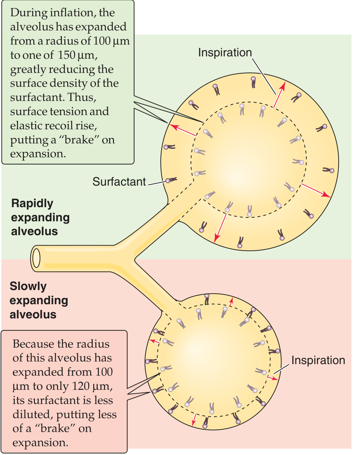
During inspiration: the rapidly expanding alveolus (top) dilutes its surface surfactant, raising surface tension - which brakes further expansion. The slowly expanding alveolus (bottom) retains denser surfactant - less braking - so it catches up. This dynamic self-regulation equalizes ventilation across alveoli of different sizes.
This variable behavior is the reason surfactant stabilizes alveoli of different radii and prevents the Laplace-predicted scenario where small alveoli empty into large ones.
5. Three Downstream Consequences of Reduced Surface Tension
| Effect | Explanation |
|---|---|
| Prevents atelectasis | Low surface tension keeps small alveoli open at end-expiration |
| Increases lung compliance | Lower elastic recoil force at any given volume = less work of breathing during inspiration |
| Prevents pulmonary edema | Without surfactant, unopposed surface tension (~20 mmHg force) would draw fluid from interstitium into alveoli, impairing gas diffusion; surfactant keeps this force low |
6. Historical Note
In 1929, von Neegaard first discovered that lungs inflated with air required more pressure than those inflated with saline - suggesting a surface tension component. In the 1950s, Clements and Pattle demonstrated that phospholipids in lung extracts were responsible for reducing surface tension. Avery and Mead then showed surfactant deficiency was the cause of neonatal RDS - leading to modern surfactant replacement therapy.
Sources: Guyton & Hall Medical Physiology, p. 495 | Ganong's Review of Medical Physiology, 26th Ed., p. 631 | Murray & Nadel's Textbook of Respiratory Medicine, pp. 81-86 | Medical Physiology (Boron & Boulpaep), p. 900 | Fishman's Pulmonary Diseases, p. 123
Airway resistance in pulmonary tree
Your credits are exhausted. You can purchase a top-up or subscribe to a plan to continue using Orris. If you're already subscribed, your daily credits will reset tomorrow.
This is a shared conversation. Sign in to Orris to start your own chat.