
Explain air blast injuries
Reading File
Finding Sources
Searching PubMed
"blast injury" pathophysiology management
Reading File
Reading File
Reading File
Reading File
Reading File
Reading File
Reading File
Reading File
Reading File
Reading File
Reading File
Reading File
Loading Image
I now have all the material needed for a thorough answer. Here it is:
Air Blast Injuries
An air blast injury (explosion/blast injury) refers to the constellation of trauma sustained when a person is exposed to the pressure wave generated by an explosion. The injuries are systematically classified into five categories based on mechanism.
Physics of the Blast Wave
An explosion is a near-instantaneous chemical transformation of solid or liquid into gas, releasing enormous energy. This creates a supersonic positive-pressure wave (>4,500 m/s for high-order explosives) that moves radially outward, followed immediately by a negative-pressure (suction) wave. As a result, victims feel the blast wave before they hear the explosion.
Factors that amplify blast injury:
- Distance: Blast overpressure obeys the inverse cube law. A person 3 m from the source experiences 8 times more overpressure than one 6 m away.
- Enclosed spaces: Blast waves reflect off walls, floors, and ceilings, superimposing on the outgoing wave to amplify it - a phenomenon called mach stem formation. Explosions in corners can be up to 8 times more lethal than in open air.
- Surface reflection: Standing adjacent to a wall significantly increases primary blast exposure.
- Quantity and type of explosive: High-order explosives (detonation velocity >4,500 m/s) are far more damaging than low-order explosives (e.g. black powder, <1,000 m/s).
- Embedded shrapnel: Deliberately placed nails, bolts, or ball bearings multiply the secondary injury burden.
Classification of Blast Injuries
| Category | Mechanism | Key Examples |
|---|---|---|
| Primary | Blast overpressure wave | Tympanic membrane rupture, blast lung, intestinal contusion/perforation |
| Secondary | Penetrating - energized fragments | All ballistic/shrapnel wounds |
| Tertiary | Blunt - victim displaced by blast wind | Blunt trauma, traumatic amputation, crush injury |
| Quaternary | Miscellaneous effects | Burns, inhalation injury, exacerbation of existing disease |
| Quinary | Device additions | Radiation sickness, infection from biological material |
(Bailey and Love's Short Practice of Surgery, 28th Ed.)
Primary Blast Injuries
These are unique to blast - they occur without any physical contact. Damage is concentrated at air-tissue interfaces, where the blast wave encounters sudden density changes.
Mechanisms at the Cellular Level
Three distinct physical forces cause injury at these interfaces:
- Spalling forces - the blast wave displaces and fragments high-density tissue into low-density tissue (e.g. blood forced from capillaries into alveoli, causing alveolar hemorrhage).
- Implosion forces - compressed gas at the leading edge of the wave rapidly compresses and reexpands air-filled structures (e.g. alveoli), causing barotrauma and air embolism.
- Inertia forces - different tissue densities absorb energy differently, moving at different velocities, causing massive shearing and avulsion injuries.
(Murray & Nadel's Textbook of Respiratory Medicine)
Blast Lung (Primary Blast Lung Injury)
This is the most common fatal primary blast injury and the most researched blast phenomenon. The injury depends on energy propagation from the shockwave into lung tissue, with the severity determined by:
- the blast wave strength
- distance from detonation
- the surrounding environment (enclosed > open)
Pathophysiology:
- Immediate vagally mediated bradycardia and apnoea (variable duration)
- Alveolar-capillary rupture with intrapulmonary bleeding and oedema
- Free hemoglobin in alveoli generates free radicals, driving further edema
- Leukocyte accumulation and inflammatory cytokine release over 12-24 hours cause further epithelial cell damage, followed by endothelial cell damage over 24-56 hours
- The resulting syndrome closely resembles ARDS
Clinical features:
- Progressive hypoxia - may be absent initially (only 28% hypoxic on first presentation in one series)
- Shortness of breath, cough, chest pain, haemoptysis, tachypnoea, cyanosis
- External skin injuries may be absent ("occult" blast)
- Associated injuries: pneumothorax, haemothorax, pneumomediastinum, subcutaneous emphysema, air embolism
Imaging:
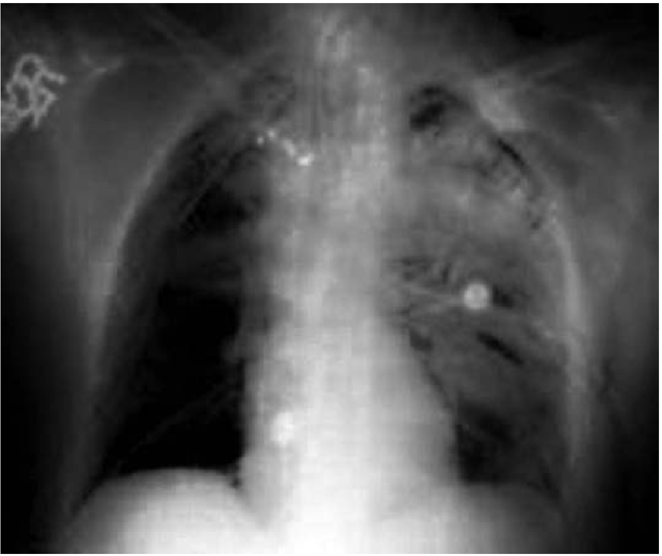
- Classic "bat-wing" bilateral pulmonary infiltrates on chest radiograph (shown below)
- CT helps distinguish central blast lung infiltrates from more peripheral contusional changes
Tympanic Membrane (TM) Rupture
The TM ruptures at 1-8 psi dynamic overpressure and is the single most common primary blast injury overall. Patients may be asymptomatic, have transient hearing loss, or have otorrhoea. Importantly:
- TM rupture is not a reliable severity marker - it has poor sensitivity/specificity for predicting serious visceral blast injury
- Intact TMs miss up to 50% of primary blast lung injuries
- Reliable markers of severe blast injury are: burns >10% TBSA, skull/facial fractures, penetrating head or torso injuries
Ossicular dislodgement may also occur. Follow-up audiology is required.
Gastrointestinal Blast Injury
Abdominal primary blast injury affects ~3% of cases but carries significant morbidity if missed. Damage is most marked at tissue-air interfaces, making hollow organs the primary targets:
- Caecum and terminal ileum are most commonly injured
- Presentation may be delayed relative to blast lung - initial symptoms absent, progressing to pain and peritonitis with perforation
- The most common operative finding is subserosal haemorrhage
- Mucosal tears, mural haematomas (which can progress to perforation), and full-thickness perforations all occur
- Solid organs are more resistant but can be injured at very high overpressures via attachment distraction
Management: serial clinical examinations over 24-48 hours; CT abdomen/pelvis if near the explosion; surgical principles as for conventional blunt/penetrating trauma.
Secondary Blast Injuries
Caused by fragments energized by the explosion - from the device casing, deliberately embedded projectiles (nails, bolts, ball bearings), surrounding debris (glass, stones), or even biological material (bone from the bomber or other victims - a documented route of hepatitis B transmission).
Secondary fragments obey the inverse square law rather than the cube law, so they can wound at much greater distances than primary blast overpressure.
- Wound patterns are highly variable (no two the same) due to irregular fragment geometry, yaw, and tumble
- Both permanent and temporary wound cavities are produced
- Management: formal debridement, all wounds treated as contaminated/dirty, serial debridement with delayed primary closure preferred
- Fragment removal indicated if easily accessible, within a joint, or adjacent to vital structures at risk of erosion
Tertiary Blast Injuries
Caused by the blast wind physically propelling people into hard surfaces, or from falling debris crushing victims. These are analogous to conventional blunt trauma and may injure any organ system. Traumatic amputation is typically classified here (though primary blast brisance on bone may contribute).
Quaternary and Quinary Injuries
Quaternary: Burns (thermal or chemical), inhalation injury, exacerbation of pre-existing conditions (e.g. cardiovascular disease), psychological trauma.
Quinary: Injury from deliberate addition of radioactive or biological material to the device - radiation sickness, unusual infections.
Special Scenarios
Enclosed Blast (e.g. bus, train, building)
Higher rates of blast lung and TM rupture; secondary fragment injuries may be lower (some shielding from walls). Mortality is significantly greater than open-air blast.
Underbody Blast (vehicle-borne IED)
The shockwave propagates through a solid substrate. Injuries include severe foot/ankle and pelvic injuries from upward acceleration, plus head injury and non-compressible torso haemorrhage (aortic disruption, liver laceration) as the main causes of death.
Dismounted IED Casualty
Characteristic pattern: lower limb amputation, pelvic fracture, perineal/genital/rectal injuries. Hemorrhage is the leading cause of death. Management priorities:
- Immediate tourniquet application and proximal vascular control
- REBOA (resuscitative endovascular balloon occlusion of the aorta) is under evaluation
- Damage control resuscitation (DCR) followed by serial debridement
- Rectal examination and proctoscopy; early faecal diversion if indicated
- Careful urological assessment and catheterization
CNS and Vascular Injuries
- Traumatic brain injury (TBI): Penetrating shrapnel is the most common cause; blast overpressure may also compromise the blood-brain barrier. Small shrapnel entry wounds are easily missed under the hair. Early neuroimaging is essential.
- Vascular injury: Small entry wounds mask significant vascular damage. Document and monitor distal pulses and perfusion carefully. Measure compartment pressures if compartment syndrome is suspected. Early angiography and intervention for absent pulses.
- Air embolism: A recognized complication of blast lung, causing cardiac dysfunction and sudden death. Treat by positioning the patient in the left lateral decubitus (head down, feet up) position, minimizing ventilator peak pressures, and considering hyperbaric therapy.
Management Summary
Immediate Priorities
- ATLS framework - airway, breathing, circulation
- Hemorrhage control - direct pressure, tourniquets for extremity bleeding (effective up to 6 hours in field conditions), angiographic occlusion where available
- Blood and blood product resuscitation (massive transfusion may be required)
Blast Lung Specific Management
- Limit IV fluids to avoid worsening pulmonary edema
- Lung-protective ventilation: tidal volume 6-7 mL/kg ideal body weight, plateau pressures <30 cmH₂O, permissive hypercapnia
- Neuromuscular blockade may be needed
- Inverse I:E ratio ventilation may help
- Bilateral prophylactic pleural decompression is practiced in some centres (limited evidence)
- Rescue strategies for refractory ARDS: inhaled nitric oxide, inhaled prostaglandins, open-lung ventilation, ECMO (use with caution due to coagulopathy risk in setting of alveolar hemorrhage)
- Avoid high PEEP that may worsen pneumothorax or air embolism
Observation and Discharge
- Asymptomatic patients with normal CXR and normal room-air SpO₂ may be observed for 4-6 hours and discharged if no deterioration
- Significant blast lung or other injuries warrant ICU admission
- Long-term pulmonary outcomes are generally excellent in survivors
Key Points
- Primary blast injuries are unique to explosions and may have no external signs
- TM rupture is neither sensitive nor specific for severe primary blast injury
- Blast lung may be initially silent - delayed hypoxemia is characteristic
- Abdominal injuries may present days later with peritonitis
- Always assume multiple injury mechanisms are present simultaneously (primary + secondary + tertiary)
- Enclosed spaces dramatically worsen all blast injury categories
- Hemorrhage is the most common preventable cause of death
Sources:
- Bailey and Love's Short Practice of Surgery, 28th Ed., pp. 489-490
- Tintinalli's Emergency Medicine: A Comprehensive Study Guide, pp. 72-73
- Murray & Nadel's Textbook of Respiratory Medicine, pp. 2411-2413
I didn't understand air blast wave
Finding Sources
Finding Sources
Reading File
Reading File
Loading Image
I have everything needed for a clear, step-by-step explanation with the actual diagram from the textbook.
The Air Blast Wave - Explained Simply
Think of it this way: when an explosion happens, it does not just make a loud noise and a fireball. It sends a powerful wall of compressed air outward at supersonic speed. That wall is the blast wave, and it is what makes explosions medically unique.
Step 1 - What Happens at the Moment of Detonation
When a high explosive detonates, the reaction is almost instantaneous (within 1 microsecond). The solid or liquid explosive converts entirely into extremely hot, high-pressure gas:
- Local pressures reach ~25 million atmospheres
- Temperatures hit 2,000-6,000°C
This superheated gas has nowhere to go except outward, expanding radially at 3-4 km/s and violently pushing the surrounding air out of its way.
Step 2 - How the Blast Wave Forms
As the expanding gas pushes outward, it compresses the air in front of it into a dense, thin shell - like a bubble of compressed air that travels outward faster than the speed of sound. That compressed-air shell is the blast wave.
A helpful analogy: imagine pushing your palm through a swimming pool. The water bunches up in front of your hand and pushes outward as a wave. The blast wave is the same concept, but in air, at supersonic speed.
Step 3 - The Friedlander Wave (The Shape of the Blast)
The pressure changes at any fixed point as the blast wave passes through it follow a very specific pattern, called the Friedlander curve:
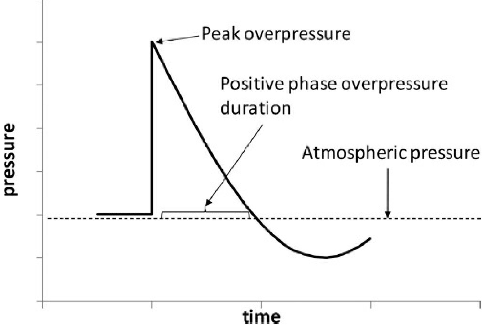
Reading the graph from left to right, three phases happen in sequence:
Phase 1 - The Positive Phase (Overpressure)
- Pressure jumps instantaneously from normal atmospheric to a massive peak (the "peak overpressure")
- This wall of high pressure hits the body like a hammer
- It then decays back down toward atmospheric pressure over a short time (the "positive phase duration")
- This is the part that causes primary blast injuries - the overpressure crushes, ruptures, and tears
Phase 2 - Return to Baseline
- Pressure briefly returns to normal atmospheric
Phase 3 - The Negative Phase (Underpressure / Suction)
- Pressure drops below atmospheric - a partial vacuum
- Air rushes back inward to fill this vacuum, generating powerful blast winds that carry debris
- This suction can worsen injuries already caused in Phase 1 (e.g. further tearing of already-disrupted alveoli)
Step 4 - The Blast Wind
The blast wave has two components traveling together:
| Component | What it is | Effect |
|---|---|---|
| Shock wave | The wall of high pressure | Crushes and ruptures tissues directly - causes primary injury |
| Blast wind | Air mass set in motion behind the shock wave | Throws people and objects - causes tertiary injury |
The blast wind is the reason victims are found far from the explosion site, and why flying debris (secondary injuries) can travel at enormous speed.
Step 5 - Why Pressure Drops With Distance
The blast wave expands spherically in all directions. As it gets bigger, the same energy is spread over a much larger surface area. The pressure falls off very rapidly - following the inverse cube law:
- Double the distance → 8 times less overpressure
- Triple the distance → 27 times less overpressure
This is why someone 3 m from an explosion experiences 8× more overpressure than someone 6 m away.
Step 6 - Why Enclosed Spaces Are So Much More Deadly
In open air, the blast wave expands outward and the energy disperses. In a closed space (bus, building, train), the wave hits the walls and bounces back. These reflected waves collide with the incoming wave and stack on top of each other - amplifying the pressure enormously. A phenomenon called Mach stem formation occurs when reflected waves meet the ground or walls at angles greater than 40°, creating a new, even more powerful composite wave. Explosions in corners can be up to 8 times more lethal than in open air.
Step 7 - Why the Body Is Injured Where Air Meets Tissue
The blast wave travels through the body and transmits energy unevenly. It moves at different speeds through different tissue densities (bone, soft tissue, air). The worst damage happens at interfaces between air and tissue, because that is where the biggest density change occurs - the energy has nowhere to go and is released destructively. That is why:
- Lungs (full of air pockets surrounded by tissue) are the most commonly injured organ
- Ears (air against the thin tympanic membrane) rupture even at low pressures
- Bowel (gas-filled hollow organ) is vulnerable
- Solid organs (liver, spleen - uniform density, no air) are much more resistant
Summary in One Sentence
The blast wave is a supersonic wall of compressed air that hits the body like a hydraulic punch, with an instant spike in pressure followed by a suction phase - and the body is most damaged wherever air and tissue meet, because that is where the energy change is most abrupt.
Source: Rockwood and Green's Fractures in Adults, 10th Ed., p. 588 | Tintinalli's Emergency Medicine, p. 72 | Murray & Nadel's Textbook of Respiratory Medicine, p. 2412
This is a shared conversation. Sign in to Orris to start your own chat.