
Pathophysiology of acute pulpal pain for 10 marks. Please include contents also
dental pulp innervation nerve fibers pain pathophysiology
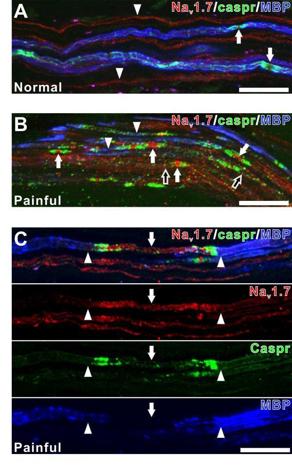
This set of confocal micrographs illustrates the spatial relationship between sodium channel Nav1.7 (red), the paranodal protein caspr (green), and myelin basic protein (MBP; blue) in human dental pulp nerve fibers. Panel A displays a normal sample where Nav1.7 expression is primarily restricted to unmyelinated fibers (arrowheads) and specific nodal sites in myelinated fibers (arrows) identified by intact MBP staining. Panels B and C represent painful clinical samples, demonstrating significant neurobiological alterations. In painful pulp, there is a prominent increase in Nav1.7 expression at caspr-identified nodal sites (white arrows) often associated with atypical nodal morphology. High-magnification single-channel images in Panel C reveal that intense Nav1.7 immunoreactivity (arrows) occurs specifically within axon segments exhibiting a focal loss or alteration of MBP staining (demyelination), whereas areas with preserved MBP expression (arrowheads) show lower Nav1.7 intensity. These diagnostic images provide visual evidence of sodium channel remodeling and demyelination associated with dental pain. Scale bars = 20 μm.
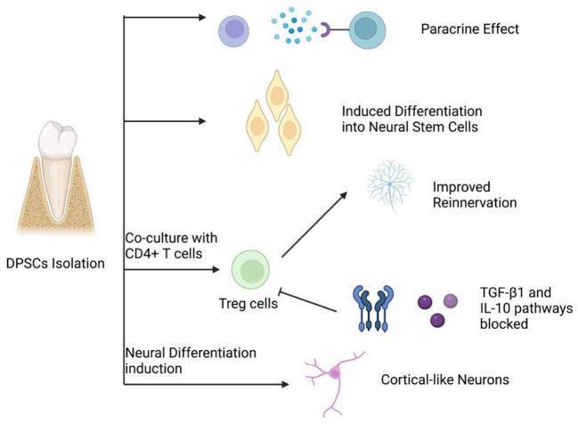
This medical pathophysiology diagram illustrates the regenerative mechanisms of Dental Pulp Stem Cells (DPSCs) in peripheral nerve repair. The flowchart begins on the left with 'DPSCs Isolation' from a human tooth, branching into four primary therapeutic pathways. 1) Paracrine Effect: Depicted by DPSCs secreting signaling molecules (blue spheres) that interact with target cell receptors. 2) Induced Differentiation into Neural Stem Cells: Shows DPSCs transforming into elongated, teardrop-shaped neural progenitor cells. 3) Immunomodulatory Pathway: Describes the co-culture of DPSCs with CD4+ T cells to produce Regulatory T cells (Treg cells), which promotes 'Improved Reinnervation' (visualized as a complex neuronal network) or blocks TGF-̢1 and IL-10 pathways to modulate the inflammatory environment. 4) Neural Differentiation Induction: Shows the direct maturation of DPSCs into 'Cortical-like Neurons' characterized by a distinct soma and branching processes. This diagram serves as a pedagogical summary of biotherapy and neuroregeneration concepts within regenerative medicine.
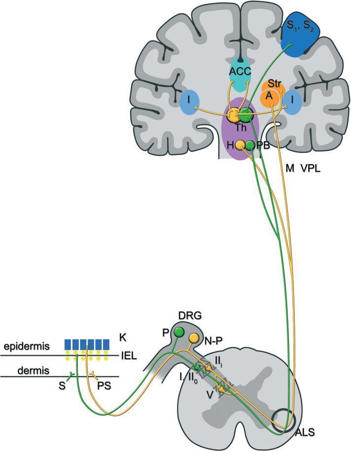
This pathophysiology diagram illustrates the lateral and medial ascending pain pathways, specifically focusing on peptidergic (green) and non-peptidergic (yellow) C-fibers involved in chemotherapy-induced peripheral neurotoxicity. The pathway begins in the skin layers (epidermis and dermis) where free nerve endings originate. Peptidergic fibers (P) and non-peptidergic fibers (N-P) have cell bodies in the dorsal root ganglion (DRG) and project to the spinal cord's dorsal horn. The peptidergic pathway synapses in laminae I and IIo, ascending via the anterolateral system (ALS) to the parabrachial nuclei (PB), thalamus (Th), and somatosensory cortex (S1, S2) and insula (I), mediating sensory-discriminative pain. The non-peptidergic pathway synapses in lamina IIi and V, ascending to the hypothalamus (H), amygdala (A), striatal nuclei (Str), and anterior cingulate cortex (ACC), mediating affective-emotional pain components. The diagram highlights anatomical structures including the keratinocytes (K), sympathetic (S) and parasympathetic (PS) fibers, and the Ventroposterolateral nucleus (VPL). This visual serves as an educational tool for understanding the neuroanatomical basis of different pain dimensions.
Pathophysiology of Acute Pulpal Pain
Contents
- Introduction and Anatomy of the Dental Pulp
- Neural Innervation of the Pulp - A-delta and C Fibers
- Initiation of Pulpal Pain - Stimuli and Triggers
- The Hydrodynamic Theory of Dentinal Pain
- Inflammatory Mediators and Peripheral Sensitization
- Neuropeptides - Substance P and CGRP
- Reversible vs. Irreversible Pulpitis
- Vascular Changes and the "Closed Compartment" Concept
- Central Sensitization and Pain Referral
- Summary
1. Introduction and Anatomy of the Dental Pulp
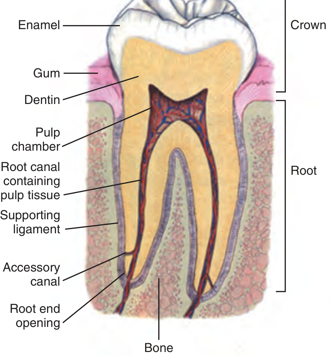
2. Neural Innervation of the Pulp
A. A-delta (Aδ) Fibers - "Sharp Pain"
- Myelinated, fast-conducting fibers
- Extend 0.2 - 0.3 mm into the dentinal tubules surrounding the pulp chamber
- Respond to heat, mechanical, and osmotic stimuli applied to the distal end of the dentinal tubules when enamel is breached
- Mediate sharp, bright, well-localized pain - the classic "dentin sensitivity"
B. C Fibers - "Dull, Burning Pain"
- Unmyelinated, slow-conducting, polymodal nociceptors
- Constitute the majority (50-75%) of pulpal innervation
- Terminate within the pulp chamber itself
- Respond to thermal stimuli AND inflammatory mediators (e.g., histamine, bradykinin)
- Contain and release neuropeptides (substance P, CGRP) upon activation
- Mediate dull, burning, poorly localized pain - the pain of established pulpitis
| Feature | A-delta Fibers | C Fibers |
|---|---|---|
| Myelination | Myelinated | Unmyelinated |
| Location | Dentinal tubules (0.2-0.3 mm) | Pulp chamber |
| Pain quality | Sharp, bright | Dull, burning |
| Stimuli | Mechanical, thermal, osmotic | Thermal, inflammatory mediators |
| Proportion | Minority | 50-75% of pulpal innervation |
| Theory | Hydrodynamic theory | Peripheral sensitization |
3. Initiation of Pulpal Pain - Stimuli and Triggers
- Dental caries: Streptococcus mutans and plaque bacteria produce acids from fermentation of dietary carbohydrates, dissolving hydroxyapatite of enamel. Once dentin is involved, caries spreads along dentinal microtubules, establishing direct communication between the oral environment and the vital dental pulp.
- Thermal stimuli: Hot liquids indicate pulpal inflammation; cold sensitivity can indicate early decay or exposed dentin.
- Osmotic stimuli: Sweet or acidic foods draw fluid through dentinal tubules.
- Mechanical stimuli: Biting, percussion, or microcracks in tooth structure.
- Dental procedures: Mechanical instrumentation of the tooth or direct pulpal exposure.
4. The Hydrodynamic Theory of Dentinal Pain (Brannstrom's Theory)
- The dentinal tubules are filled with dentinal fluid (odontoblastic processes and extracellular fluid).
- When external thermal, mechanical, or osmotic stimuli act on exposed dentin, they create a rapid shift of this fluid within the tubules.
- This fluid movement mechanically activates A-delta nerve endings located at the pulpal end (proximal end) of the dentinal tubules.
- This generates a sharp, immediate pain response - the classic dentin hypersensitivity.
- Cold stimuli (which contract fluid, causing inward flow) produce pain even before bacteria reach the pulp.
- Drying dentin (osmotic stimulus) also causes sharp pain.
- Once enamel is lost, even air temperature changes suffice to generate pain.
5. Inflammatory Mediators and Peripheral Sensitization
- Bacteria and toxins reach the pulp - activating resident immune cells (macrophages, mast cells, dendritic cells).
- Release of inflammatory mediators: histamine, bradykinin, prostaglandins, interleukins, serotonin, and leukotrienes.
- These mediators directly activate and sensitize C fibers and A-delta fibers in the pulp.
- Bradykinin is particularly potent - it lowers the activation threshold of nociceptors (peripheral sensitization), so stimuli that would normally be painless now produce pain (allodynia) and normally painful stimuli produce exaggerated pain (hyperalgesia).
- Prostaglandins (especially PGE2) further sensitize nociceptors - this is the basis for using NSAIDs to treat pulpal pain.
6. Neuropeptides - Substance P and CGRP
-
Substance P (SP): Released from C fiber terminals in the pulp. Directly measured at higher levels in irreversible pulpitis vs. normal teeth using microdialysis studies. SP causes:
- Vasodilation of pulpal blood vessels
- Increased vascular permeability - edema
- Mast cell degranulation - more histamine release
- Recruitment of inflammatory cells
-
Calcitonin Gene-Related Peptide (CGRP): Also released from C fibers. Potent vasodilator that amplifies the local inflammatory response.
Nociceptor activation → SP/CGRP release → local vasodilation → increased pulpal pressure → increased C fiber activation → more SP/CGRP → further pressure rise (in a closed compartment)
7. Reversible vs. Irreversible Pulpitis
| Feature | Reversible Pulpitis | Irreversible Pulpitis |
|---|---|---|
| Duration of pain | Short - seconds | Long - minutes to hours |
| Spontaneous pain | Absent | May be present |
| Stimulus required | Yes (thermal, sweet) | May be spontaneous |
| Pulpal status | Vitality maintained | Healing capacity lost |
| Histology | Mild inflammation | Severe inflammation, necrosis |
| Outcome without treatment | May recover | Progresses to necrosis |
8. The "Closed Compartment" Concept - Vascular Changes
- The pulp is enclosed in a rigid, non-compliant dentinal chamber. Unlike soft tissues elsewhere in the body, the pulp has NO room to expand.
- Inflammation → vasodilation and increased vascular permeability → edema accumulates.
- In a closed compartment, this edema cannot dissipate - intrapulpal pressure rises markedly.
- Elevated intrapulpal pressure directly stimulates C fibers and A-delta fibers, even in the absence of the original stimulus.
- Rising pressure can eventually exceed perfusion pressure → ischemia and ischemic necrosis of pulpal tissue.
- Necrotic tissue further sustains the inflammatory process, leading to eventual spread via the apical foramen to periapical tissues.
9. Central Sensitization and Pain Referral
- Continuous barrage of nociceptive signals from the pulp reaches the trigeminal nucleus caudalis in the brainstem (the dental equivalent of the spinal dorsal horn).
- With prolonged activation, central sensitization occurs - wide dynamic range neurons in the trigeminal nucleus become hyperexcitable.
- This results in:
- Referred pain - pain perceived in adjacent teeth, ear, jaw, or temples rather than the truly affected tooth (making clinical localization difficult).
- Hyperalgesia - increased sensitivity to painful stimuli.
- Allodynia - normally innocuous stimuli (e.g., mild temperature change) become painful.
10. Summary
Enamel breach (caries/trauma)
        ↓
Dentinal fluid movement → Hydrodynamic activation of Aδ fibers → Sharp, short pain
        ↓
Bacterial/irritant access to pulp
        ↓
Inflammatory mediator release (bradykinin, histamine, PGE2)
        ↓
Activation & sensitization of C fibers → Dull, burning persistent pain
        ↓
Neuropeptide release (Substance P, CGRP)
        ↓
Neurogenic inflammation → Vasodilation → Increased intrapulpal pressure
        ↓
Closed compartment → Pressure builds → Ischemia → Necrosis
        ↓
Central sensitization → Spontaneous pain, referral, hyperalgesia
- Cummings Otolaryngology Head and Neck Surgery - Chapter 86 (Dental Pain, Trigeminal System), pp. 1558-1559
- Tintinalli's Emergency Medicine: A Comprehensive Study - Dental Caries and Pulpitis, p. 1843-1845
- Roberts and Hedges' Clinical Procedures in Emergency - Acute Toothache in the ED, p. 1564
Please give me conclusion in 2 sentences
Not satisfied with conclusion . Should be in only 2 sentences . Please give me