
Sex determination from bone FMT
Reading File
Sex determination from bone is a topic in forensic medicine. "FMT" likely refers to Forensic Medicine & Toxicology. Let me search the medical library for this topic.
Searching sources...
Finding Sources
Finding Sources
Finding Sources
Finding Sources
I found the key sources. Let me read the FMT textbook section and the Forensic Anthropology sections in parallel.
Reading File
Reading File
Reading File
Excellent content found. Let me now read the core sections from the Forensic Anthropology book covering pelvis, skull, and metric methods, plus the FMT skeletal sex determination content.
Reading File
Reading File
Reading File
Reading File
Reading File
I now have comprehensive content. Let me compile the complete answer.
Loading Image
I now have all the material needed for a comprehensive answer. Here it is:
Sex Determination from Bone - Forensic Medicine & Toxicology
Sex determination from skeletal remains is a core component of forensic identification. The aim is to assign a biological sex (male or female) to an unknown individual based on morphological and metric skeletal features. The pelvis and skull are the two primary sites of analysis.
Why It Is Needed
Sex determination from bone is required in:
- Identification of dismembered body parts
- Skeletal remains found at crime scenes
- Mass disasters and decomposed bodies
- Archaeological/medicolegal investigations
Sex determination is not attempted on children under 12 years (SWGANTH consensus) because sexual dimorphism in the skeleton becomes pronounced only after puberty. Methods for adults may be cautiously applied to individuals older than 14 years if pelvic fusion is evident.
General Principle: Sexual Dimorphism
The male skeleton is larger and more robust with more prominent muscular markings. Females are smaller and more gracile. Males can be 20-30% larger than females in certain skeletal dimensions (e.g., femoral and humeral head diameter). However, there is overlap between small males and large females, which is the main source of error.
Accuracy by region:
- Pelvis: 90-95% (best indicator)
- Skull: 70-high 80%
- Postcranial skeleton: lower accuracy; useful as supplemental data
1. PELVIS (Most Reliable - 90-95% Accuracy)
The pelvis is the most sexually dimorphic bone due to reproductive function.
| Feature | Male | Female |
|---|---|---|
| Overall shape | Narrow, deep, funnel-shaped | Wide, shallow, basin-shaped |
| Pelvic inlet | Heart-shaped | Round/oval |
| Pelvic outlet | Narrower, more constricted | Wider |
| Pelvic blades (iliac wings) | Narrow, less flared | Broad, more flared |
| Sacrum | Narrow, more anteriorly curved | Wider, less curved |
| Sacroiliac joint | Smooth, even contour | Forms a low plateau above surrounding bone |
| Subpubic angle | Acute (<90°) | Obtuse (>90°) |
| Obturator foramen | Oval | Triangular |
| Acetabulum | Large (faces laterally) | Smaller (faces more anterolaterally) |
Phenice Method (95%+ accuracy)
Developed in 1969 on 275 specimens - evaluates three traits of the subpubic region:
- Ventral arc - A slightly elevated ridge running inferiorly and laterally across the ventral pubis surface. Present in females; absent in males.
- Subpubic concavity - The medial surface of the pubic ramus is concave in females and straight/convex in males.
- Medial aspect of the ischiopubic ramus - Appears as a sharp/narrow ridge in females; broad/flat in males.
Scoring: Each trait scored 0-2; total score determines sex.
2. SKULL (70-85% Accuracy)
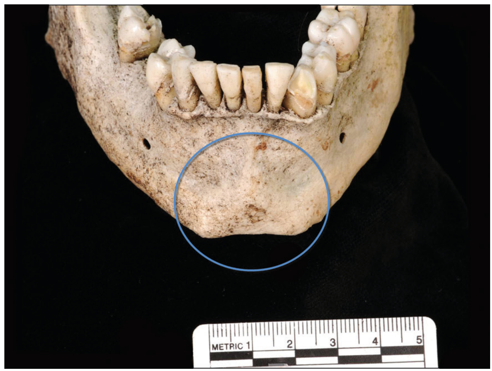
Five main cranial traits are assessed on a 1-5 scale (Walker 2005, 2008), where 1 = most female-like and 5 = most male-like:
| Feature | Male (Score 4-5) | Female (Score 1-2) |
|---|---|---|
| Nuchal crest | Large, prominent, rugose | Small, smooth |
| Mastoid process | Large, bulbous | Small, slender |
| Supraorbital margin | Blunt, thick, rounded | Sharp, thin |
| Glabella | Large, prominent protuberance; sloping frontal bone | Flat/absent; vertical frontal bone |
| Mental eminence (chin) | Large, square, prominent | Small, pointed, V-shaped |
Additional features:
- Orbital shape: Rectangular in males; square/round in females
- Zygomatic arch: Extends beyond external acoustic meatus in males
- Forehead: More sloping in males; more vertical/rounded in females
- Palate: Larger and broader in males
3. POSTCRANIAL SKELETON
Though less reliable, these bones provide useful supplementary data:
| Bone | Male Features |
|---|---|
| Femur | Head diameter >47.5 mm (usually male); <43.5 mm (usually female); vertical diameter of femoral head is the most useful single metric |
| Humerus | Head diameter >47 mm = male; <43 mm = female |
| Sternum | Manubrium:body ratio >1:1 in males; body relatively longer in females |
| Scapula | Larger glenoid fossa and maximum length in males |
| Long bones | Generally longer and more robust in males |
Order of accuracy (most to least):
- Pelvis
- Skull
- Long bones (femur, humerus)
- Sternum, scapula, clavicle
4. METRIC APPROACHES (Discriminant Function Analysis)
Statistical methods use measurements of skeletal elements to classify sex. These are population-specific - standards developed from one population should not be applied to another.
- Measurements are entered into a discriminant function equation
- A sectioning point is defined; values above = male, below = female
- These methods can achieve >90% accuracy for certain bones (e.g., femoral head, humeral head)
- Commonly used bones: skull (multiple dimensions), femur, humerus, tibia, radius
5. DNA ANALYSIS
A definitive method: testing for the presence or absence of the Y chromosome from bone marrow or dental pulp. Particularly useful for:
- Juvenile remains (where morphological methods are unreliable)
- Fragmentary or highly weathered bone
Limitations: expensive, time-consuming (weeks to months), requires bone destruction, may require special permissions in archaeological contexts.
6. SEX CHROMATIN FROM BONE MARROW/DENTAL PULP
Sex can be determined from:
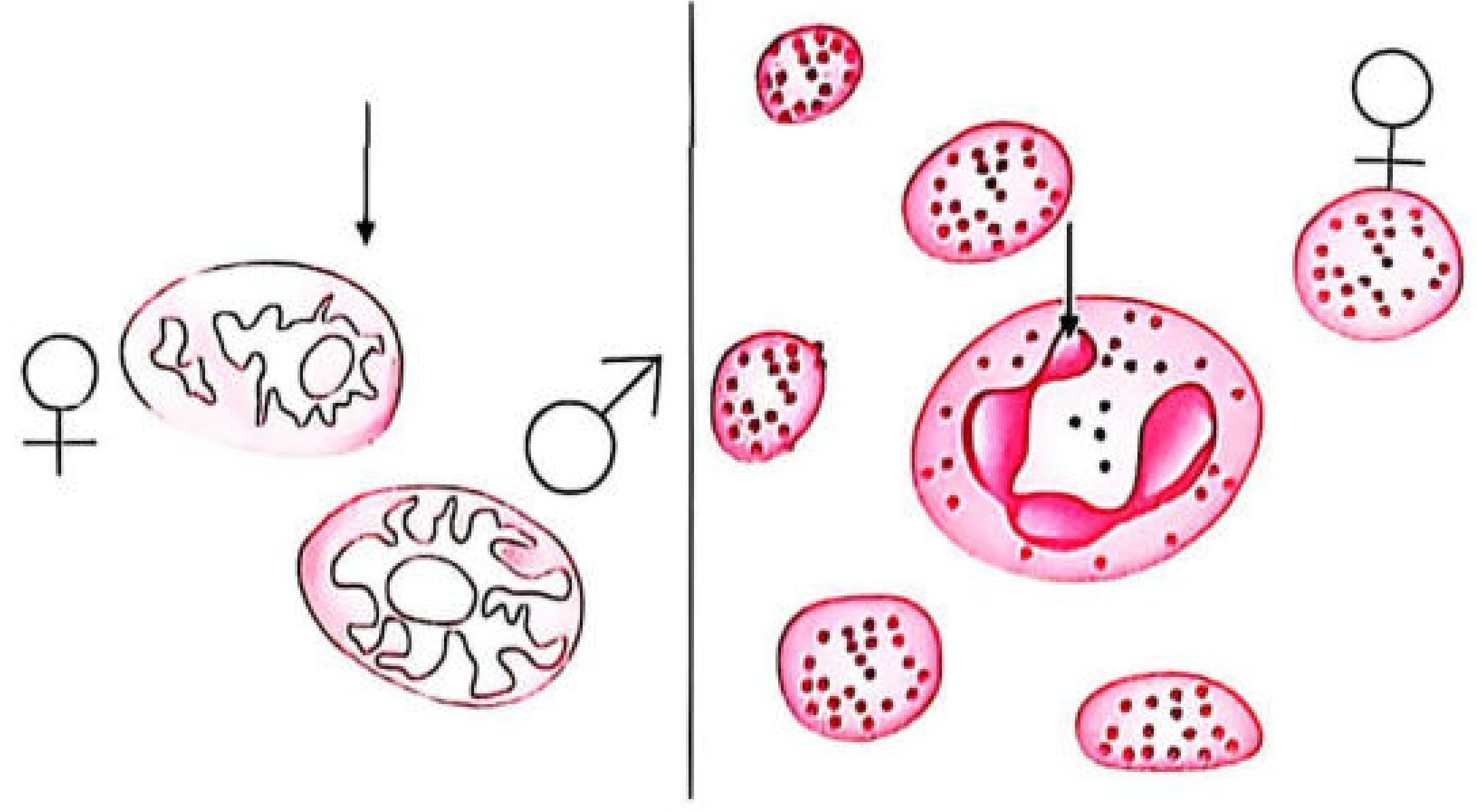
- Bone marrow - Barr bodies (condensed inactive X chromosome) visible in nuclei of female cells
- Dental pulp - Y chromosome demonstrated using fluorescent dyes (F-bodies) for up to 1 year post-mortem
- Males: 30-70% of cells contain F-bodies
- Females: 0-4% of cells contain F-bodies
Summary Table: Skeletal Sex Determination
| Method | Bone | Accuracy | Notes |
|---|---|---|---|
| Phenice method | Pubis | 95%+ | Best morphological method |
| Morphological | Full pelvis | 90-95% | First-line assessment |
| Morphological | Skull (5 traits) | 70-85% | Walker scoring system |
| Metric/DFA | Femur, humerus | >90% | Population-specific |
| DNA testing | Any bone/tooth | ~100% | Costly, time-consuming |
| Sex chromatin | Dental pulp/marrow | High | F-bodies or Barr bodies |
Key exam point: The pelvis is the best single indicator of sex. The Phenice method (ventral arc + subpubic concavity + ischiopubic ramus shape) gives the highest morphological accuracy at 95%+. Sex determination from bone is unreliable before puberty and always probabilistic - not definitive without DNA.
Sources: The Essentials of Forensic Medicine and Toxicology, 36th edition (2026); Forensic Anthropology: A Comprehensive Introduction, 2nd edition; Brogdon's Forensic Radiology
This is a shared conversation. Sign in to Orris to start your own chat.