
Thyroid Development The thyroid diverticulum arises from the floor of the primitive pharynx and descends into the neck. It is connected to the tongue by the thyroglossal duct, which normally disappears but may persist as cysts or the pyramidal lobe of the thyroid. The foramen cecum is a normal remnant of the thyroglossal duct. The most common site for ectopic thyroid tissue is the tongue, known as a lingual thyroid. Removal of this tissue may result in hypothyroidism if it is the only thyroid tissue present. Thyroglossal Duct Cyst A thyroglossal duct cyst presents as an anterior midline neck mass that moves with swallowing or protrusion of the tongue. This is in contrast to a persistent cervical sinus, which leads to a pharyngeal cleft cyst in the lateral neck. Thyroid and Parafollicular Cels • Thyroid follicular cells are derived from endoderm. • Parafollicular cells arise from the 4th pharyngeal pouch. Make an visual image to understand better. With proper explanation in an image
Educational medical diagram of thyroid development showing: 1) A sagittal view of the head and neck with the primitive pharynx at the top, showing the thyroid diverticulum arising from the floor of the pharynx, 2) The foramen cecum marked on the tongue as a small pit, 3) The thyroglossal duct as a dotted descending line from the foramen cecum down the midline neck to the final thyroid position, 4) The final thyroid gland in the lower neck shown as a butterfly-shaped gland, 5) A lingual thyroid shown on the tongue base labeled as "most common ectopic site", 6) A thyroglossal duct cyst shown as a rounded mass on the anterior midline neck with an arrow indicating it moves with swallowing and tongue protrusion, 7) A separate inset panel showing thyroid follicular cells labeled "derived from endoderm" and parafollicular C-cells labeled "from 4th pharyngeal pouch", 8) A lateral neck panel showing a pharyngeal cleft cyst from persistent cervical sinus for contrast. Use clear anatomical labels, color coding (blue for duct, red for ectopic tissue, green for normal thyroid, orange for cyst), arrows, and clean educational illustration style suitable for medical students
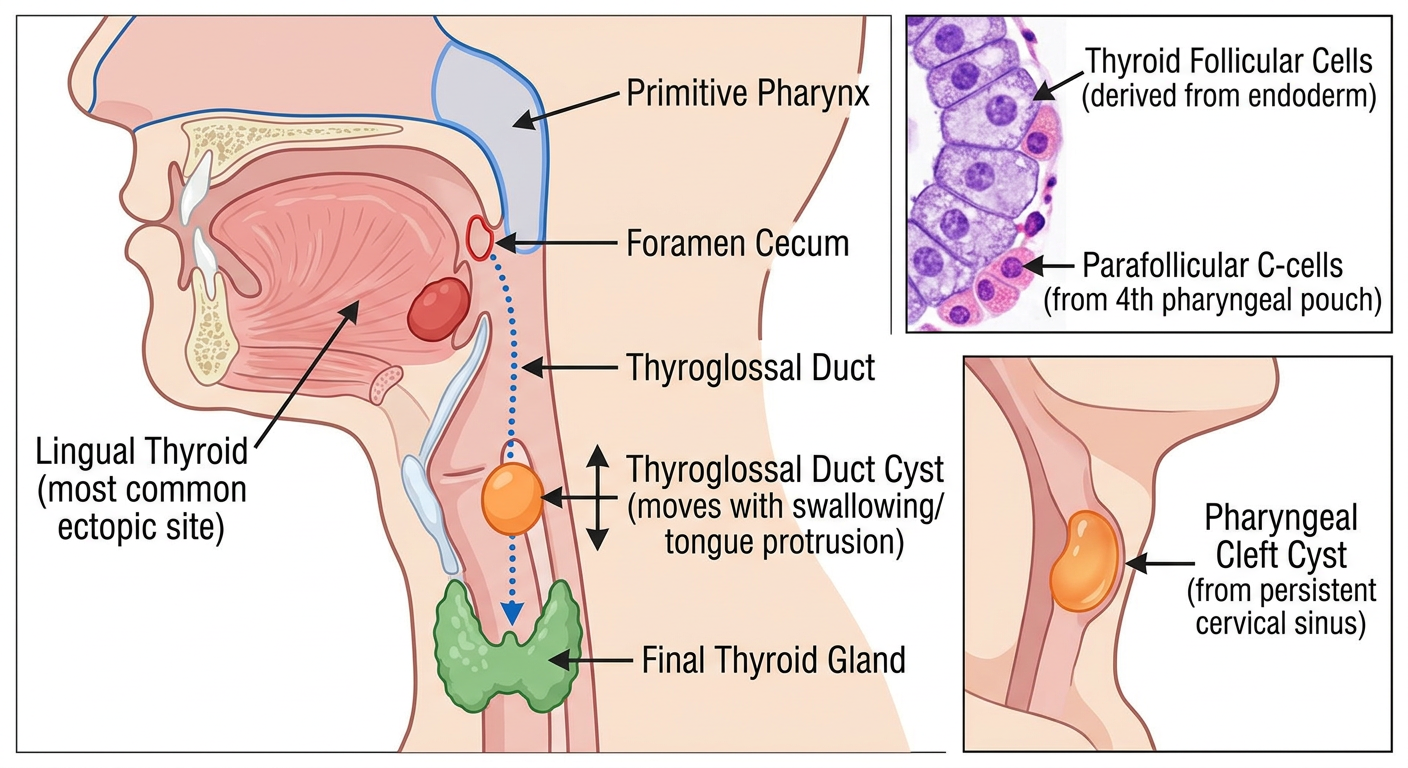
Thyroid Development - Visual Explanation
1. Origin of the Thyroid
- The thyroid diverticulum buds from the floor of the primitive pharynx (endodermal origin) at the base of the tongue.
- It then descends in the midline of the neck to its final position anterior to the trachea.
2. Thyroglossal Duct
- During descent, the thyroid remains connected to the tongue by the thyroglossal duct.
- This duct normally involutes and disappears.
- Remnants can persist as:
- Thyroglossal duct cyst (most common)
- Pyramidal lobe of the thyroid
- Foramen cecum - a small pit at the base of the tongue, the normal permanent remnant
3. Thyroglossal Duct Cyst
- Presents as an anterior midline neck mass
- Key clinical feature: moves upward with swallowing AND with tongue protrusion (distinguishes it from other neck masses)
- Compare with pharyngeal cleft cyst (from persistent cervical sinus) which appears in the lateral neck
4. Ectopic Thyroid Tissue
- The most common site = lingual thyroid (at the base of the tongue)
- Important: if this is the only thyroid tissue in the body, removing it causes hypothyroidism
- Always do a thyroid scan before removing a lingual thyroid to confirm normal tissue is present elsewhere
5. Cell Origins
| Cell Type | Embryological Origin |
|---|---|
| Thyroid follicular cells | Endoderm (floor of pharynx) |
| Parafollicular (C) cells | 4th pharyngeal pouch (neural crest cells) |
Make again
A detailed, clean, colorful medical education poster titled "Thyroid Development" for medical students. Layout: vertical poster with multiple clearly labeled sections. TOP SECTION - "Embryological Origin": Sagittal cross-section of a fetal head showing the tongue with a small pit labeled "Foramen Cecum (permanent remnant)" at its base, and a dotted arrow path descending down the midline neck labeled "Thyroglossal Duct (normally disappears)" leading to a butterfly-shaped thyroid gland in the lower neck labeled "Final Thyroid Position (anterior to trachea)". The thyroid is colored teal/green. MIDDLE-LEFT SECTION - "Ectopic Thyroid & Cysts": Show a lingual thyroid as a red mass at the tongue base labeled "Lingual Thyroid = Most Common Ectopic Site. Warning: may be only thyroid tissue — removal causes hypothyroidism". Below it, show a rounded yellow cyst on the anterior midline neck labeled "Thyroglossal Duct Cyst: Moves UP with swallowing AND tongue protrusion. Anterior midline mass." MIDDLE-RIGHT SECTION - "Lateral Neck Contrast": Show a blue cyst on the side of the neck labeled "Pharyngeal Cleft Cyst (persistent cervical sinus) = Lateral neck. Different from thyroglossal cyst!" BOTTOM SECTION - "Cell Origins" table with two colored boxes: Left box (orange): "Thyroid Follicular Cells → Endoderm (floor of pharynx) → Produce T3/T4" Right box (purple): "Parafollicular C-Cells → 4th Pharyngeal Pouch (neural crest) → Produce Calcitonin" Use bright, distinct colors for each section, bold labels, clinical notes in small italic text, arrows showing movement directions, and a clean white background. Style: high-quality medical illustration poster.
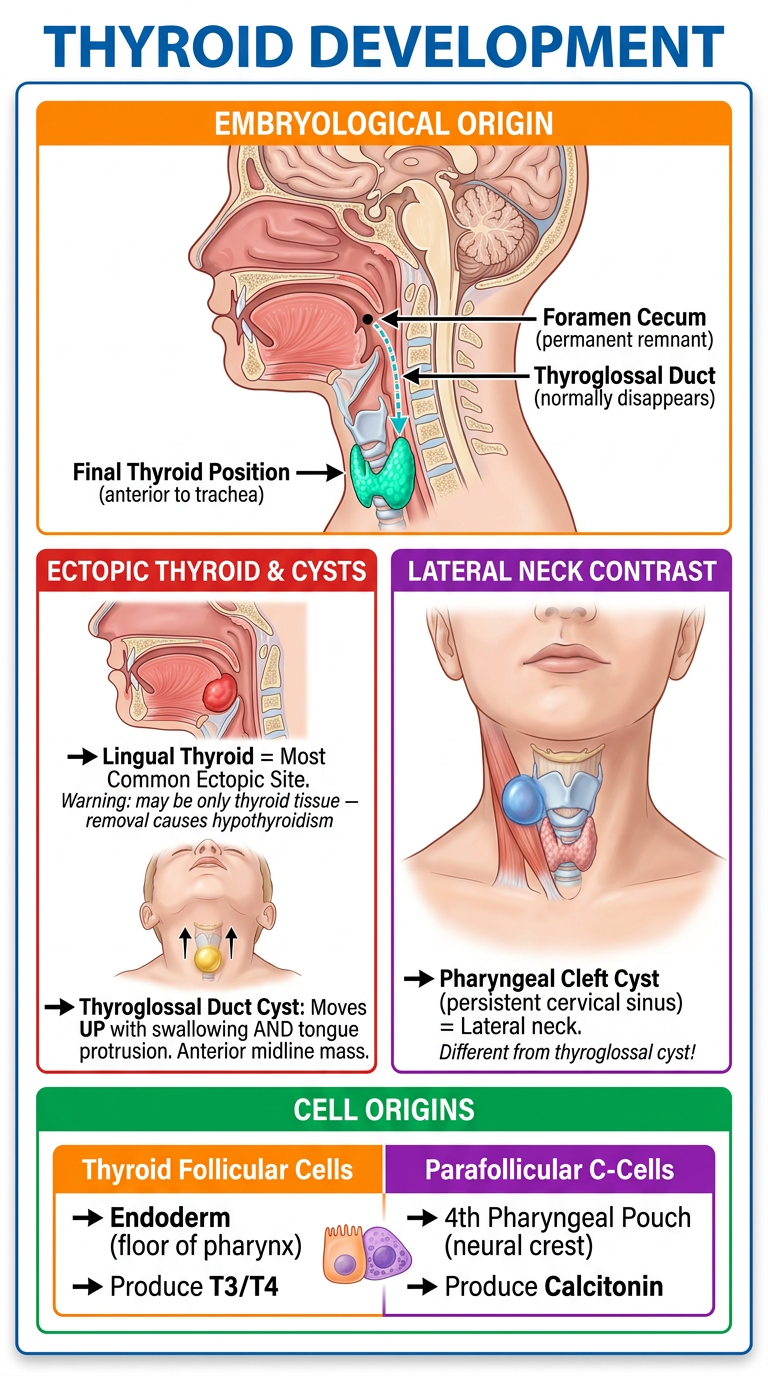
- Foramen cecum at the tongue base - the permanent normal remnant
- Thyroglossal duct descent path down the midline neck to the final thyroid position
- Lingual thyroid highlighted as the most common ectopic site, with the hypothyroidism warning
- Thyroglossal duct cyst vs. pharyngeal cleft cyst - anterior midline vs. lateral neck contrast
- Cell origins table at the bottom distinguishing follicular cells (endoderm) from parafollicular C-cells (4th pharyngeal pouch/neural crest)
Hi can we make the notes of it in a story mode style pages by pages let's start and it should be like explaining the concept in depth. And what the lines means in storytelling we have to connect it chain by chain can we do such like that.what I need a concept better. Let's start 1 10 pages
~/
What should Pages 1-10 cover first?
Thyroid Development - Visual Explanation
1. Origin of the Thyroid
- The thyroid diverticulum buds from the floor of the primitive pharynx (endodermal origin) at the base of the tongue.
- It then descends in the midline of the neck to its final position anterior to the trachea.
2. Thyroglossal Duct
- During descent, the thyroid remains connected to the tongue by the thyroglossal duct.
- This duct normally involutes and disappears.
- Remnants can persist as:
- Thyroglossal duct cyst (most common)
- Pyramidal lobe of the thyroid
- Foramen cecum - a small pit at the base of the tongue, the normal permanent remnant
3. Thyroglossal Duct Cyst
- Presents as an anterior midline neck mass
- Key clinical feature: moves upward with swallowing AND with tongue protrusion (distinguishes it from other neck masses)
- Compare with pharyngeal cleft cyst (from persistent cervical sinus) which appears in the lateral neck
4. Ectopic Thyroid Tissue
- The most common site = lingual thyroid (at the base of the tongue)
- Important: if this is the only thyroid tissue in the body, removing it causes hypothyroidism
- Always do a thyroid scan before removing a lingual thyroid to confirm normal tissue is present elsewhere
5. Cell Origins
| Cell Type | Embryological Origin |
|---|---|
| Thyroid follicular cells | Endoderm (floor of pharynx) |
| Parafollicular (C) cells | 4th pharyngeal pouch (neural crest cells) |
- Foramen cecum at the tongue base - the permanent normal remnant
- Thyroglossal duct descent path down the midline neck to the final thyroid position
- Lingual thyroid highlighted as the most common ectopic site, with the hypothyroidism warning
- Thyroglossal duct cyst vs. pharyngeal cleft cyst - anterior midline vs. lateral neck contrast
- Cell origins table at the bottom distinguishing follicular cells (endoderm) from parafollicular C-cells (4th pharyngeal pouch/neural crest)I can write it as a connected, story-style learning journey: each page explains what happens, why it happens, and how it leads to the next concept. The uploaded reproductive book and the earlier thyroid topic point to two different starting points.Please reply with one option so I can begin the story-style notes:
- Thyroid development
- Pages 1-10 of the uploaded Reproductive Book PDF
- A specific reproductive topic
- Choose the best topic for me
Go through pdf pages mention the lines of pdf and page number and connect it like story telling a nd explain concept easier
python -c "from pypdf import PdfReader; p='/tmp/workspace/attachments/70d38fbc-0efb-4bc6-ab5d-ee392143a440/Reproductive Book 2023 (B&B) [Medicalstudyzone.com].pdf'; r=PdfReader(p); print('PAGES',len(r.pages)); [print('\\n===== PDF PAGE '+str(i+1)+' =====\\n'+(r.pages[i].extract_text() or '[NO EXTRACTABLE TEXT]')[:12000]) for i in range(min(10,len(r.pages)))]"
embryology human reproduction fertilization implantation development overview official medical education
- PDF page number
- Important original line/statement from the page
- Plain-language meaning
- A connected story that links each idea to the next
- Concept checkpoints and memory hooks
Page 1
Use this page-by-page storytelling format for the whole book
Format for every PDF page
PDF Page X | Topic: _______
“Exact important line or short statement from the page.”
What the line is saying in ordinary language.
A chain-style explanation:
- What happened before this?
- Why does this event/concept need to happen?
- What does it cause next?
- How does it connect to the next page?
Why the concept matters in patients, diagnosis, or exams.
A short visual analogy, sequence, or phrase to retain it.
One or two lines that make the next page feel like the natural continuation of the story.
How we will cover the book
- Foundation and anatomy
- Physiology and hormonal control
- Gamete formation
- Fertilization and early embryonic development
- Implantation and placenta
- Menstrual cycle and ovulation
- Pregnancy changes
- Labour and puerperium
- Contraception and infertility
- Reproductive pathology and clinical concepts
human reproductive embryology teaching overview NCBI Bookshelf
~/attachments/70d38fbc-0efb-4bc6-ab5d-ee392143a440/
python -c "from pypdf import PdfReader; p='/tmp/workspace/attachments/70d38fbc-0efb-4bc6-ab5d-ee392143a440/Reproductive Book 2023 (B&B) [Medicalstudyzone.com].pdf'; r=PdfReader(p); print('TOTAL_PDF_PAGES', len(r.pages));\nfor i in range(min(10,len(r.pages))):\n print('\\n===== PDF PAGE '+str(i+1)+' ====='); print((r.pages[i].extract_text() or '[NO EXTRACTABLE TEXT]')[:14000])"
python -c "from pypdf import PdfReader; p='/tmp/workspace/attachments/70d38fbc-0efb-4bc6-ab5d-ee392143a440/Reproductive Book 2023 (B&B) [Medicalstudyzone.com].pdf'; r=PdfReader(p); print('TOTAL_PDF_PAGES',len(r.pages)); exec(\"for i in range(min(10,len(r.pages))):\\n print('\\\\n===== PDF PAGE '+str(i+1)+' =====')\\n print((r.pages[i].extract_text() or '[NO EXTRACTABLE TEXT]')[:14000])\")"
embryogenesis AND gastrulation
Reproductive Book Story Notes: PDF Pages 1-10
The big story before we begin
- Where should each body part form?
- How does one fertilized cell become a three-layered body?
PDF Page 1 | Cover Page
“Boards & Beyond: Reproductive Slides”
“Color slides for USMLE Step 1 preparation”
This is a slide companion for reproductive and embryology learning. It is not arranged as a story by default, so we will create the story ourselves.
Our journey starts before reproduction itself. To understand how reproductive organs, embryos, placentas, and congenital disorders arise, first understand the embryo’s basic instruction system: genes that tell cells where to go and what to become.
The table of contents shows the journey moves from embryonic genes to embryogenesis, germ layers, genital development, pregnancy, and pathology.
PDF Page 2 | Publisher / Blank Information Page
Think of this as the doorway. The actual scientific journey begins with the map on the next page.
PDF Page 3 | Table of Contents
“Embryonic Genes”
“Embryogenesis”
“Germ Layers”
“Genital Embryology”
“Spermatogenesis/Oogenesis”
“Placenta”
“Pregnancy”
The book follows a logical biological timeline:
Imagine building a city:
- First, you need the master plan: embryonic genes.
- Then, land is divided into major zones: germ layers.
- Then, roads and buildings are constructed: organs and body systems.
- Later, the reproductive system develops, produces gametes, permits fertilization, and supports fetal growth through the placenta.
Chapter 1: Embryonic Genes
PDF Page 4 | Printed Slide 1
Key lines from the page
“Patterning: Development of body pattern”
“Sonic Hedgehog Gene”
“Formation forebrain”
“Signaling separates right and left brain”
“Mutations: Holoprosencephaly”
Simple meaning
- which direction is head versus tail,
- which side is right versus left,
- where an arm or leg should develop,
- what part will become brain, eye, finger, or spine.
The story
- holo = whole
- prosencephalon = forebrain
Clinical connection
- cleft lip or palate
- closely spaced eyes
- cyclopia in the most severe forms
Memory hook
Bridge to the next page
PDF Page 5 | Printed Slide 2
Key lines from the page
“Apical Ectodermal Ridge”
“Critical for proximal to distal development”
“Removal: Limb stops growing”
“Humerus → radius → wrist”
“Dorsal: Extensors”
“Ventral: Flexors”
Simple meaning
- Proximal to distal: shoulder to fingers, or hip to toes
- Dorsal to ventral: back of limb to palm/sole side
- Anterior to posterior: thumb side to little-finger side
The story
- Early loss of AER causes a more severe limb truncation.
- Later loss allows more proximal structures to form but disrupts distal structures.
Dorsal and ventral surfaces
- Dorsal surface: extensor side
- back of hand
- top of foot
- Ventral surface: flexor side
- palm
- sole
Holoprosencephaly connection
“Failure of cleavage of prosencephalon”
“Left/right hemispheres fail to separate”
Memory hook
Bridge to the next page
PDF Page 6 | Printed Slide 3
Key lines from the page
“HOX Genes: Code for transcription factors”
“Regulators of AP axis development”
“Homeosis = transformation of one structure into another”
“Wnt-7a key for dorsal development”
“Gene deletion: Two ventral sides to limb”
“Key transcription factor: Fibroblast Growth Factor”
Part A: Homeobox genes and anterior-posterior patterning
Simple meaning
- anterior = toward the head
- posterior = toward the tail
The story
Part B: SHH and the thumb-to-little-finger axis
Key line
“Zone of polarizing activity”
“Posterior limb (near little finger)”
“Major signaling molecule: SHH”
The zone of polarizing activity (ZPA) is located on the posterior side of the developing limb, near the future little finger.
Memory hook
Part C: Wnt-7a and dorsal-ventral patterning
Key lines
“Wnt-7a key for dorsal development”
“Activates LMX-1 gene in mesoderm”
“Dorsalizes mesoderm”
“Ventral side: Engrailed1 represses Wnt-7”
Simple meaning
- Wnt-7a present dorsally → dorsal identity develops
- Engrailed-1 on the ventral side → suppresses Wnt-7a there, preserving ventral identity
Part D: FGF and AER-driven growth
Key lines
“Ridge removed, replaced with FGF: Normal growth”
“Influences underlying mesodermal growth”
The AER works largely through fibroblast growth factor (FGF) signaling. FGF signals the underlying mesoderm to proliferate and continue limb growth.
One integrated picture
| Limb question | Main developmental signal/region |
|---|---|
| How does the limb grow shoulder to fingers? | AER and FGF |
| Which side is thumb versus little finger? | ZPA, SHH, HOX genes |
| Which side is back of hand versus palm? | Wnt-7a dorsally, Engrailed-1 ventrally |
Bridge to the next page
PDF Page 7 | Printed Slide 4
Key lines from the page
“Sonic Hedgehog”
“Limb AP axis: zone of polarizing activity”
“FGF: Limb proximal-distal axis”
“Wnt-7a: Limb dorsal-ventral axis”
“Polydactyly”
“Syndactyly”
Simple meaning
The three limb axes, as one story
-
AER + FGF say:
“Keep growing away from the shoulder.”
This creates the proximal-distal axis. -
ZPA + SHH + HOX-related patterning say:
“This side is the thumb side, and that side is the little-finger side.”
This creates the anterior-posterior axis. -
Wnt-7a says:
“This surface will become the back of the hand.”
This creates the dorsal-ventral axis.
Abnormal digit formation
“Polydactyly (extra fingers/toes)”
“Syndactyly (fused fingers/toes)”
- Polydactyly: extra digits
- Syndactyly: digits fail to separate properly
High-yield memory chain
FGF = limb length
Wnt-7a = back of hand
HOX = positional identity
Bridge to the next page
Chapter 2: Embryogenesis
PDF Page 8 | Printed Slide 5
Key lines from the page
“Haploid mature spermatozoon (1N, 1C)”
“Haploid ovum (1N, 1C)”
“Forms zygote (2N, 2C)”
“Morula = ball of cells”
“Two cell stage: first 1-2 days after fertilization”
Part A: Fertilization makes the zygote
Simple meaning
- 1N = one set of chromosomes
- 1C = one chromatid-equivalent amount of DNA before replication
- 2N = diploid, two chromosome sets
- 2C = DNA amount before the first S phase
The story
Part B: DNA replication and cleavage
Key lines
“2N, 2C → DNA synthesis → chromatids → 2N, 4C”
“Zygote divides into two cells (2N, 2C)”
Simple meaning
- Before DNA synthesis: 2N, 2C
- After DNA synthesis: 2N, 4C
- After mitosis: two daughter cells, each 2N, 2C
Part C: Cleavage and morula
Key lines
“Cells continue to divide”
“Morula = ball of cells”
Story
Memory hook
Bridge to the next page
PDF Page 9 | Printed Slide 6
Key lines from the page
“In humans, blastula called blastocyst”
“Outer cells: trophoblast”
“Inner cell mass ... give rise to all tissues of body”
“Blastocyst implants in uterus about day 6-10”
“β-hCG secretion begins”
“Epiblast and hypoblast”
Part A: Morula becomes blastocyst
Simple meaning
- Trophoblast: outer cell layer
- Inner cell mass (embryoblast): inner group of cells
The story
- a system that helps it attach to and interact with the mother, and
- the actual cells that will build the new body.
Trophoblast
Inner cell mass
Part B: Implantation and beta-hCG
Key lines
“Blastocyst implants in uterus about day 6-10”
“β-hCG secretion begins”
Simple meaning
The story connection
“Pregnancy has begun. Keep the corpus luteum functioning. Do not shed the endometrium.”
Part C: Bilaminar disc
Key lines
“Inner cell mass → bilaminar disc”
“Epiblast and hypoblast”
Simple meaning
- Epiblast
- Hypoblast
Memory hook
Epiblast is the future embryo.
Bridge to the next page
PDF Page 10 | Printed Slide 7
Key lines from the page
“Primitive Streak”
“Formed by invagination of epiblast cells”
“Presence indicates start of gastrulation”
“Epiblast → three germ layers”
“Ectoderm, endoderm, mesoderm”
Part A: Gastrulation
Simple meaning
- Ectoderm
- Mesoderm
- Endoderm
The story
“Primitive streak ... creates a visible line (‘streak’) in blastocyst.”
- Some replace the hypoblast and form definitive endoderm
- Some spread between layers and form mesoderm
- Cells remaining on top form ectoderm
Part B: Why the primitive streak matters
Preview: the three germ layers
| Germ layer | Easy image | Examples of major derivatives |
|---|---|---|
| Ectoderm | Outer covering | Skin, nervous system |
| Mesoderm | Middle support and movement layer | Muscle, bone, blood, heart, kidneys |
| Endoderm | Inner lining | Epithelial lining of gut and respiratory tract, many associated organs |
Final chain for Pages 4-10
Embryonic genes provide a body plan
        ↓
SHH, FGF, Wnt-7a, and HOX genes establish position and limb pattern
        ↓
Sperm + ovum fuse at fertilization
        ↓
Zygote undergoes cleavage
        ↓
Morula forms
        ↓
Blastocyst forms: trophoblast outside, inner cell mass inside
        ↓
Blastocyst implants and begins β-hCG secretion
        ↓
Inner cell mass forms epiblast and hypoblast
        ↓
Epiblast invaginates through primitive streak
        ↓
Gastrulation creates ectoderm, mesoderm, and endoderm