
Hodgkins and non Hodgkin’s lymphoma take only 5 top references and give there outline in Ent And treatment
"Hodgkin lymphoma" AND "head and neck"
"non-Hodgkin lymphoma" AND treatment
Quick answer
Hodgkin's & Non-Hodgkin's Lymphoma - ENT Outline and Treatment
Top 5 References
- Cummings Otolaryngology - Head and Neck Surgery, 7th ed. - Hodgkin Lymphoma, Neoplasms chapters
- Bailey and Love's Short Practice of Surgery, 28th ed. - "Lymphoma of the head and neck"
- Robbins & Kumar Basic Pathology - Hodgkin Lymphoma chapter
- Goldman-Cecil Medicine, International Edition - Hodgkin/Non-Hodgkin Lymphoma chapters
- Kowalska M, et al. "Evidence for external beam radiotherapy in mediastinal Hodgkin and non-Hodgkin lymphoma - systematic review." Ann Agric Environ Med. 2024. PMID: 38549476
I. Introduction / Relevance to ENT
- Lymphoma is the most common head and neck malignancy in children (>50% of pediatric H&N malignancies) and the third most common H&N malignancy overall after squamous cell carcinoma and thyroid cancer.
- Lymphomas of the head and neck arise in nodal sites (cervical lymph nodes - most common presentation to an ENT surgeon) or extranodal sites (Waldeyer's ring - tonsil, nasopharynx, base of tongue; also thyroid, salivary glands, sinonasal tract, larynx, orbit).
- Both Hodgkin lymphoma (HL) and non-Hodgkin lymphoma (NHL) commonly present as cervical lymphadenopathy, making differentiation from reactive/infective and metastatic nodal disease a core ENT diagnostic skill.
II. Hodgkin Lymphoma (HL)
- Bimodal age distribution: peak in young adults (15-35 yrs) and again after age 55.
- Associated with EBV in a significant proportion of cases (especially mixed cellularity subtype).
- Painless, firm, rubbery cervical/supraclavicular lymphadenopathy - classically the lower neck/supraclavicular nodes.
- Nodes may wax and wane in size early on, which can mimic reactive adenopathy.
- Rarely presents as a primary extranodal H&N mass (e.g., tonsil, nasopharynx) - these are atypical and often reported as case reports.
- B symptoms: fever, night sweats, weight loss, pruritus.
- Nodal architecture usually preserved; contiguous spread pattern (unlike NHL).
- Defined by Reed-Sternberg cells in a background of reactive inflammatory cells.
- Classical HL subtypes: nodular sclerosis (most common, especially in young adults, often has mediastinal/lower cervical disease), mixed cellularity, lymphocyte-rich, lymphocyte-depleted.
- Nodular lymphocyte-predominant HL is a distinct, less aggressive variant.
- Excisional lymph node biopsy is preferred over FNA (architecture needed for diagnosis).
- Staging: Ann Arbor/Lugano system with PET-CT, CT neck/chest/abdomen/pelvis, bone marrow biopsy if indicated.
- Early stage (I-II), favorable: ABVD chemotherapy (doxorubicin, bleomycin, vinblastine, dacarbazine) x 2-4 cycles + involved-site radiotherapy (ISRT).
- Early stage, unfavorable / Advanced stage (III-IV): ABVD x 4-6 cycles (or escalated BEACOPP in high-risk/bulky disease) +/- consolidation radiotherapy guided by interim PET response.
- Radiotherapy to cervical/mediastinal fields is used selectively to minimize long-term toxicity (secondary malignancy, cardiac/thyroid effects) - PET-adapted "response-adapted" strategies now standard.
- Relapsed/refractory disease: salvage chemotherapy, autologous stem cell transplant, brentuximab vedotin (anti-CD30), checkpoint inhibitors (nivolumab/pembrolizumab).
- Prognosis is excellent - 5-year survival >85-90% for early-stage disease.
III. Non-Hodgkin Lymphoma (NHL)
- NHL has the highest incidence of any lymphoma in the head and neck and is more common than HL overall in adults.
- Most H&N NHLs are B-cell in origin (T-cell/NK-cell types like extranodal NK/T-cell lymphoma, nasal type, are less common but important in the sinonasal tract).
- Rapid-onset, non-contiguous, often multifocal lymphadenopathy - can appear in unpredictable nodal groups (unlike the orderly spread of HL).
- Extranodal H&N sites frequently involved:
- Waldeyer's ring (tonsil, nasopharynx, base of tongue) - most common extranodal H&N site; presents as unilateral tonsillar enlargement, nasal obstruction, or a nasopharyngeal mass.
- Sinonasal tract - extranodal NK/T-cell lymphoma, nasal type, presents with midline destructive lesions, epistaxis, nasal obstruction, sometimes mimicking granulomatosis with polyangiitis.
- Thyroid - MALT lymphoma or diffuse large B-cell lymphoma, often arising in Hashimoto thyroiditis; presents as a rapidly enlarging goiter.
- Salivary glands (parotid) - MALT lymphoma, associated with Sjogren syndrome; also reported as primary parotid DLBCL.
- Orbit, larynx - less common sites.
- B symptoms less prominent than in HL but can occur, especially in aggressive subtypes.
- Diffuse large B-cell lymphoma (DLBCL) is the most common subtype in H&N.
- Others: follicular lymphoma, MALT (marginal zone) lymphoma, mantle cell lymphoma, Burkitt lymphoma (children), extranodal NK/T-cell lymphoma.
- WHO classification based on cell lineage (B vs T/NK), grade, and molecular markers (CD20, CD3, CD30, EBV status, etc.).
- Excisional biopsy preferred; FNA can suggest lymphoma but core/excisional biopsy needed for subtyping and flow cytometry/immunohistochemistry.
- Imaging (CT/MRI, PET-CT) and staging as per HL; bone marrow biopsy often part of staging given extranodal spread tendency.
- Indolent NHL (e.g., follicular, MALT lymphoma): "watch and wait" if asymptomatic; localized MALT lymphoma may respond to radiotherapy alone or, if H. pylori/Chlamydia-associated (not typical for H&N), antibiotic therapy; rituximab +/- chemotherapy for symptomatic/advanced disease.
- Aggressive NHL (e.g., DLBCL): R-CHOP (rituximab, cyclophosphamide, doxorubicin, vincristine, prednisone) is standard first-line; involved-site radiotherapy added for bulky or extranodal disease.
- Burkitt lymphoma: intensive short-course multi-agent chemotherapy (e.g., CODOX-M/IVAC or similar) with CNS prophylaxis given high proliferation rate.
- Extranodal NK/T-cell lymphoma, nasal type: combined chemoradiotherapy; radiotherapy is particularly important for localized disease given relative chemoresistance.
- Relapsed/refractory disease: salvage chemotherapy, autologous/allogeneic stem cell transplant, and increasingly CAR-T cell therapy - a 2025 systematic review/meta-analysis found CAR-T therapy achieves meaningful response rates in relapsed/refractory NHL, though with notable rates of cytokine release syndrome and neurotoxicity requiring careful patient selection (PMID: 39924687). Newer targeted agents (polatuzumab vedotin, an anti-CD79b antibody-drug conjugate) are also showing efficacy in relapsed/refractory NHL per recent meta-analysis data (PMID: 40725080).
- Prognosis is more variable than HL and depends heavily on subtype, stage, and molecular/genetic features (IPI score for aggressive lymphomas).
IV. Key ENT Clinical Pearls
- Any persistent (>2-4 weeks), painless, firm cervical node without an identifiable infective source warrants biopsy to exclude lymphoma, especially in a young adult (favor HL) versus older patient with rapid extranodal H&N mass (favor NHL).
- Radiotherapy for mediastinal/cervical HL and NHL has shifted toward more conformal techniques (including proton therapy) to reduce cardiac, pulmonary, and thyroid toxicity - supported by recent systematic review evidence (PMID: 38549476).
- Waldeyer's ring lymphoma can mimic tonsillar squamous cell carcinoma or infectious mononucleosis-related tonsillar hypertrophy; unilateral, asymmetric tonsillar enlargement in an adult should raise suspicion and prompt biopsy rather than simple tonsillectomy assumption of benign hypertrophy.
Hodgkins and non Hodgkin’s lymphoma take only 5 top references and give there outline in Ent And treatment
Top 5 References
- Cummings Otolaryngology - Head and Neck Surgery, 7th ed. - Hodgkin Lymphoma, Neoplasms chapters
- Bailey and Love's Short Practice of Surgery, 28th ed. - "Lymphoma of the head and neck"
- Robbins & Kumar Basic Pathology - Hodgkin Lymphoma chapter
- Goldman-Cecil Medicine, International Edition - Hodgkin/Non-Hodgkin Lymphoma chapters
- Kowalska M, et al. "Evidence for external beam radiotherapy in mediastinal Hodgkin and non-Hodgkin lymphoma - systematic review." Ann Agric Environ Med. 2024. PMID: 38549476
I. Relevance to ENT
II. Hodgkin Lymphoma (HL)
- Painless, firm, rubbery cervical/supraclavicular lymphadenopathy - classically lower neck nodes, with orderly, contiguous spread.
- Rarely primary extranodal H&N disease (tonsil, nasopharynx) - reported mainly as case reports.
- B symptoms: fever, night sweats, weight loss, pruritus.
- Early stage, favorable: ABVD (doxorubicin, bleomycin, vinblastine, dacarbazine) x 2-4 cycles + involved-site radiotherapy.
- Early stage unfavorable / Advanced stage: ABVD x 4-6 cycles (or escalated BEACOPP for high-risk/bulky disease) +/- consolidation radiotherapy, guided by interim PET response ("response-adapted" approach).
- Relapsed/refractory: salvage chemotherapy, autologous stem cell transplant, brentuximab vedotin (anti-CD30), checkpoint inhibitors (nivolumab/pembrolizumab).
- Prognosis excellent - 5-year survival >85-90% for early-stage disease.
III. Non-Hodgkin Lymphoma (NHL)
- Rapid, non-contiguous, multifocal lymphadenopathy (unlike HL's orderly spread).
- Extranodal H&N sites are common:
- Waldeyer's ring (tonsil, nasopharynx, base of tongue) - unilateral tonsillar enlargement or nasopharyngeal mass.
- Sinonasal tract - extranodal NK/T-cell lymphoma, nasal type: midline destructive lesion, epistaxis, nasal obstruction (can mimic granulomatosis with polyangiitis).
- Thyroid - MALT lymphoma or DLBCL, often in Hashimoto thyroiditis; rapidly enlarging goiter.
- Parotid/salivary glands - MALT lymphoma, associated with Sjogren syndrome.
- Indolent NHL (follicular, MALT): watch-and-wait if asymptomatic; localized radiotherapy for MALT; rituximab +/- chemotherapy if symptomatic/advanced.
- Aggressive NHL (DLBCL): R-CHOP (rituximab, cyclophosphamide, doxorubicin, vincristine, prednisone) first-line +/- involved-site radiotherapy for bulky/extranodal disease.
- Burkitt lymphoma: intensive short-course multi-agent chemotherapy with CNS prophylaxis.
- Extranodal NK/T-cell lymphoma, nasal type: combined chemoradiotherapy - radiotherapy especially important given relative chemoresistance.
- Relapsed/refractory: salvage chemo, stem cell transplant, CAR-T cell therapy, and newer agents like polatuzumab vedotin (anti-CD79b antibody-drug conjugate).
- Prognosis more variable than HL, depends on subtype, stage, and IPI score.
IV. Key ENT Pearls
- Persistent (>2-4 weeks), painless, firm cervical node without infective cause warrants biopsy - young adult favors HL, older patient with rapid extranodal mass favors NHL.
- Radiotherapy for cervical/mediastinal lymphoma has shifted toward conformal/proton techniques to reduce cardiac, pulmonary, and thyroid toxicity, per recent systematic review evidence (PMID: 38549476).
- Unilateral, asymmetric tonsillar enlargement in an adult should prompt biopsy rather than assumption of benign hypertrophy, to exclude Waldeyer's ring lymphoma.
Create a comparison poster: HL vs NHL presentation, diagnosis, and treatment
A medical comparison poster titled "Hodgkin Lymphoma (HL) vs Non-Hodgkin Lymphoma (NHL)" organized as a side-by-side two-column table with three row sections: Presentation, Diagnosis, and Treatment. Presentation - HL: Painless, firm, rubbery cervical/supraclavicular lymphadenopathy; orderly contiguous nodal spread; bimodal age peak (15-35 yrs and >55 yrs); B symptoms (fever, night sweats, weight loss, pruritus); rarely extranodal. Presentation - NHL: Rapid, non-contiguous, multifocal lymphadenopathy; frequent extranodal head and neck sites (Waldeyer's ring/tonsil, sinonasal tract, thyroid, parotid gland); highest incidence of any lymphoma in the head and neck. Diagnosis - HL: Excisional lymph node biopsy showing Reed-Sternberg cells in reactive background; subtypes nodular sclerosis, mixed cellularity, lymphocyte-rich, lymphocyte-depleted; Ann Arbor/Lugano staging with PET-CT. Diagnosis - NHL: Excisional or core biopsy with flow cytometry and immunohistochemistry (CD20, CD3, CD30); subtypes diffuse large B-cell lymphoma, follicular, MALT, mantle cell, Burkitt, extranodal NK/T-cell; PET-CT staging plus bone marrow biopsy. Treatment - HL: ABVD chemotherapy plus involved-site radiotherapy for early stage; escalated BEACOPP for advanced/high-risk disease; brentuximab vedotin and checkpoint inhibitors for relapse; excellent prognosis over 85 percent five-year survival. Treatment - NHL: Watch-and-wait for indolent lymphoma; R-CHOP chemoimmunotherapy for diffuse large B-cell lymphoma; intensive regimens with CNS prophylaxis for Burkitt lymphoma; chemoradiotherapy for extranodal NK/T-cell lymphoma; CAR-T cell therapy and antibody-drug conjugates for relapsed/refractory disease; prognosis more variable, depends on subtype and IPI score. Include small icons representing a lymph node, a microscope/biopsy slide, and a treatment/IV drip symbol.
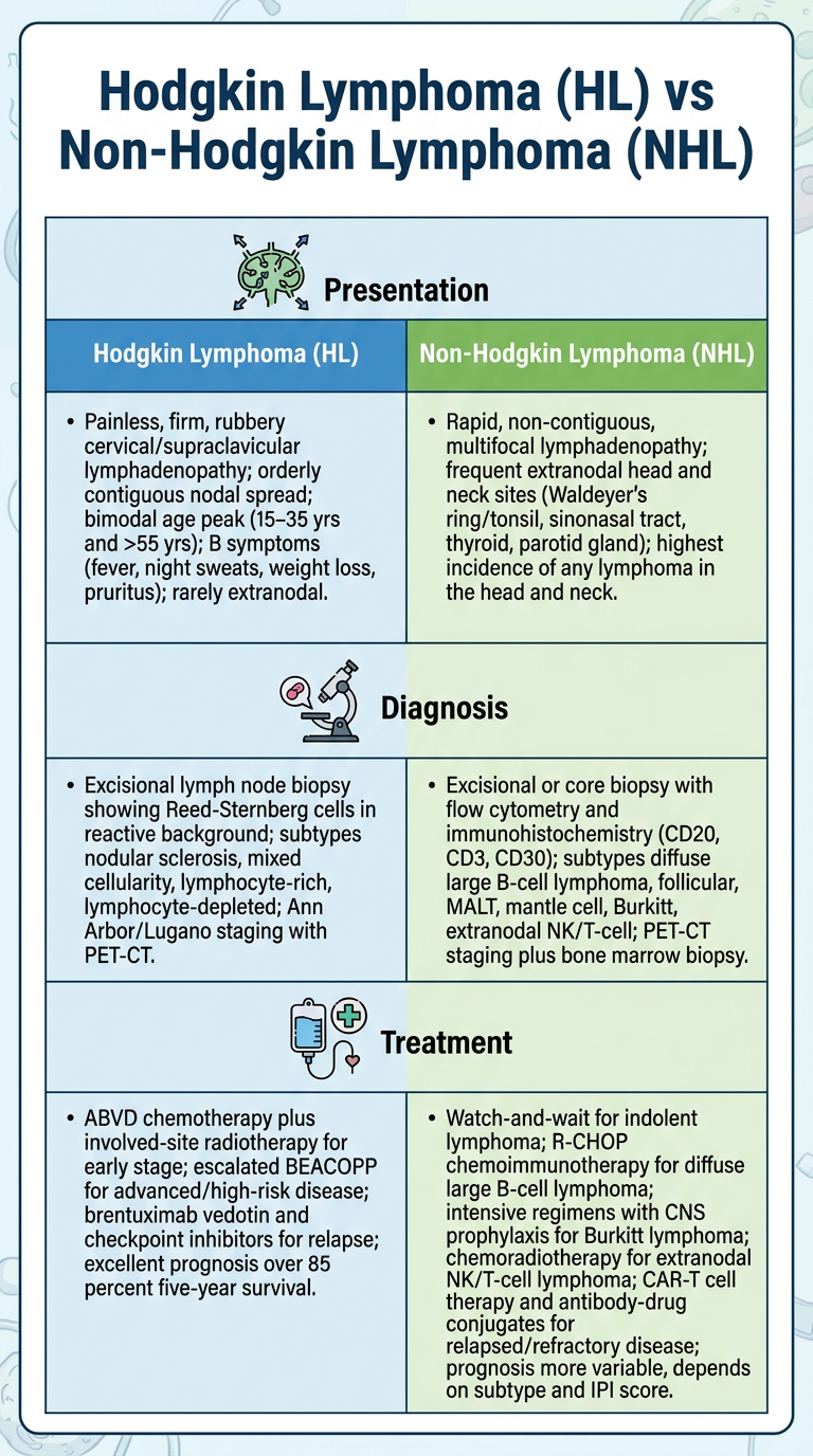
| Hodgkin Lymphoma | Non-Hodgkin Lymphoma | |
|---|---|---|
| Presentation | Painless, rubbery cervical/supraclavicular nodes; orderly contiguous spread; bimodal age (15-35, >55); B symptoms common | Rapid, multifocal, non-contiguous spread; frequent extranodal H&N sites (Waldeyer's ring, sinonasal, thyroid, parotid) |
| Diagnosis | Excisional biopsy showing Reed-Sternberg cells; Ann Arbor/Lugano staging with PET-CT | Biopsy with flow cytometry/IHC (CD20, CD3, CD30); PET-CT + bone marrow biopsy |
| Treatment | ABVD + involved-site RT (early stage); escalated BEACOPP (advanced); brentuximab vedotin/checkpoint inhibitors for relapse | R-CHOP for DLBCL; watch-and-wait for indolent disease; CAR-T/antibody-drug conjugates for relapse |