
Define choledocholithiasis
Reading File
Now I'll search the medical library for choledocholithiasis content in parallel:
Finding Sources
Finding Sources
Good results. Now let me read the most comprehensive sections from Sleisenger & Fordtran (the gold-standard GI textbook) and Sabiston Surgery:
Reading File
Reading File
Reading File
I now have rich content from multiple authoritative textbooks. Let me also fetch the intraoperative cholangiogram image and get the ASGE risk stratification table, then compile the answer:
Loading Image
Reading File
I now have comprehensive content to deliver a thorough answer. Here is the full definition and overview:
Choledocholithiasis
Choledocholithiasis is the presence of stones within the common bile duct (CBD). It occurs in approximately 5-20% of patients with symptomatic gallstones (cholelithiasis).
Classification
There are two main types, based on how the stones arise:
| Type | Description | Stone Type | Population |
|---|---|---|---|
| Primary | De novo stone formation within the CBD itself | Brown pigment stones (precipitated bile pigments + cholesterol) | More common in Asian populations; associated with bacterial infection |
| Secondary | Stones formed in the gallbladder that migrate into the CBD | Cholesterol or black pigment stones | More common in the United States |
A subset called retained stones refers to secondary stones identified in the CBD within 2 years after cholecystectomy; these occur in 1-2% of post-cholecystectomy patients.
- Sabiston Textbook of Surgery, p. 1832
Clinical Presentation
Choledocholithiasis may be asymptomatic (incidental finding) or may present with a spectrum of manifestations:
- Biliary colic - right upper quadrant (RUQ) or epigastric pain
- Obstructive jaundice - scleral icterus, dark urine, pale/acholic stools; onset is typically painful (distinguishing it from malignant obstruction, which is painless)
- Ascending cholangitis (Charcot's triad: fever, jaundice, RUQ pain) - due to ascending bacterial infection from the duodenum; bacteremia is present in ~74% of cases
- Gallstone pancreatitis - if the stone obstructs the pancreatic duct at the ampulla of Vater
Complications are frequently the presenting manifestation.
- Symptom to Diagnosis: An Evidence Based Guide, p. 50
Diagnosis
Laboratory Findings
No lab test is pathognomonic. Key findings include:
- Elevated total bilirubin - low sensitivity, high specificity for choledocholithiasis; >4 mg/dL is a very strong predictor
- Elevated AST, ALT, ALP - early in the disease, transaminases rise first; later, an obstructive pattern (elevated ALP, bilirubin) dominates
- Leukocytosis - raises concern for cholangitis
- Lipase >3x upper limit of normal - diagnostic for concomitant pancreatitis
- When elevated bilirubin is combined with an abnormal ultrasound, pre-test probability for choledocholithiasis approaches 90%; a patient with normal LFTs and normal ultrasound has <5% probability
Imaging
| Modality | Notes |
|---|---|
| Transabdominal ultrasound | Sensitivity ~80%, specificity ~90% for CBD stones; CBD dilation >5-6 mm is suggestive; only ~25% of cases show a dilated CBD |
| CT scan | Only ~75% sensitive - not the test of choice |
| MRCP | Highly accurate, non-invasive; preferred for intermediate-risk patients |
| EUS (Endoscopic Ultrasound) | Highly sensitive and specific; preferred for low-to-intermediate risk patients to avoid unnecessary ERCP |
| ERCP | Gold standard - both diagnostic and therapeutic; proceed directly in high-risk patients |
- Sabiston Textbook of Surgery, p. 1832
ASGE Risk Stratification (Predictors of Choledocholithiasis)
| Predictor Strength | Criteria |
|---|---|
| Very strong | CBD stone on transabdominal US; ascending cholangitis; bilirubin >4 mg/dL |
| Strong | Dilated CBD (>6 mm) on US; bilirubin 1.8-4 mg/dL |
| Moderate | Abnormal liver biochemical tests (other than bilirubin); age >55; clinical gallstone pancreatitis |
-
Any very strong predictor = high likelihood
-
Both strong predictors present = high likelihood
-
No predictors = low likelihood
-
All other patients = intermediate likelihood
-
Mulholland and Greenfield's Surgery, p. 3073-3074
Intraoperative Cholangiogram
Below is an intraoperative cholangiogram demonstrating choledocholithiasis - note the filling defect (stone, indicated by arrow) in the CBD with no filling of the duodenum:
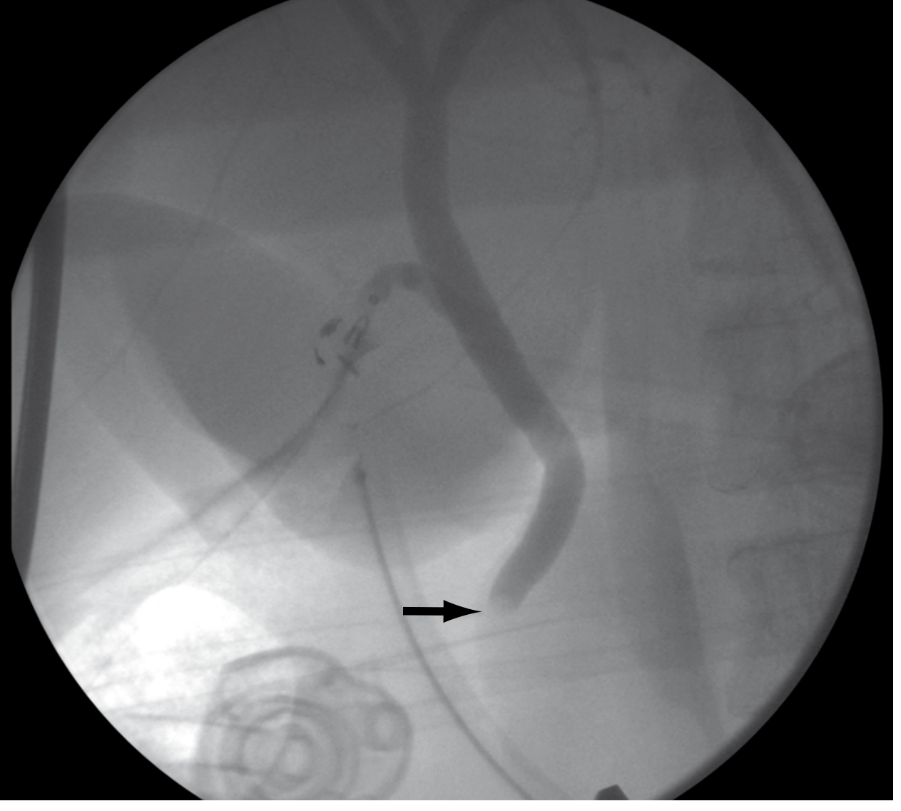
Intraoperative cholangiogram showing choledocholithiasis in an asymptomatic patient (arrow). - Sabiston Textbook of Surgery, p. 1832
Management
Management depends on the clinical context (jaundice, cholangitis, pancreatitis, or incidental finding), gallbladder status, and available expertise. The main options are:
1. Endoscopic Approach (First-line in most cases)
- ERCP with endoscopic sphincterotomy and stone extraction - the primary therapy; success rate ~84-89% for complete duct clearance in a single session
- Recommended in high-risk patients and for acutely ill patients with cholangitis requiring biliary decompression
- Recommended for stones identified before cholecystectomy: clear duct endoscopically, then proceed with laparoscopic cholecystectomy
2. Surgical Approach
- Laparoscopic bile duct exploration (transcystic or transcholedochal approach) - stone clearance rates average 95%, operative mortality ~0.5%
- Preferred when choledocholithiasis is discovered intraoperatively and surgeon expertise allows it
- Open bile duct exploration - reserved for large impacted stones, multiple failed endoscopic attempts, or anatomy precluding endoscopy (e.g., prior Roux-en-Y gastric bypass)
3. Percutaneous Approach
- Used when endoscopic and surgical options are not feasible
Summary of Timing Decisions
| Scenario | Preferred Approach |
|---|---|
| High risk + cholangitis | ERCP first, then cholecystectomy |
| Severe gallstone pancreatitis with persistent CBD stones | ERCP first |
| Stone found preoperatively, no cholangitis | ERCP + laparoscopic cholecystectomy OR single-stage laparoscopic cholecystectomy + CBD exploration |
| Stone found during laparoscopic cholecystectomy | Laparoscopic CBD exploration OR complete cholecystectomy + postoperative ERCP |
| Large/impacted stones or failed ERCP | Open CBD exploration |
- Sleisenger and Fordtran's Gastrointestinal and Liver Disease, p. 1267
- Mulholland and Greenfield's Surgery, p. 3074-3075
In summary, choledocholithiasis is the presence of gallstones in the common bile duct, most commonly arising from migration of gallbladder stones (secondary type). It can range from asymptomatic to life-threatening cholangitis. Risk stratification using clinical, laboratory, and imaging findings guides the choice between ERCP, laparoscopic CBD exploration, or open surgery, and the sequencing of these procedures relative to cholecystectomy.
Give me the answer on biliary struc- strictures. Describe the cause and risk factor of the biliary strictures. Explain the pathophysiology of the same and their effect on bile flow and liver function. Describe the clinical manifestation of biliary strictures.
Finding Sources
Finding Sources
Now reading all the key sections in parallel:
Reading File
Reading File
Reading File
Reading File
Excellent content found. Now let me read further into Maingot's for pathogenesis details and search for pathophysiology and clinical manifestations sections:
Reading File
Finding Sources
Finding Sources
Now reading the pathophysiology sections from Robbins Pathology, Schwartz's Surgery, and Yamada's in parallel:
Reading File
Reading File
Reading File
Good rich data so far. Let me now fetch the cholangiogram image showing biliary stricture from Schwartz's, and also get the PSC section from Bailey & Love and the cholestasis section from Schwartz's:
Loading Image
Reading File
Reading File
Excellent. Also fetching the PSC ERCP image and reading Maingot's clinical presentation section:
Loading Image
Reading File
I now have the Bismuth and Strasberg classification tables plus rich content on clinical presentation. Let me fetch the Bismuth classification diagram and read the clinical presentation section from Maingot's:
Loading Image
Reading File
I now have comprehensive content from multiple authoritative textbooks. Let me compile the full answer.---
Biliary Strictures
A biliary stricture is an abnormal narrowing of the bile duct lumen - either the intrahepatic or extrahepatic biliary tree - that impairs or obstructs the flow of bile from the liver to the duodenum. Strictures can be benign or malignant in origin.
1. Causes and Risk Factors
Biliary strictures are broadly categorized as benign or malignant.
A. Benign Biliary Strictures
i. Iatrogenic / Postoperative (Most Common)
Operative injury is the leading cause of benign biliary stricture, accounting for the vast majority of cases.
- Cholecystectomy - responsible for >90% of postoperative biliary strictures in the US; laparoscopic cholecystectomy carries a 2-3x higher rate of major bile duct injury (0.4-1.3%) compared to the open technique (0.2-0.3%)
- Choledochotomy (bile duct exploration)
- Gastrectomy
- Hepatic resection
- Liver transplantation - hepatic artery thrombosis post-transplant is a recognised cause of ischemic biliary stricture
The classic laparoscopic injury involves misidentification of the common bile duct (CBD) as the cystic duct, leading to clipping and division of the CBD.
Risk factors for intraoperative bile duct injury:
| Category | Specific Factors |
|---|---|
| Pathologic | Acute cholecystitis, cholangitis, gallstone pancreatitis, severe portal hepatis inflammation |
| Anatomic | Short cystic duct, low cystic duct insertion, long common wall between cystic duct and CBD, aberrant right sectoral duct |
| Technical | Limited laparoscopic field of view, excessive clip placement, injudicious electrocautery, bleeding obscuring anatomy, obesity |
- Maingot's Abdominal Operations, p. 1057-1058
ii. Inflammatory / Chronic
- Choledocholithiasis (CBD stones) - recurrent stone impaction and cholangitis produce fibrosis and stricture formation
- Chronic pancreatitis - fibrosis of the pancreatic head compresses the intrapancreatic portion of the CBD
- Cholangitis (acute/recurrent) - ascending bacterial infection causes ductal wall inflammation and subsequent scarring
- Primary Sclerosing Cholangitis (PSC) - autoimmune disease, more common in males, strongly associated with inflammatory bowel disease (especially ulcerative colitis); causes multifocal fibro-inflammatory stricturing of intra- and extrahepatic bile ducts
- IgG4-related cholangitis - immune-mediated segmental or diffuse narrowing; responds to corticosteroids
- Parasitic infections - liver flukes (Clonorchis sinensis, Opisthorchis viverrini), Ascariasis; mechanical obstruction plus chronic ductal inflammation leads to fibrosis
- AIDS cholangiopathy - opportunistic infections (CMV, Cryptosporidium) in immunocompromised patients
iii. Congenital
- Biliary atresia - fibro-inflammatory obliteration of the bile ducts in neonates
iv. Radiation-induced
- Radiotherapy to the upper abdomen can cause late-onset ductal fibrosis and stricture
v. Traumatic
- Blunt abdominal trauma; penetrating injury to extrahepatic bile ducts
vi. Mirizzi's Syndrome
- External compression of the common hepatic duct by a large stone impacted in the cystic duct or gallbladder neck, causing a functional biliary stricture
B. Malignant Biliary Strictures
-
Cholangiocarcinoma - most common primary malignancy causing biliary stricture; patients with PSC have significantly elevated risk
-
Pancreatic carcinoma - compresses the intrapancreatic CBD; typically presents as painless progressive jaundice
-
Gallbladder carcinoma - causes obstruction by direct invasion or secondary stricture
-
Ampullary carcinoma - obstruction at the ampulla of Vater
-
Metastatic lymphadenopathy - periportal nodes compressing the hilum
-
Frameworks for Internal Medicine, p. 9376-9381; Bailey and Love's Short Practice of Surgery, p. 1275
2. Pathophysiology - Effect on Bile Flow and Liver Function
A. Mechanical Obstruction of Bile Flow
A biliary stricture creates a fixed mechanical barrier to the antegrade flow of bile. The sequence of events is:
- Increased luminal pressure proximal to the stricture - bile continues to be secreted by hepatocytes but cannot drain freely
- Proximal ductal dilation - the bile ducts upstream of the stricture dilate as back-pressure builds
- Biliary stasis - stagnant bile predisposes to stone formation, infection, and colonization by enteric bacteria (ascending cholangitis)
- Cholestasis - impaired bile flow leads to accumulation of bile constituents (bilirubin, bile acids, cholesterol) in the hepatic parenchyma and bloodstream
"Cholestasis occurs with impaired bile flow, leading to accumulation of bile pigment in the hepatic parenchyma. Large bile duct obstruction is most commonly associated with gallstones and carcinomas; ascending cholangitis may develop. Chronic obstruction can lead to secondary sclerosing cholangitis and cirrhosis."
- Robbins, Cotran & Kumar Pathologic Basis of Disease
B. Effects on Liver Function
Prolonged biliary obstruction has a cascade of hepatic consequences:
| Effect | Mechanism |
|---|---|
| Conjugated (direct) hyperbilirubinemia | Conjugated bilirubin accumulates in hepatocytes and is secreted into the circulation; being water-soluble, it is filtered in the urine (bilirubinuria) |
| Elevated Alkaline Phosphatase (ALP) and GGT | Biliary obstruction stimulates increased synthesis and release of ALP from bile duct epithelium; GGT is an early, sensitive marker |
| Mild transaminase elevation | Secondary hepatocellular injury from bile acid toxicity |
| Bile acid retention | Bile acids accumulate in the blood, depositing in skin and causing pruritus |
| Fat malabsorption | Deficient bile delivery to the duodenum impairs emulsification and absorption of dietary fats and fat-soluble vitamins (A, D, E, K) |
| Coagulopathy | Vitamin K malabsorption leads to deficient production of clotting factors II, VII, IX, X; PT/INR prolonged |
| Secondary biliary fibrosis | Persistent cholestasis activates hepatic stellate cells via bile acid-mediated and inflammatory pathways, driving periportal and periductal fibrosis |
| Secondary biliary cirrhosis | Untreated, long-standing obstruction leads to progressive fibrosis, loss of hepatic architecture, and cirrhosis within approximately 5 years |
| Portal hypertension | Cirrhosis-related increased resistance in the portal circulation leads to portal hypertension with its sequelae (varices, ascites, splenomegaly) |
| Liver failure | End-stage consequence of unrelieved obstruction |
"Inflammation of the biliary tree results in fibrosis and stricture formation. Due to stricture formation, biliary stasis occurs, which predisposes patients to infection and hepatic abscess formation. Long-term complications include cirrhosis and liver failure."
- Yamada's Textbook of Gastroenterology
"Bile duct strictures that go unrecognized or are improperly managed can lead to severe complications such as recurrent cholangitis, secondary biliary cirrhosis, and portal hypertension."
- Schwartz's Principles of Surgery, p. 1446
C. Inflammatory Pathways (PSC)
In PSC and inflammatory strictures, the mechanism is distinct: periductal inflammation and fibrosis develop concentrically around the bile ducts ("onion-skin" pattern on histology), progressively obliterating the ductal lumen. This leads to the same downstream consequences of cholestasis but also to multisegmental stricturing and beading of the biliary tree.
3. Classification of Biliary Strictures (Bismuth Classification)
Biliary strictures are classified by anatomical level, which guides surgical planning:
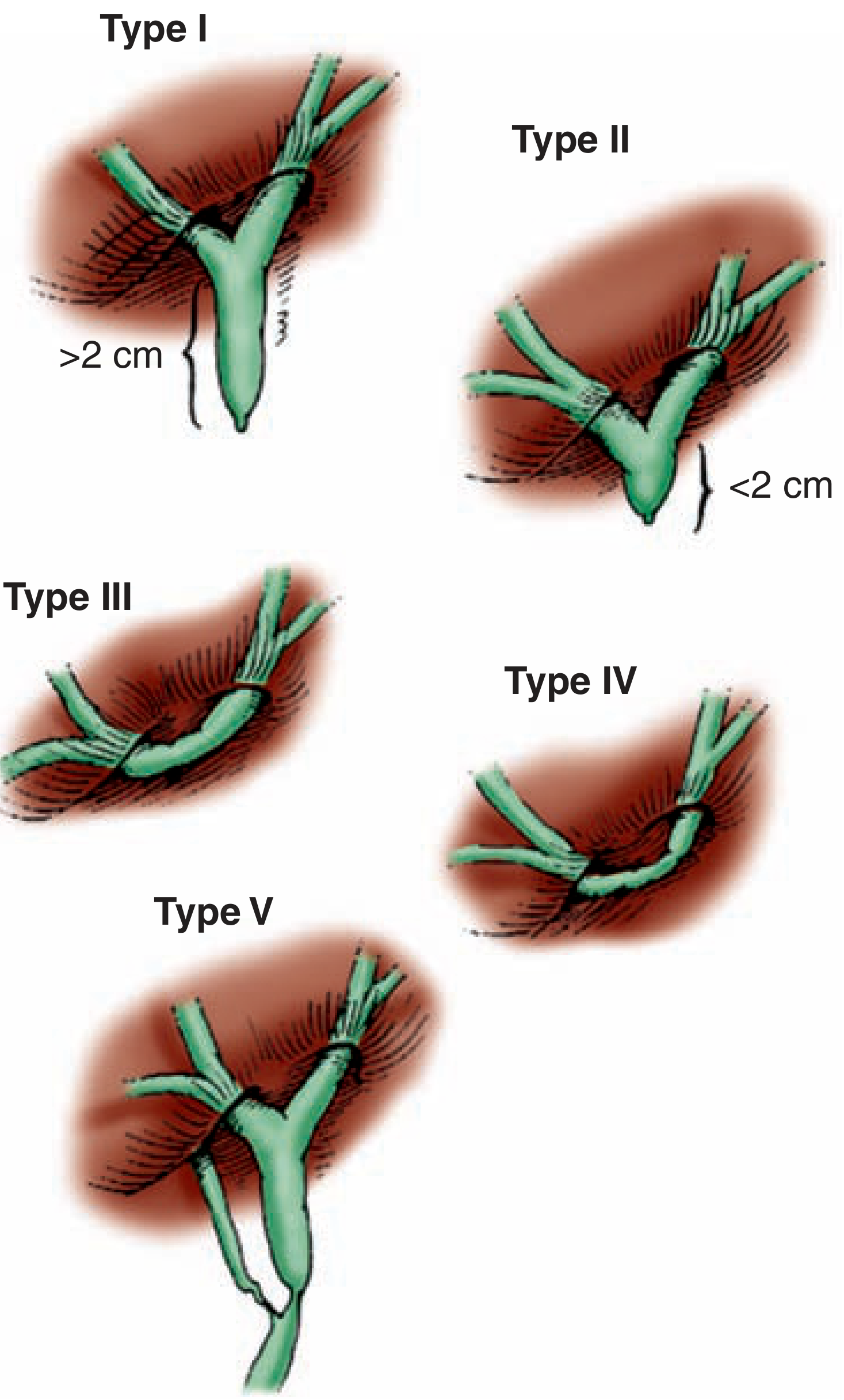
Bismuth classification of bile duct strictures based on level in relation to the confluence of hepatic ducts. Types III-V are considered complex. - Maingot's Abdominal Operations
| Bismuth Type | Location |
|---|---|
| I | Stricture >2 cm below the hepatic confluence |
| II | Stricture <2 cm below the hepatic confluence |
| III | Stricture at the hepatic confluence; hepatic ducts remain in continuity |
| IV | Stricture involving right and left hepatic ducts; ducts not in continuity |
| V | Stricture involving a right sectoral duct |
The Strasberg classification expands this to include types A-E, with E subtypes corresponding to Bismuth types I-V.
4. Clinical Manifestations
A. Early / Acute Presentation (Bile Duct Injury / Leak)
When a bile duct injury is unrecognized at surgery and results in a bile leak:
- Abdominal pain and distension - typically within the first week post-operatively
- Nausea and vomiting
- Fever and signs of sepsis - tachycardia, hypotension in severe cases
- Bilious drainage from incision sites or operative drains
- Biloma - loculated bile collection, presenting as a low-grade fever and localized pain
- Chemical peritonitis or frank biliary peritonitis if bile drains freely into the peritoneum
- Biliary ascites in severe leaks
B. Subacute and Chronic Presentation (Established Stricture)
Bile duct strictures may present months to years after the inciting event:
| Symptom/Sign | Description |
|---|---|
| Jaundice | Yellow discoloration of skin, sclera, and mucous membranes; detectable when serum bilirubin >2.5-3 mg/dL; may be the sole presenting feature |
| Pruritus | Intense itching due to bile acid deposition in skin; often preceding or accompanying jaundice; patients may have multiple excoriations on examination |
| Right upper quadrant (RUQ) pain | Dull aching or biliary colic-type pain from ductal distension |
| Dark urine (bilirubinuria) | Water-soluble conjugated bilirubin filtered by kidneys |
| Pale/acholic stools | Absence of bile pigment (urobilinogen) from stool |
| Cholangitis (Charcot's Triad) | Fever + jaundice + RUQ pain; the most common presentation of benign biliary strictures; may progress to sepsis with hypotension and altered consciousness (Reynolds' Pentad) |
| Steatorrhoea | Fatty, malodorous stools from fat malabsorption due to deficient intraluminal bile |
| Weight loss | Malabsorption and anorexia |
| Hepatomegaly | Palpable enlarged liver due to intrahepatic bile duct dilation or underlying hepatocellular damage |
| Splenomegaly | Suggests portal hypertension from secondary biliary cirrhosis |
| Unexplained fever / malaise | Particularly in isolated right sectoral duct injuries; episodes of mild cholangitis |
"Bile duct strictures most commonly result in recurrent episodes of cholangitis but may present with isolated jaundice without infection. Liver function tests usually show evidence of cholestasis with elevations of bilirubin and alkaline phosphatase."
- Schwartz's Principles of Surgery, p. 1446
"Patients with a slowly evolving stricture may have nonspecific abdominal complaints, jaundice, pruritus, cholangitis, or derangements in liver function tests... patients can present with painless jaundice, which can be confused with a malignant stricture."
- Maingot's Abdominal Operations, p. 1061
ERCP Imaging of Biliary Strictures
Below: ERCP showing a stricture of the common hepatic duct (arrow) in a patient following laparoscopic cholecystectomy. Clips from the operation are visible projected over the CBD.
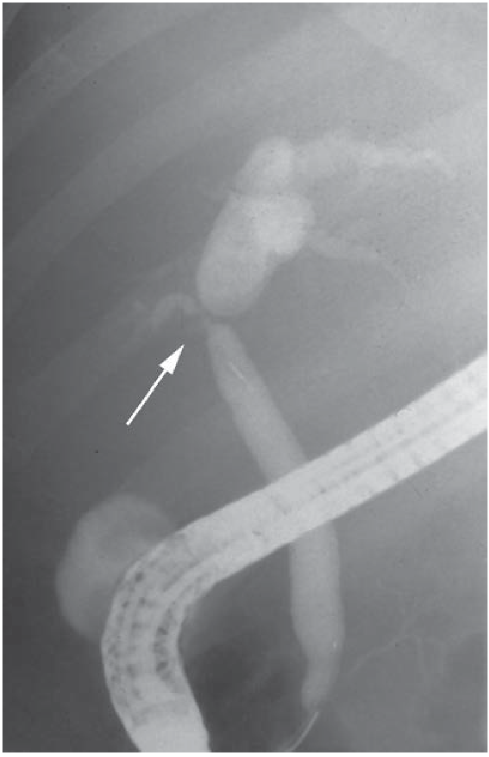
ERCP showing stricture of the common hepatic duct (arrow) - Schwartz's Principles of Surgery
PSC: Stricturing and Beading Pattern on ERCP
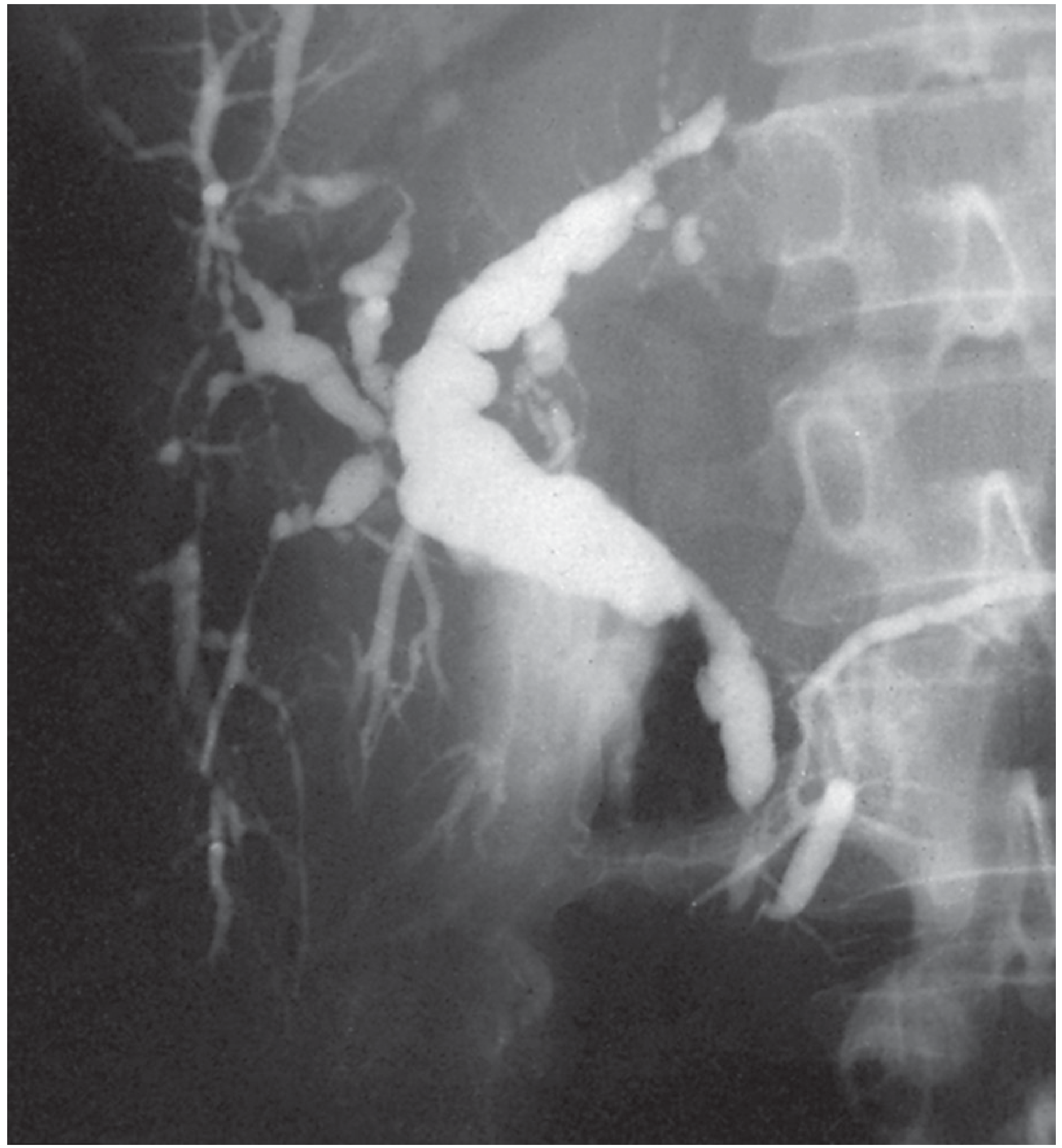
ERCP demonstrating the characteristic stricturing and beading pattern of PSC in a patient with ulcerative colitis - Bailey & Love's Short Practice of Surgery, p. 1276
5. Laboratory Findings Summary
| Test | Expected Finding | Significance |
|---|---|---|
| Serum bilirubin (conjugated) | Elevated | Cholestasis from obstruction |
| Alkaline phosphatase (ALP) | Markedly elevated | Biliary epithelial synthesis/release |
| GGT | Elevated | Sensitive early marker of hepatobiliary obstruction |
| AST / ALT | Mildly elevated | Secondary hepatocellular injury |
| Prothrombin time (PT/INR) | Prolonged | Vitamin K malabsorption |
| WBC | Elevated (leukocytosis) | Suggests superimposed cholangitis |
| IgG4 levels | May be elevated | Suggests IgG4-related cholangiopathy |
| Lipase | Elevated >3x ULN | Concomitant pancreatitis |
In summary, biliary strictures arise most commonly from surgical injury (especially laparoscopic cholecystectomy), but also from inflammatory, autoimmune, parasitic, malignant, and congenital processes. The core pathophysiology is mechanical obstruction of bile flow, producing upstream ductal hypertension and biliary stasis, which triggers cholestasis, hepatocellular injury, fibrosis, and eventually secondary biliary cirrhosis if untreated. Clinically, they manifest as recurrent cholangitis, obstructive jaundice with its systemic consequences (pruritus, steatorrhoea, coagulopathy), and ultimately liver failure - with the clinical picture varying depending on whether the presentation is acute (bile leak) or chronic (established stricture).
Pay
Pathophysiology of surgical jaundice in flow chart
Finding Sources
Finding Sources
Finding Sources
Finding Sources
Finding Sources
Finding Sources
Reading File
Finding Sources
Finding Sources
Finding Sources
I have sufficient foundational content from the previous session (Schwartz's, Robbins, Yamada's, Bailey & Love) combined with what I've now retrieved. I have enough to build a comprehensive, accurate flowchart-style answer on the pathophysiology of surgical jaundice.
Reading File
I now have all the textbook content needed to construct a thorough flowchart-style answer. Here it is:
Pathophysiology of Surgical (Obstructive) Jaundice - Flowchart
Surgical jaundice = posthepatic (obstructive) jaundice caused by mechanical blockade of bile outflow from an extrahepatic cause. The liver conjugates bilirubin normally, but the conjugated product cannot be excreted.
Master Flowchart
CAUSE OF OBSTRUCTION
│
▼
┌─────────────────────────────────────────────────────────────────┐
│ SURGICAL / MECHANICAL CAUSES │
│ • Choledocholithiasis (CBD stones) │
│ • Malignancy (pancreatic Ca, cholangiocarcinoma, ampullary Ca) │
│ • Biliary stricture (post-op, PSC, chronic pancreatitis) │
│ • Mirizzi's syndrome │
│ • Biliary atresia (neonatal) │
│ • Choledochal cyst, parasitic obstruction │
└─────────────────────────────────────────────────────────────────┘
│
▼
MECHANICAL OBSTRUCTION OF EXTRAHEPATIC BILE DUCT
│
▼
┌──────────────────────────────────┐
│ INCREASED INTRADUCTAL PRESSURE │
│ Bile secretion continues but │
│ cannot drain distally │
└──────────────────────────────────┘
│
├──────────────────────────────────────────────────┐
▼ ▼
PROXIMAL BILE DUCT DILATION BILIARY STASIS
(Upstream ducts, intrahepatic ducts, Stagnant bile pools
gallbladder distension if patent) proximal to obstruction
│ │
▼ ▼
INTRAHEPATIC DUCTAL HYPERTENSION BACTERIAL COLONIZATION
Bile forced back into hepatocytes (Ascending from duodenum)
│ │
▼ ▼
DISRUPTION OF CANALICULAR TRANSPORT ──→ ACUTE CHOLANGITIS
(Failure of bile salt export pumps) (Fever + Jaundice + RUQ pain)
│ = Charcot's Triad
▼
REGURGITATION OF CONJUGATED BILIRUBIN
INTO SINUSOIDS → BLOODSTREAM
Downstream Systemic Cascade
REGURGITATION OF CONJUGATED BILIRUBIN INTO BLOOD
│
├──────────────────────────────────────────────────────────────────────┐
▼ ▼
CONJUGATED HYPERBILIRUBINEMIA BILE ACIDS RETAINED IN BLOOD
(Direct bilirubin ↑↑; water-soluble) (Cholemia)
│ │
├──────────────┐ ├──────────────────────┐
▼ ▼ ▼ ▼
JAUNDICE BILIRUBINURIA PRURITUS BRADYCARDIA
(Skin + sclera (Dark/tea-coloured (Bile acid deposition (Bile acids sensitize
icteric when urine; "coca-cola in skin; intense cardiac conduction
bili > 2.5-3 urine") generalised itching) system)
mg/dL)
│
▼
ABSENT BILIRUBIN IN GUT
│
├──────────────────────────────────┐
▼ ▼
PALE / ACHOLIC STOOLS FAT MALABSORPTION
(No stercobilinogen) (No bile for micelle formation)
│
┌───────────┴────────────────────┐
▼ ▼
STEATORRHOEA FAT-SOLUBLE VITAMIN
(Fatty, bulky, DEFICIENCY
foul-smelling stools) (Vit A, D, E, K)
│
▼
VITAMIN K DEFICIENCY
(Clotting factors
II, VII, IX, X ↓)
│
▼
COAGULOPATHY
(Prolonged PT/INR)
Risk of bleeding
Effects on Liver Parenchyma
SUSTAINED BILIARY OBSTRUCTION
│
▼
BILE ACID ACCUMULATION IN HEPATOCYTES
(Hepatotoxic at high concentrations)
│
▼
HEPATOCELLULAR INJURY
(AST ↑, ALT ↑ — mild)
│
▼
PERIDUCTAL & PERIPORTAL INFLAMMATION
│
▼
ACTIVATION OF HEPATIC STELLATE CELLS
(Fibrogenesis: TGF-β, PDGF)
│
▼
PROGRESSIVE HEPATIC FIBROSIS
│
▼
SECONDARY BILIARY CIRRHOSIS
(~5 years of untreated obstruction)
│
├──────────────────────────────────────────────┐
▼ ▼
PORTAL HYPERTENSION HEPATOCELLULAR INSUFFICIENCY
│ │
┌────┴────────────────────┐ ┌──────────┴──────────────┐
▼ ▼ ▼ ▼ ▼
OESOPHAGEAL ASCITES SPLENOMEGALY SYNTHETIC HEPATIC
VARICES (+ SBP risk) (Hypersplenism) FAILURE ENCEPHALOPATHY
(Bleeding) (↓ Albumin,
↓ Clotting factors,
↓ Gluconeogenesis)
Renal Complications
OBSTRUCTIVE JAUNDICE
│
├─────────────────────────────────────────────────────────┐
▼ ▼
BILE ACIDS IN SYSTEMIC CIRCULATION SEPSIS (from cholangitis)
(Direct renal tubular toxicity) │
│ ▼
▼ REDUCED RENAL PERFUSION
RENAL TUBULAR DYSFUNCTION (Hypotension, ↓ cardiac output)
│ │
└──────────────────────────┬──────────────────────────────┘
▼
ACUTE KIDNEY INJURY (AKI)
("Obstructive nephropathy")
- Oliguria
- Rising creatinine
Risk is highest in malignant/
complete obstruction + cholangitis
Immune / Endotoxin Pathway
BILIARY STASIS + BACTERIAL COLONISATION
│
▼
ENDOTOXIN ABSORPTION FROM GUT (Reduced bile in duodenum
│ → ↓ Kupffer cell function
│ → Increased gut permeability)
▼
SYSTEMIC ENDOTOXAEMIA
│
├──────────────────────────────────┐
▼ ▼
SYSTEMIC INFLAMMATORY IMPAIRED KUPFFER CELL
RESPONSE (SIRS) PHAGOCYTOSIS
│ (Kupffer cells normally
▼ detoxify endotoxin)
SEPSIS / SEPTIC SHOCK
(Especially if cholangitis present)
│
▼
MULTI-ORGAN DYSFUNCTION (MOD)
(Kidneys, lungs, heart, brain)
Haematological Effects
OBSTRUCTIVE JAUNDICE
│
├─────────────────────────────────────────────────┐
▼ ▼
VITAMIN K MALABSORPTION ENDOTOXIN-MEDIATED
│ PLATELET DYSFUNCTION
▼ │
↓ Factors II, VII, IX, X ▼
│ DISSEMINATED INTRAVASCULAR
▼ COAGULATION (DIC) in severe
COAGULOPATHY / BLEEDING RISK sepsis / MOD
(↑ PT/INR, ↑ APTT)
Summary Table: Consequences of Surgical Jaundice
| System | Effect | Mechanism |
|---|---|---|
| Skin/Sclera | Jaundice, pruritus, excoriations | Conjugated bilirubin + bile acid deposition |
| Urine | Dark (bilirubinuria) | Water-soluble conjugated bilirubin filtered renally |
| Stool | Pale/acholic | No bilirubin/urobilinogen reaching gut |
| GI Tract | Steatorrhoea, malabsorption | No bile acids for fat emulsification |
| Nutrition | Fat-soluble vitamin deficiency (A, D, E, K) | Impaired absorption |
| Coagulation | Coagulopathy, bleeding | Vitamin K deficiency → ↓ Factors II, VII, IX, X |
| Liver | Fibrosis → secondary biliary cirrhosis | Bile acid toxicity + stellate cell activation |
| Portal circulation | Portal hypertension, varices, ascites, splenomegaly | Cirrhosis-related obstruction |
| Kidney | AKI ("obstructive nephropathy") | Bile acid nephrotoxicity + sepsis-related hypoperfusion |
| Cardiovascular | Bradycardia, hypotension | Bile acid sensitisation of cardiac conduction; sepsis |
| Immune | Endotoxaemia, SIRS, sepsis | ↓ bile in gut → ↑ gut permeability + ↓ Kupffer cell function |
| CNS | Encephalopathy (late) | Hepatocellular failure in secondary biliary cirrhosis |
Core concept: Surgical jaundice is not just a cosmetic discoloration - it is a multi-system disease. The initiating event (mechanical bile duct obstruction) triggers a cascade involving cholestasis, bile acid retention, fat malabsorption, coagulopathy, hepatocellular injury, and - if unrelieved - secondary biliary cirrhosis, renal failure, and sepsis. Early decompression (ERCP, surgery, or percutaneous drainage) is the only way to interrupt this cascade.
- Schwartz's Principles of Surgery, pp. 1381-1382; Robbins & Kumar Basic Pathology, p. 619; Schwartz's Principles of Surgery, p. 1446

17839494616702858331946428672592.jpg
Can make answer of all 4 at once or else will do one by one ?
I can absolutely answer all 4 topics at once! The image shows questions 23-26 covering:
- Q23 - Splenomegaly (MSLV)
- Q24 - Splenic Artery Aneurysm (MSOV)
- Q25 - Splenic Infarction (MSOV)
- Q26 - Splenic Abscess (MSOV)
Let me search the medical library for all four topics simultaneously.
Finding Sources
Finding Sources
Finding Sources
Finding Sources
Excellent hits across all 4 topics. Now reading all key sections in parallel:
Reading File
Reading File
Reading File
Reading File
Good content flowing in. Now reading the Sabiston SAA section, Goldman-Cecil splenomegaly diagnosis, Robbins splenomegaly, and Maingot SAA in parallel:
Reading File
Reading File
Reading File
Reading File
Excellent data from all 4 topics. Let me now fetch the key images and get the Sabiston splenic abscess and splenomegaly anatomy sections:
Loading Image
Reading File
Reading File
I now have comprehensive content for all 4 questions. Let me fetch the CT splenomegaly image and compile the full answer:
Loading Image
Here are all 4 complete answers:
Q23 - Splenomegaly (MSLV)
Anatomy and Physiology of the Spleen Relevant to Splenomegaly
The spleen is the largest lymphatic organ in the body, situated in the left upper quadrant, posterior to the stomach, and protected by the 9th-11th ribs. It has three functional compartments:
| Compartment | Structure | Function |
|---|---|---|
| White pulp | Periarterial lymphatic sheath + lymphoid follicles | Immune response to blood-borne antigens; B and T lymphocyte activation; removes microorganisms and foreign proteins |
| Marginal zone | Transition between white and red pulp | Interface for antigen processing |
| Red pulp | Splenic cords + sinusoids; >50% of splenic volume | Filtration and destruction of senescent/defective red blood cells (mechanical + immunologic phagocytosis); platelet and leukocyte sequestration |
Additional functions include:
- Blood volume regulation - splenic contraction and the splenorenal reflex promotes fluid/sodium retention
- Phagocytosis of encapsulated bacteria - absence (post-splenectomy) predisposes to Streptococcus pneumoniae, Haemophilus influenzae, Neisseria meningitidis infections
- Platelet pooling - normally stores 30-40% of total platelet mass
The spleen must enlarge 3-fold before it becomes palpable on clinical examination. By ultrasound, splenomegaly is defined as length ≥13 cm and thickness >5 cm.
Causes of Splenomegaly
| Category | Examples |
|---|---|
| Infectious | Infectious mononucleosis (EBV), malaria, leishmaniasis, tuberculosis, brucellosis, syphilis, endocarditis, HIV, CMV, histoplasmosis, typhoid |
| Congestive (vascular) | Liver cirrhosis with portal hypertension, portal vein thrombosis, splenic vein thrombosis, congestive cardiac failure, Budd-Chiari syndrome |
| Haematological malignancies | CML (massive splenomegaly), lymphoma (Hodgkin/Non-Hodgkin), acute/chronic leukaemia, myeloproliferative neoplasms, hairy cell leukaemia, myelofibrosis |
| Immune-mediated / autoimmune | Rheumatoid arthritis (Felty syndrome), SLE, autoimmune haemolytic anaemia, ITP, drug reactions (e.g., phenytoin) |
| Non-malignant haematological | Hereditary spherocytosis, thalassaemia, sickle cell disease (early), haemolytic anaemias, extramedullary haematopoiesis |
| Storage diseases | Gaucher disease, Niemann-Pick disease |
| Others | Sarcoidosis, amyloidosis, splenic cysts/pseudocysts, metastatic solid tumours |
Clinical Manifestations
- Incidental finding on imaging (most common presentation due to reliance on CT/US)
- Left upper quadrant (LUQ) fullness or pain - particularly with rapid enlargement
- Left shoulder tip pain - from diaphragmatic irritation (referred via phrenic nerve); suggests splenic infarct or capsular stretch
- Early satiety - from gastric compression by the enlarged spleen
- Unexplained cytopenias from hypersplenism (see below)
- Catastrophic presentation - splenic rupture (spontaneous or from minor trauma)
Hypersplenism
A clinical syndrome of:
- Splenic enlargement
- Any combination of anaemia, leukopenia, or thrombocytopenia (platelets most commonly and severely affected - sequestration in red pulp interstices)
- Compensatory bone marrow hyperplasia
- Improvement after splenectomy
Diagnostic Workup
Initial Diagnostic Workup
History: Age, travel history, infectious exposures, medications, family history, systemic symptoms (fever, weight loss, night sweats)
Physical Examination:
- Patient in right lateral decubitus position, knees slightly flexed
- Palpation begins in the pelvis and moves upward to LUQ (to avoid missing massive splenomegaly)
- Note size, tenderness, consistency, and any splenic rub
- Look for jaundice, lymphadenopathy, hepatomegaly, signs of portal hypertension (ascites, caput medusae, varices)
Laboratory Investigations (CBC + Liver Function + Imaging)
| Test | Finding | Significance |
|---|---|---|
| CBC + peripheral smear | Anaemia, thrombocytopenia, leukopenia | Hypersplenism; peripheral smear may show abnormal lymphocytes (lymphoma/leukaemia), spherocytes (hereditary spherocytosis), sickle cells, malaria parasites, Howell-Jolly bodies (asplenic), blast cells |
| Liver function tests | Elevated bilirubin, ALP, ALT, low albumin | Chronic liver disease with portal hypertension |
| LDH and uric acid | Elevated | Lymphoid/myeloid malignancies |
| Bone marrow biopsy | Hyperplasia, infiltration by malignant cells | Haematological malignancies, storage diseases |
| Flow cytometry | Abnormal cell populations | Lymphoma, leukaemia |
| Thick blood smear | Parasites | Malaria |
| Monospot test / EBV serology | Positive heterophile antibodies | Infectious mononucleosis |
| Splenic biopsy | Rare; sensitivity ~94%, specificity ~79% | Differentiate benign vs. malignant splenic masses |
Imaging
| Modality | Utility |
|---|---|
| Ultrasound (US) | First-line; measures spleen dimensions accurately; detects portal hypertension features; allows serial follow-up |
| CT scan | Provides comprehensive abdominal imaging; shows whether diffuse or nodular enlargement; identifies aetiology (e.g., liver cirrhosis, lymphoma); preferred for most clinical evaluations |
| MRI | Superior soft tissue characterisation; used for further evaluation of focal lesions or indeterminate CT findings |
| Splenic biopsy | Reserved when diagnosis cannot be established otherwise |
| FDG-PET | Not routinely useful - cannot distinguish between malignancy, infection, and other causes |
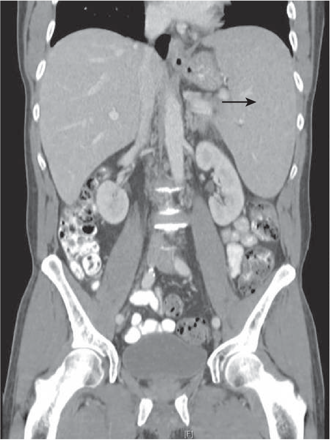
CT showing splenomegaly (arrow) - Goldman-Cecil Medicine
Therapeutics
-
Treat the underlying cause (majority of cases)
-
Splenectomy is indicated only when:
- Hypersplenism causing clinically significant cytopenias
- Haematological conditions where splenectomy is therapeutic (e.g., hereditary spherocytosis, ITP refractory to medical therapy, hairy cell leukaemia, some lymphomas)
- Massive symptomatic splenomegaly (e.g., myelofibrosis)
- Splenic complications (abscess, rupture, infarction with sepsis)
-
Pre-splenectomy: Vaccinate against S. pneumoniae, H. influenzae type b, N. meningitidis ≥2 weeks before elective splenectomy
-
Goldman-Cecil Medicine, pp. 1777-1778; Robbins & Kumar Basic Pathology; Bailey & Love's Short Practice of Surgery
Q24 - Splenic Artery Aneurysm (MSOV)
Overview
Splenic artery aneurysm (SAA) is the most common visceral artery aneurysm (60% of all splanchnic artery aneurysms) and the third most common intra-abdominal arterial aneurysm after the aorta and iliac artery. Prevalence: 0.1-0.8% general population. Female predominance (4:1; some series 2-4:1).
Risk Factors
| Risk Factor | Details |
|---|---|
| Female sex / multiparity | 2-4x more common in women; associated with increased parity; hormonal and haemodynamic changes during pregnancy weaken arterial walls |
| Portal hypertension / cirrhosis | Found in 14% of patients awaiting liver transplant; increased portal flow → hyperdynamic splenic circulation |
| Arterial dysplasia / medial degeneration | Leading pathological mechanism (Stanley et al.) |
| Hypertension | Present in >50% of patients with SAA; a common comorbidity |
| Pancreatitis | Chronic/acute pancreatitis causes perivascular inflammation and erosion of the arterial wall, especially pseudoaneurysm formation |
| Atherosclerosis | More common in elderly patients; degenerative aneurysms |
| Autoimmune arteritis | Polyarteritis nodosa, SLE, other connective tissue disorders |
| Intra-abdominal sepsis | Mycotic aneurysm from contiguous infection |
| IV drug use | Haematogenous bacterial seeding |
| Prior tobacco use | Risk factor for rupture |
| Connective tissue disorders | Marfan syndrome, Ehlers-Danlos syndrome |
Most SAAs arise at the mid- and distal main splenic artery branch points and are solitary and saccular. 90% of patients with portal HTN will have multiple SAAs.
Common Clinical Manifestations
- Asymptomatic (>90%) - incidental finding on CT scan or plain abdominal X-ray (calcified "signet ring" shadow in LUQ)
- Vague LUQ or epigastric discomfort - when symptomatic without rupture
- Bruit on auscultation over the LUQ (rare)
- GI bleeding - if aneurysm erodes into the colon, stomach, or intestine
- Rupture presentations (most feared):
- Sudden severe abdominal pain
- "Double rupture" phenomenon - initial haemorrhage is contained in the lesser sac (temporary stabilisation), followed by catastrophic free intraperitoneal haemorrhage → cardiovascular collapse
- Mimics splenic rupture clinically
Rupture in Pregnancy
- Occurs in 20-50% of ruptures, typically in the third trimester (69% of cases)
- Maternal mortality >75%; fetal death nearly inevitable
- Rupture risk is higher even with aneurysms <2 cm in pregnant women
Diagnostic Approach
| Modality | Findings |
|---|---|
| Plain abdominal X-ray | Calcified "signet ring" opacity in LUQ (curvilinear calcification) |
| CT angiography (CTA) | Gold standard for preoperative planning; shows size, location, morphology, relationship to splenic hilum; distinguishes true aneurysm from pseudoaneurysm |
| MRI/MRA | Alternative to CTA; avoids radiation; useful for follow-up |
| Selective coeliac angiography | Confirms diagnosis; enables endovascular treatment in the same session |
| Doppler ultrasound | Initial screening; may detect pulsatile flow in the aneurysm sac |
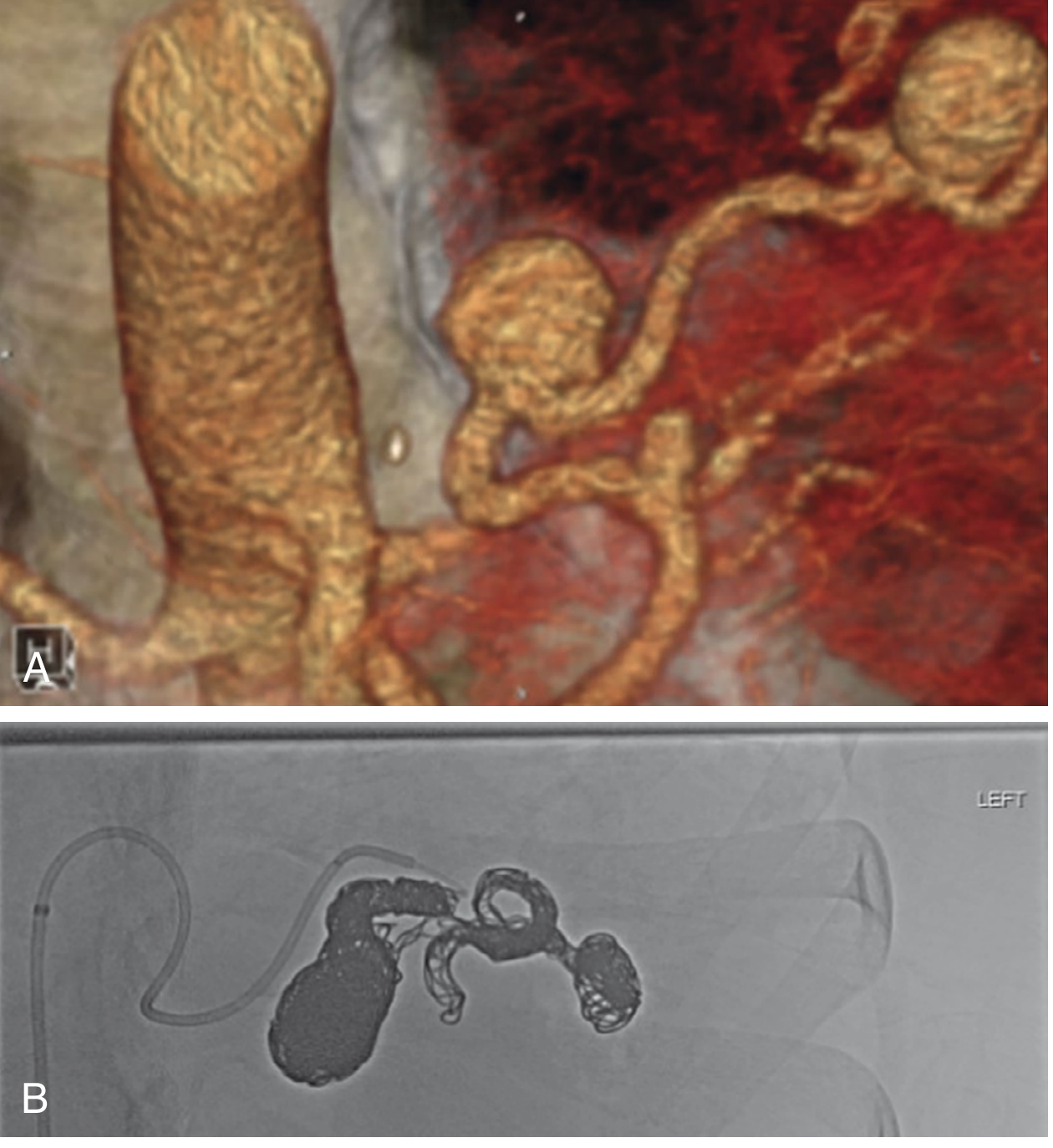
Reformatted CT angiogram showing splenic artery aneurysms (A) and post-coil embolisation angiogram (B) - Sabiston Textbook of Surgery
Diagnostic (Operative) Indications and Non-Surgical Management
Indications for Repair
| Indication | Priority |
|---|---|
| Ruptured aneurysm (any size) | Emergency repair |
| Pseudoaneurysm (any size) | All should be repaired |
| Any size SAA in females of childbearing age | Elective repair due to rupture risk in pregnancy |
| SAA ≥3 cm with demonstrated growth or symptoms | Elective repair |
| Portal hypertension, especially pre-liver transplant | Elective repair (risk of haemorrhage post-transplant) |
| Rapid growth (>0.5 cm/year) | Repair |
| Non-atherosclerotic/non-degenerative SAA | Consider repair |
Observation
- Asymptomatic, small (<3 cm), stable SAA in elderly, high-risk patients with degenerative/calcified aneurysms - annual surveillance CT or US
Non-Surgical Management
- Endovascular coil embolisation (most common current approach) - via selective splenic artery angiography; particularly for mid-splenic artery aneurysms
- Endovascular stent grafting - for aneurysms at the hilum or where embolisation risks splenic infarction
Surgical Management
-
Open splenectomy + resection of diseased artery - traditional approach
-
Laparoscopic splenectomy - for distal aneurysms
-
Aneurysmorrhaphy or aneurysm exclusion with vascular reconstruction - for proximal aneurysms with spleen preservation
-
Sabiston Textbook of Surgery, pp. 1962-1984; Bailey & Love's Short Practice of Surgery; Maingot's Abdominal Operations, p. 1264-1265
Q25 - Splenic Infarction (MSOV)
Common Causes and Risk Factors
Splenic infarction results from occlusion of the splenic artery or its branches, causing ischaemic necrosis of splenic parenchyma.
| Category | Specific Causes |
|---|---|
| Thromboembolism (most common) | Atrial fibrillation (emboli from the left atrium), bacterial endocarditis, left ventricular thrombus, cardiac valvular disease, atherosclerosis |
| Myeloproliferative disorders | CML, polycythaemia vera, myelofibrosis, essential thrombocythaemia - massively enlarged spleen with sluggish blood flow predisposes to thrombosis |
| Haemoglobinopathies | Sickle cell disease - repeated sickling episodes occlude splenic microvessels; results in progressive "autosplenectomy" |
| Haematological malignancies | Lymphoma, leukaemia |
| Vascular / local causes | Splenic vein thrombosis, portal hypertension, spleen-preserving distal pancreatectomy (surgical intervention disrupting splenic blood flow) |
| Pancreatic disease | Pancreatitis causing perivascular inflammation and thrombosis |
| Hypercoagulable states | Antiphospholipid syndrome, protein C/S deficiency, Factor V Leiden mutation |
| Trauma | Blunt trauma to LUQ |
Clinical Signs and Symptoms
- Asymptomatic - small infarcts may be entirely silent (incidental finding on CT)
- LUQ pain - sudden onset, may be severe; sharp or dull aching character
- Left shoulder tip pain - referred pain via the left phrenic nerve from diaphragmatic irritation (Kehr's sign)
- Fever - especially with septic infarction
- Nausea and vomiting
- Splenic rub - pleuritic/peritoneal friction rub heard on auscultation over LUQ (from peritoneal reaction to the infarcted segment)
- Rapid increase in spleen size due to oedema
Diagnostic Approaches
| Test | Expected Finding |
|---|---|
| CBC | May show findings of underlying disease (e.g., leukocytosis in myeloproliferative disorder, sickle cells) |
| Blood cultures | If endocarditis or septic emboli suspected |
| Echocardiography | Identify cardiac source of emboli (thrombus, vegetation) |
| Contrast-enhanced CT (CECT) | Investigation of choice - shows characteristic wedge-shaped (triangular) hypodense perfusion defect with the base at the splenic capsule; can detect haemorrhagic infarction or complications |
| Doppler ultrasound | Can show absence of flow in infarcted segments; less sensitive than CT |
| MRI | Alternative to CT; superior characterisation of haemorrhagic changes |
| Angiography | Rarely needed diagnostically; may be therapeutic (embolisation) |
Surgical Indications in Splenic Infarction
| Indication | Management |
|---|---|
| Uncomplicated infarction | Conservative management - analgesia, anticoagulation (if embolic source identified), treat underlying cause |
| Septic infarction (abscess formation) | Splenectomy indicated when septic infarct causes an abscess not amenable to percutaneous drainage |
| Haemorrhagic infarction with haemoperitoneum | Emergency splenectomy |
| Persistent pain / failed conservative management | Splenectomy |
| Massive or total splenic infarction | Splenectomy (infected tissue, risk of rupture) |
| Spontaneous rupture | Emergency splenectomy |
"Treatment is conservative and splenectomy should be considered only when a septic infarct causes an abscess."
- Bailey & Love's Short Practice of Surgery
- Bailey & Love's Short Practice of Surgery, p. 3806; Goldman-Cecil Medicine
Q26 - Splenic Abscess (MSOV)
Common Causes and Risk Factors
Splenic abscess is rare - the spleen's robust immune function (phagocytic macrophages and active immune surveillance) resists infection. Mortality: 15-20% in immunocompetent patients; up to 80% in immunocompromised patients.
Pathways of Infection
- Haematogenous spread (~70%) - most common route
- Bacterial endocarditis (complicates 5% of cases)
- Osteomyelitis
- IV drug use
- Bacteraemia from any source
- Contiguous spread - from adjacent structures
- Pancreatic necrosis / peripancreatic abscess
- Left subphrenic abscess
- Colonic, renal, or other intra-abdominal infections
-
Splenic infarction with superinfection - especially in sickle cell disease (thrombosis → infarction → colonisation)
-
Trauma - haematoma formation with secondary bacterial seeding
Risk Factors
| Risk Factor | Mechanism |
|---|---|
| IV drug use | Direct haematogenous seeding |
| HIV/AIDS and immunocompromised states | Reduced immune surveillance; fungal and mycobacterial infections (Candida, MAI, TB) |
| Sickle cell disease | Repeated infarctions create nidus for infection; more common in tropics |
| Malignancy | Polycythaemia vera, leukaemia, lymphoma - altered immunity + splenomegaly |
| Endocarditis | Septic emboli to splenic artery branches |
| Haemoglobinopathies | Structural vessel occlusion → infarction → abscess |
| Splenic trauma | Haematoma or splenic laceration serves as culture medium |
| Pancreatic necrosis | Direct contiguous spread to spleen |
| Puerperal sepsis, typhoid, paratyphoid | Bacteraemia with splenic seeding |
Organisms
Most abscesses are polymicrobial:
- Gram-positive: Staphylococcus, Streptococcus (group D), Enterococcus
- Gram-negative: E. coli, Klebsiella pneumoniae, Salmonella, Proteus mirabilis
- Anaerobes: Bacteroides, Fusobacterium, Peptostreptococcus, Clostridium
- Fungi: Candida albicans (especially in immunocompromised)
- Mycobacteria: M. tuberculosis, M. avium (in HIV)
Pathophysiology of Abscess Formation
Bacteraemia / Septic Emboli / Contiguous Spread
↓
Bacteria lodge in splenic parenchyma
(especially in areas of prior infarction or haematoma)
↓
Local tissue necrosis + neutrophil infiltration
↓
Abscess cavity forms with liquefactive necrosis
↓
Encapsulation OR rupture
↓
Rupture → Left subphrenic abscess / Diffuse peritonitis
Clinical Manifestations
| Feature | Description |
|---|---|
| Fever | Persistent, often high-grade |
| LUQ pain / abdominal pain | Vague, may radiate to left shoulder |
| Malaise and weight loss | Systemic sepsis features |
| Pleuritic chest pain | From diaphragmatic involvement |
| Leukocytosis | Neutrophilia; elevated CRP/ESR |
| Splenomegaly | Variable; not typical |
| Peritonism | If abscess has ruptured |
Note: Many patients have multiple abscesses in other organs (liver, kidneys), as splenic abscess is often part of overwhelming systemic sepsis.
Diagnostic Investigations
| Test | Finding |
|---|---|
| CBC | Leukocytosis |
| Blood cultures | May identify causative organism; bacteraemia present in many cases |
| Ultrasound | First-line; shows hypoechoic or complex cystic lesion in the spleen; guides percutaneous drainage |
| CT scan (CECT) | Preferred modality - shows single or multiple low-density splenic lesions; characterises whether unilocular or multilocular; identifies contiguous spread; guides drainage |
| MRI | Used for equivocal cases or further characterisation |
| Culture of aspirate | Definitive microbiological diagnosis; guides antibiotic therapy |
Treatment
| Scenario | Management |
|---|---|
| Unilocular abscess | Percutaneous drainage (radiologically guided US or CT) + broad-spectrum IV antibiotics - high success rates; first-line approach |
| Multilocular or multiple abscesses | Splenectomy (laparoscopic or open) + LUQ drainage + IV antibiotics |
| Failed percutaneous drainage | Laparoscopic or open splenectomy |
| Ruptured abscess | Emergency laparotomy, splenectomy, and peritoneal lavage |
| Immunocompromised patients | Often require splenectomy due to fungal/mycobacterial aetiology not amenable to drainage alone |
Antibiotic therapy should be continued until all drains/catheters have been removed. Coverage should be broad-spectrum (gram-positive, gram-negative, and anaerobic organisms), with antifungal added if immunocompromised.
- Sabiston Textbook of Surgery, pp. 2294-2300; Maingot's Abdominal Operations, pp. 1266-1267; Bailey & Love's Short Practice of Surgery; Current Surgical Therapy 14e
Quick Reference Summary Table
| Topic | Key Point |
|---|---|
| Splenomegaly | Spleen 3x enlargement before palpable; massive splenomegaly = CML / myelofibrosis; treat cause; splenectomy for hypersplenism |
| Splenic Artery Aneurysm | Most common visceral aneurysm; F>M; risk in pregnancy; "double rupture" into lesser sac then peritoneum; repair if ≥3 cm, symptomatic, or in woman of childbearing age; endovascular embolisation preferred |
| Splenic Infarction | Wedge-shaped defect on CECT; causes: AF, myeloproliferative disease, sickle cell; conservative unless septic infarct/haemorrhage |
| Splenic Abscess | Rare but life-threatening (80% mortality in immunocompromised); hematogenous spread most common; unilocular → percutaneous drainage + antibiotics; multilocular → splenectomy |
This is a shared conversation. Sign in to Orris to start your own chat.