
Clinical feature, investigation and management of acute labyrinthitis
"labyrinthitis"[MeSH Terms] OR "vestibular neuritis"[MeSH Terms]
PMID: 37339059
Acute labyrinthitis
Types and causes
| Type | Cause | Hearing |
|---|---|---|
| Viral (most common) | Presumed viral, similar to sudden sensorineural hearing loss (SNHL). Presents as sudden SNHL with acute vertigo (Cummings) | SNHL, variable recovery |
| Serous (toxic) | Bacterial toxins or mediators enter through the oval or round window or a fistula during acute or chronic otitis media (OM). Most common in children. The labyrinth has no bacteria or virus inside it. Endolymphatic hydrops is the principal finding (K J Lee's) | Mild to severe SNHL, often reversible |
| Suppurative (bacterial) | Otogenic: acute OM, or chronic OM with cholesteatoma and a lateral semicircular canal fistula. Meningogenic: via the cochlear aqueduct or internal auditory canal. Congenital labyrinthine anomalies (Mondini, enlarged vestibular aqueduct) and prior stapes surgery are risk factors | Total, permanent loss of hearing and vestibular function |
Clinical features
- Sudden-onset severe rotatory vertigo with nausea, vomiting, pallor and sweating.
- Tinnitus and hearing loss, which distinguish labyrinthitis from vestibular neuritis.
- In suppurative disease, tinnitus and dizziness progress rapidly to whirling vertigo. Otalgia, otorrhoea and fever reflect the underlying OM (Cummings).
- Spontaneous, direction-fixed horizontal-torsional nystagmus. It is irritative (beating toward the affected ear) early and becomes paralytic (beating toward the opposite ear) later.
- The nystagmus is suppressed by visual fixation. It is stronger when looking in the direction of the fast phase (Alexander's law).
- The patient falls or veers toward the affected side, and the Romberg and Fukuda tests are abnormal.
- Head impulse test (HIT) is positive, with a catch-up saccade toward the affected side (Rosen's Emergency Medicine).
- Otoscopy may show OM, a cholesteatoma or a perforation. A positive fistula sign suggests a canal fistula.
- Suppurative labyrinthitis is rare in the antibiotic era (K J Lee's).
- After the first several hours, vertigo and nystagmus abate. Central compensation over 2 to 3 weeks restores near-normal balance.
- In suppurative disease the tinnitus often fades, but hearing is lost permanently (Cummings).
- Recovery reflects contralateral compensation, not return of function in the affected ear (Scott-Brown's).
- Vestibular neuritis (no hearing loss).
- Posterior circulation stroke or cerebellar infarct.
- Meniere's disease.
- Perilymph fistula.
- Ramsay Hunt syndrome.
- Autoimmune inner ear disease.
- Vestibular migraine.
- Vestibular schwannoma.
- Meningitis.
- Labyrinthitis ossificans (fibrous or bony replacement of the labyrinth).
- Permanent unilateral SNHL.
- Chronic imbalance.
Investigations
-
Bedside examination. Check for nystagmus, HIT, and test of skew. In acute continuous vertigo with nystagmus, use HINTS (head impulse, nystagmus, test of skew) to exclude a central cause. These findings suggest a central lesion:
- a normal HIT;
- direction-changing or vertical nystagmus;
- skew deviation.
A peripheral picture shows an abnormal HIT, unidirectional nystagmus and no skew (Symptom to Diagnosis). Also look for new severe headache, neck pain, focal neurology, dysarthria, ataxia or inability to sit. -
Pure tone audiometry. This confirms SNHL, which separates labyrinthitis from neuritis and is the baseline for steroid decisions. A conductive component may be present from middle ear fluid. Repeat audiometry to track recovery.
-
Culture. Swab any ear discharge or middle ear effusion. Take blood cultures if febrile. Consider lumbar puncture only if meningitis is suspected and imaging is safe.
-
Imaging.
- No imaging is needed for a typical viral picture, or for a typical suppurative picture with OM and a predisposing condition (Cummings).
- Imaging is indicated when complications, a central cause or an anatomical defect is suspected.
- CT temporal bone shows mastoiditis, cholesteatoma, fistula and labyrinthine anomalies. It does not diagnose labyrinthitis itself (Scott-Brown's).
- MRI with gadolinium shows enhancement within the labyrinth (vestibule and cochlea) in acute viral and bacterial labyrinthitis. This is thought to reflect breakdown of the labyrinth-blood barrier (Shambaugh). In suppurative cases MRI also looks for meningeal and intracranial extension.
- In the ED, MRI is advised for bacterial labyrinthitis, and none is needed for viral cases (Tintinalli's).
-
Vestibular tests (caloric testing, vHIT, VEMPs) confirm a unilateral deficit but are not needed acutely.
-
Other tests when relevant. Syphilis serology, and autoimmune screening if hearing loss is progressive or bilateral.
Management
- Bed rest in the first 24 to 72 hours.
- Vestibular suppressants only short term (a few days) for severe vertigo and vomiting: prochlorperazine, cyclizine, or a benzodiazepine such as diazepam.
- IV fluids and antiemetics if vomiting is severe.
- Prolonged use delays central compensation.
- Start as soon as symptoms allow, within days.
- A 2024 meta-analysis of 12 RCTs (536 patients) found rehabilitation comparable to steroids for dizziness handicap in vestibular neuritis. Rehabilitation plus steroids was better than steroids alone (PMID 37339059). The evidence is for vestibular neuritis and is extrapolated to labyrinthitis.
- Viral labyrinthitis or sudden SNHL: a short oral prednisolone course, for example 1 mg/kg (about 60 mg) daily for 5 to 7 days, tapered. Intratympanic steroids are an option for salvage or when systemic steroids are contraindicated. Evidence for hearing recovery is limited, and the textbooks describe benefit as suggested but based on small studies (Scott-Brown's). Dosing here is from general practice, not the retrieved sources.
- Antivirals have no proven added benefit. Valacyclovir was not helpful in the Strupp trial.
- Serous labyrinthitis: treat the OM source. Oral antibiotics are typically effective, and steroids may improve hearing outcomes but data are lacking. Do a myringotomy if the infection does not resolve. Do a tympanomastoidectomy if cholesteatoma is present (K J Lee's). Adjuvant corticosteroids should be considered when labyrinthitis complicates acute OM (Shambaugh).
- Antibiotics. Give IV broad-spectrum antibiotics with good CSF penetration, such as a third-generation cephalosporin. Recommended duration is about 10 days. This does not reverse the labyrinthine damage but prevents meningeal spread (Cummings).
- Surgery. Manage the underlying OM or cholesteatoma. Options range from ventilation tube insertion to tympanomastoidectomy under antibiotic cover. Labyrinthectomy is unnecessary in labyrinthitis secondary to acute OM (Cummings).
- Steroids. Early steroids may help prevent labyrinthitis ossificans (Scott-Brown's).
- Monitoring. Watch for meningitis, facial palsy and intracranial extension.
- Long-term. The prognosis for recovery of hearing and vestibular function is poor. Unilateral vestibular loss is managed with physical therapy. Unilateral deafness can be managed with a bone-anchored or implantable hearing device (Shambaugh). Prompt CT or MRI is advised in children with deafness, because cochlear ossification can affect later cochlear implantation.
- Repeat audiometry at 2 to 4 weeks.
- Continue rehabilitation if imbalance persists.
- Refer if symptoms are recurrent or the hearing loss is fluctuating.
Recent evidence check
Clinical feature, investigation and management of acute labyrinthitis
labyrinthitis MRI contrast enhancement of labyrinth
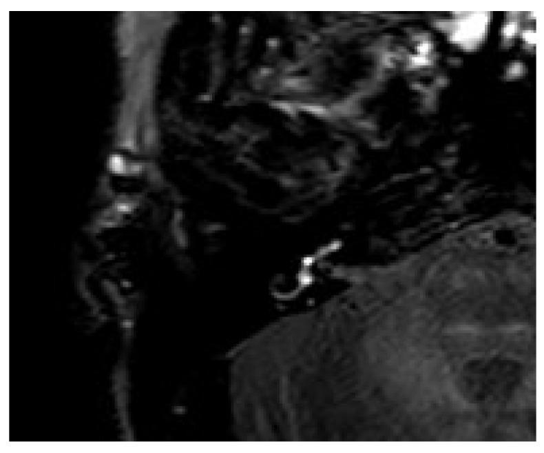
This diagnostic image is a 4-hour delayed post-contrast 3D Fluid-Attenuated Inversion Recovery (3D-FLAIR) MRI sequence of the temporal bone and inner ear. The axial view highlights focal areas of hyperintense signal within the right inner ear structures, specifically localized to the vestibulum and the basal turn of the cochlea. This pathological contrast enhancement indicates increased blood-labyrinth barrier permeability or fluid accumulation, often associated with conditions such as labyrinthitis or perilymphatic fistula (PLF). The surrounding anatomical regions, including the adjacent brainstem and cerebellum, appear with normal signal suppression characteristic of the FLAIR technique, emphasizing the high-intensity signals in the vestibular and cochlear compartments. This modality is highly sensitive for detecting subtle inner ear pathology in patients presenting with acute sensorineural hearing loss and vertigo.
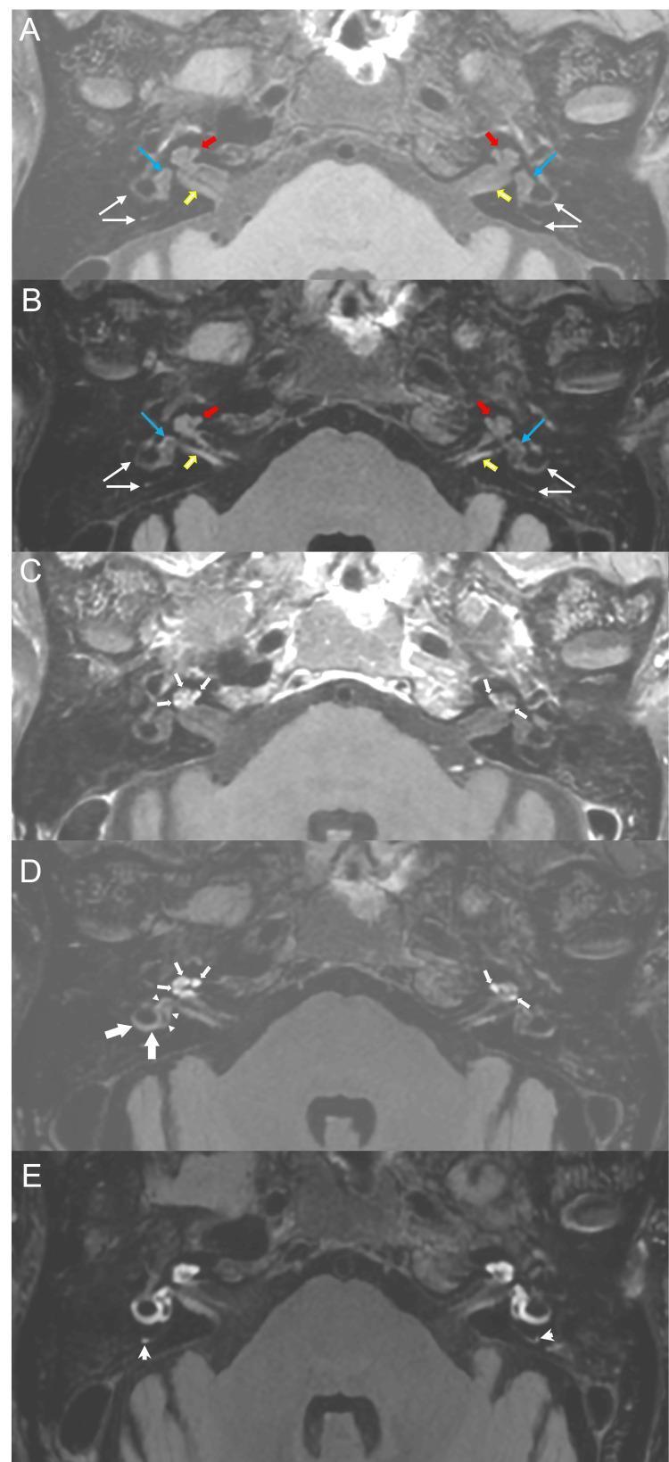
This diagnostic imaging sequence presents a series of axial Magnetic Resonance Imaging (MRI) scans of the internal auditory canal (IAC), comparing pre-contrast and post-contrast enhancement patterns in the inner ear. (A) Pre-contrast T1-weighted and (B) 3D Fluid-Attenuated Inversion Recovery (FLAIR) images establish baseline normal signal intensity in the bilateral cochlea, vestibules, and semicircular canals. (C) Post-contrast T1-weighted and (D) 10-minute delayed 3D FLAIR images show initial enhancement of the bilateral cochlea and subtle uptake in the right vestibule. (E) 4-hour delayed 3D FLAIR image demonstrates profuse, widespread hyperintense signal across the bilateral cochlea, vestibules, and lateral and posterior semicircular canals. This progressive contrast accumulation over time indicates a breakdown of the blood-labyrinth barrier (BLB). The image illustrates a key neuroradiological protocol for assessing inner ear inflammatory conditions, labyrinthitis, or endolymphatic hydrops, highlighting the clinical utility of delayed post-contrast FLAIR sequences in detecting subtle barrier permeability changes.
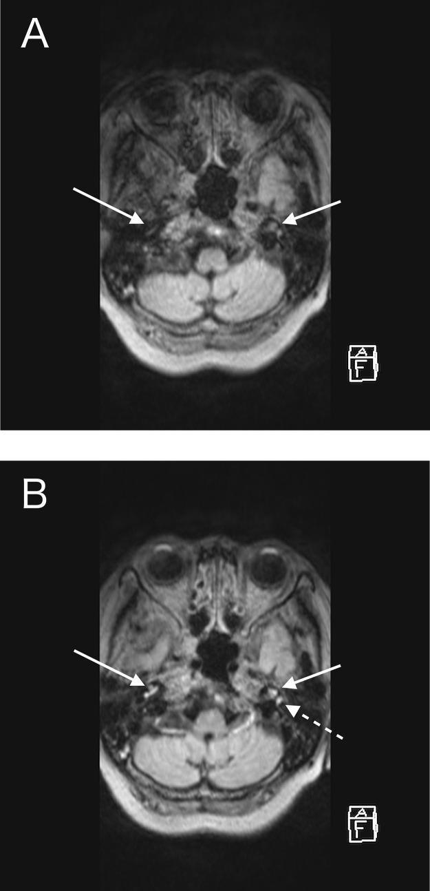
This diagnostic image consists of two axial magnetic resonance imaging (MRI) views of the head at the level of the inner ear. Panel A shows a non-enhanced Fluid-Attenuated Inversion Recovery (FLAIR) sequence, where the cochleae and semicircular canals (indicated by white solid arrows) appear isointense, blending with the surrounding petrous temporal bone structures. Panel B displays a contrast-enhanced FLAIR (CE-FLAIR) image, revealing prominent bilateral pathological enhancement within the cochleae (solid arrows) and the semicircular canals (dashed arrow). This comparative finding is clinically significant for identifying labyrinthitis or inflammatory processes associated with systemic conditions such as granulomatosis with polyangiitis (GPA). The imaging demonstrates the high sensitivity of the CE-FLAIR modality in detecting blood-labyrinth barrier breakdown compared to standard non-enhanced sequences.
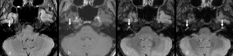
This set of axial MRI images displays the inner ear structures, specifically comparing different sequences for detecting cochlear pathology. (a) Pre-contrast 2D FLAIR shows a subtle, faintly increased signal intensity in the right cochlea (arrow). (b) Post-gadolinium 3D T1-weighted image demonstrates faint enhancement in the middle turn of the right cochlea. (c, d) Post-gadolinium 3D FLAIR images, including multiplanar reconstructions, show significantly more conspicuous bilateral enhancement. The right cochlea exhibits enhancement from the apical to the basal turn, while the left cochlea shows enhancement localized primarily to the apical turn (arrows in d). These findings illustrate the superior sensitivity of Gd-enhanced 3D FLAIR over 3D T1-weighted sequences in identifying blood-labyrinth barrier breakdown and inflammatory processes in the inner ear. The images are highly relevant for diagnosing conditions like Vogt-Koyanagi-Harada (VKH) disease, labyrinthitis, or sudden sensorineural hearing loss, highlighting the importance of sequence selection in neuro-otologic imaging.
acute labyrinthitis diagnosis and management viral bacterial steroids vestibular rehabilitation
Acute labyrinthitis
Types and causes
| Type | Mechanism | Hearing outcome |
|---|---|---|
| Viral (most common) | Presumed viral, probably the same cause as sudden SNHL. Presents with sudden SNHL and acute vertigo (Cummings) | SNHL, variable recovery |
| Serous (toxic) | Bacterial toxins and mediators cross the oval or round window, or a fistula, during acute or chronic otitis media. There is no organism in the labyrinth. Endolymphatic hydrops is the main finding. It is more common in children. Organisms behind the OM: S. pneumoniae, H. influenzae, M. catarrhalis (K J Lee's) | Mild to severe SNHL, often reversible |
| Suppurative (bacterial) | Otogenic: acute OM, or chronic OM with cholesteatoma and a lateral canal fistula. Meningogenic: spread via the cochlear aqueduct or internal auditory canal. Risk factors are congenital malformations (Mondini, enlarged vestibular aqueduct), prior stapes surgery, cochlear implants and immune deficiency (Cummings; Scott-Brown's) | Total, permanent loss of hearing and vestibular function |
Clinical features
- Sudden severe rotatory vertigo with nausea, vomiting, pallor and sweating.
- Tinnitus and hearing loss.
- In suppurative disease, tinnitus and dizziness progress rapidly to whirling vertigo. Otalgia, otorrhoea and fever from the underlying OM are common (Cummings).
- Spontaneous, direction-fixed horizontal-torsional nystagmus.
- It beats toward the affected ear early (irritative).
- It beats toward the opposite ear later (paralytic).
- Visual fixation suppresses it, and it is stronger on gaze in the direction of the fast phase.
- Gait is unsteady, with falling or veering toward the affected side.
- The head impulse test is abnormal, with a corrective saccade (Rosen's).
- Otoscopy may show OM, perforation or cholesteatoma. A positive fistula sign suggests a canal fistula.
- Fever, severe headache or neck stiffness suggests meningitis.
- Vertigo and nystagmus ease after the first several hours, and improvement continues over days.
- Central compensation over 2 to 3 weeks restores near-normal balance.
- In suppurative disease hearing is lost permanently (Cummings).
- Recovery reflects contralateral compensation, not return of function in the affected ear (Scott-Brown's).
- Vestibular neuritis (no hearing loss).
- Posterior circulation stroke or cerebellar infarct.
- Meniere's disease.
- Perilymph fistula.
- Ramsay Hunt syndrome.
- Vestibular migraine.
- Vestibular schwannoma.
- Autoimmune inner ear disease.
- Meningitis.
- Labyrinthitis ossificans (fibrous or bony replacement of the labyrinth).
- Permanent unilateral deafness.
- Chronic imbalance.
- Facial palsy.
Investigations
- Rule out a central cause first. Use HINTS (head impulse, nystagmus, test of skew) in acute continuous vertigo with nystagmus.
- Central signs: a normal head impulse test, direction-changing or vertical nystagmus, or skew deviation.
- Peripheral picture: an abnormal head impulse test, unidirectional nystagmus and no skew (Symptom to Diagnosis).
- Also look for focal neurology, severe headache, ataxia or inability to sit.
- Pure tone audiometry. It confirms SNHL, which separates labyrinthitis from neuritis. It gives a baseline for steroid decisions and for follow-up. A conductive component may come from middle ear fluid.
- Microbiology. Swab ear discharge or a middle ear effusion. Take blood cultures if febrile. Do a lumbar puncture only if meningitis is suspected and imaging shows it is safe.
- Imaging, only when there is a reason for it.
- A typical viral picture, or a typical suppurative picture in a patient with OM and a predisposing condition, needs no specific studies (Cummings).
- CT temporal bone shows mastoiditis, cholesteatoma, fistula and labyrinthine malformation. It does not diagnose labyrinthitis itself (Scott-Brown's).
- MRI with gadolinium shows enhancement within the labyrinth in acute viral and bacterial labyrinthitis. This reflects breakdown of the blood-labyrinth barrier (Shambaugh). Delayed post-contrast 3D-FLAIR is the most sensitive sequence. MRI is advised when bacterial labyrinthitis is suspected and none is needed for viral cases (Tintinalli's).
- Vestibular function tests (calorics, video head impulse test, VEMPs) confirm a unilateral deficit but are not needed in the acute phase.
- Other tests as indicated. Syphilis serology, Lyme serology, and an autoimmune screen if loss is progressive or bilateral.
Management
- Rest and fluids in the first 1 to 3 days. Give antiemetics, such as promethazine or prochlorperazine, if vomiting is severe.
- Use vestibular suppressants such as meclizine or diazepam for only 3 to 5 days (Textbook of Family Medicine). Longer use delays central compensation.
- Start early mobilisation and vestibular rehabilitation as symptoms allow. A 2024 meta-analysis of 12 RCTs in vestibular neuritis found rehabilitation comparable to steroids, and rehabilitation plus steroids better than steroids alone (PMID 37339059). A Cochrane review also supports vestibular rehabilitation for peripheral vestibular dysfunction. This evidence is from vestibular neuritis and mixed populations, and is extrapolated to labyrinthitis.
- Treat the hearing loss like sudden SNHL: a short oral corticosteroid course started early, for example prednisolone about 1 mg/kg daily (up to 60 mg) for 5 to 7 days, with or without a taper. Intratympanic steroid is an option if systemic steroids are contraindicated or as salvage. Dosing is from standard practice, not the retrieved textbooks.
- Evidence for benefit is limited. The textbooks describe steroid benefit in vestibular neuritis as suggested but based on small studies (Scott-Brown's).
- Antivirals have not shown added benefit. In the Strupp trial, valacyclovir added nothing to methylprednisolone.
- Repeat audiometry at 2 to 4 weeks.
- Treat the source. Oral antibiotics are typically effective for the OM (K J Lee's).
- Do a myringotomy with a ventilation tube if the infection persists.
- Do a tympanomastoidectomy if there is cholesteatoma.
- Consider steroids (oral or intratympanic). They may improve hearing outcomes, but data are lacking.
- Untreated, serous disease can progress to bacterial labyrinthitis (Swanson's Family Medicine Review).
- Admit the patient. Give IV broad-spectrum antibiotics with CSF penetration, adjusted to culture. Treatment is about 10 days. It will not reverse the damage but is needed to stop spread to the meninges (Cummings; K J Lee's).
- Treat the underlying ear disease, under antibiotic cover.
- Use a ventilation tube if there is no cholesteatoma.
- Use tympanomastoidectomy if there is cholesteatoma.
- Labyrinthectomy is unnecessary when the cause is acute OM.
- Give steroids at diagnosis. They are associated with less labyrinthitis ossificans and, in meningitic labyrinthitis from H. influenzae and S. pneumoniae, better hearing outcomes (Cummings; K J Lee's). Evidence on preventing SNHL is still unclear.
- Watch for meningitis, facial palsy and intracranial extension.
- Follow with serial CT or MRI for labyrinthitis ossificans if SNHL persists. This matters in children who may need a cochlear implant.
- Long-term, unilateral vestibular loss is managed with physical therapy. Unilateral deafness can be managed with a bone-anchored or implantable hearing device (Shambaugh). Consider cochlear implant assessment for bilateral or profound loss.
- Audiometry at 2 to 4 weeks, then as needed.
- Continue rehabilitation until compensation is complete.
- Refer if hearing fluctuates, symptoms recur, or the loss is bilateral.