
Multiple myeloma
multiple myeloma bone marrow plasma cells histology
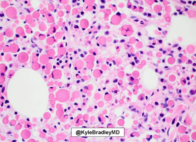
Imaging modality: light microscopy, bright-field, with hematoxylin and eosin staining of a bone marrow core biopsy. Specimen shows a cellular marrow with sheets of plasma cells occupying a large proportion of the tissue, interrupting normal hematopoietic elements. The malignant plasma cells display classic features of plasma cell neoplasia, including eccentric nuclei and coarse clumped chromatin, and abundant cytoplasm. Within many plasma cells, there are eosinophilic cytoplasmic inclusions representing Russell bodies (immunoglobulin condensations). The distribution is diffuse and sheets are interspersed with residual adipocytes and scattered karyorrhectic debris. The background shows mild interstitial fibrosis and occasional binucleated plasma cells. Notable bystander plasma cells are present but in smaller numbers, consistent with clonal proliferation. In the context of myeloma, such marrow involvement supports a diagnosis of multiple myeloma with heavy plasmacytosis and intracellular immunoglobulin accumulation. Clinically, this morphology prompts ancillary testing including serum protein electrophoresis, immunofixation, and light chain assay to determine monoclonality and disease burden. The image is valuable for educational illustration of Russell bodies in myeloma, differential diagnosis of plasmacytosis, and for correlating histology with cytology and radiology in diagnostic workups. It serves as a reference for hematopathology trainees and pathology education in plasma cell dyscrasias.

Histology, Light microscopy; bone marrow trephine biopsy stained with Hematoxylin and Eosin (H&E). The specimen shows extensive replacement of normal hematopoietic elements by sheets of immature plasma cells, consistent with marrow involvement by plasma cell myeloma. The infiltrate is markedly cellular with high nuclear-to-cytoplasmic ratio, round to oval nuclei, coarse clumped chromatin, and inconspicuous nucleoli in many cells. Nucleoli are more prominent in a subset of plasma blasts, and some cells display multinucleation. The cytoplasm is variably basophilic and often abundant, contributing to a purple to pink background. The normal marrow architecture is effaced; residual hematopoiesis is sparse. Occasional binucleated or trinucleated plasma cells may be seen; mitotic figures can be present, reflecting active proliferation. The overall pattern is diffuse, with possible focal aggregates. In non-secretory myeloma, as in this case, monoclonal plasma cell proliferation can be demonstrated by immunohistochemistry for kappa or lambda light chains and by molecular studies showing clonal rearrangements, even in the absence of detectable M protein in serum or urine. However, this image itself shows characteristic plasma cell morphology and marrow effacement; clinical correlation with serum free light chains and immunophenotyping is essential for diagnosis and staging. This histopathology supports a diagnosis of multiple myeloma with marrow involvement.
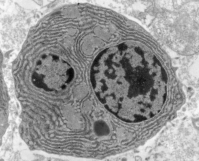
Transmission electron microscopy image of bone marrow plasma cells in multiple myeloma, highlighting ultrastructural features of malignant plasmacytosis. The specimen is a bone marrow biopsy/aspirate subjected to TEM to reveal subcellular architecture at high magnification. The primary subject is a plasma cell characterized by a prominent, eccentrically placed nucleus with chunky, irregular clumped chromatin and abundant cytoplasm. Bi- or multinucleation is frequently observed in myeloma cells. The cytoplasm shows extensive rough endoplasmic reticulum organized in parallel lamellae, reflecting heavy immunoglobulin synthesis. Light gray, round to ovoid cytoplasmic inclusions known as Russell bodies are evident within the cytoplasm, representing condensed immunoglobulin within intact endoplasmic reticulum. The cellular borders are well defined, and there is sometimes distension of the ER cisternae near the perinuclear region. The image underscores hallmark features of malignant plasma cells in multiple myeloma and demonstrates both proliferative activity and high secretory load. Clinically, these ultrastructural findings correlate with a high monoclonal immunoglobulin production and marrow infiltration, informing prognosis and therapeutic decisions when integrated with light-chain studies and routine histology. This EM view complements light microscopy by providing evidence of intracellular organelle expansion and intracellular inclusions that are not discernible with conventional staining. Potential educational use includes teaching plasmacytosis, plasma cell neoplasia morphology, and EM-based diagnostic histopathology.
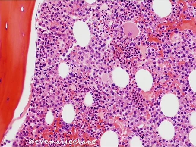
Bone marrow trephine biopsy, Hematoxylin and Eosin stained, evaluated by light microscopy. This histopathology image depicts medullary marrow with cellular admixture dominated by clonal plasma cells. Plasma cells comprise approximately 50% of nucleated cells, demonstrating interstitial (non-follicular) infiltration in a background of residual hematopoietic elements and adipocytes. The architectural pattern is interstitial rather than diffuse sheets at this stage, consistent with stage II myeloma by marrow involvement (Stage II: 20–50% replacement). The plasma cells show hyperchromatic nuclei with clumped chromatin and moderate cytoplasm; occasional binucleation may be present; cytoplasmic features not fully assessable at this magnification. Normal hematopoiesis is variably preserved in focal areas but is increasingly suppressed toward representing marrow replacement. The image correlates with increasing tumor burden and is used for staging, risk assessment, and monitoring response to therapy. In clinical practice, plasmacytosis of this magnitude suggests symptomatic multiple myeloma with potential cytopenias and immunoparesis; cytomorphology must be correlated with flow cytometry and serum/urine studies to confirm clonality. Differential considerations include reactive plasmacytosis, MGUS, and plasmacytoma; however, the extensive involvement and pattern here favor a systemic plasma cell neoplasm. This histology guides prognosis and therapeutic decisions.
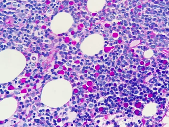
This is a bone marrow biopsy/section analyzed by light microscopy using Periodic acid–Schiff (PAS) stain. The image shows marrow with lymphoplasmacytic infiltration including malignant plasma cells. Dutcher bodies are intranuclear immunoglobulin inclusions that stain PAS-positive, appearing as rounded, eosinophilic or magenta inclusions within the nucleus of plasma cells. The plasma cells display eccentric nuclei and basophilic cytoplasm. The overall architecture shows a hypercellular marrow with scattered adipocytes; residual hematopoietic cells vary between small lymphocytes and plasmacytoid cells. The PAS stain highlights the Dutcher bodies and cytoplasmic granularity, aiding identification of plasma cell involvement. In Lymphoplasmacytic lymphoma, marrow involvement is common with clonal expansion of small mature lymphocytes, plasmacytoid cells, and plasma cells. Diagnostic significance lies in recognizing Dutcher bodies as a clue to immunoglobulin production within malignant plasma cells; the PAS positivity helps differentiate from non-plasmacytoid marrow processes. Potential clinical use cases include confirming marrow involvement in suspected Waldenström macroglobulinemia, assessing tumor burden, monitoring response to therapy, and guiding differential diagnoses such as marginal zone lymphoma or multiple myeloma. Given the PAS staining pattern and Dutcher bodies, this image is especially relevant for hematopathology education, marrow pathology case repositories, and histology atlases.
multiple myeloma lytic bone lesions skull X-ray
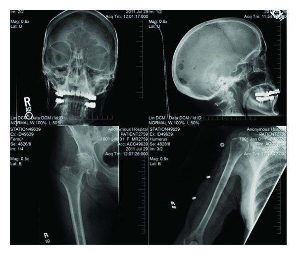
This diagnostic composite image presents a radiographic skeletal survey consisting of four X-ray views: an AP and lateral view of the skull (top row), a proximal femur/pelvis view (bottom left), and a humerus view (bottom right). The skull radiographs demonstrate classic 'punched-out' lytic lesions, particularly visible in the lateral projection as multiple, discrete, circular areas of radiolucency throughout the calvarium. Similar diffuse lytic lesions and areas of cortical thinning are evident in the diaphysis and metaphysis of the humerus and the proximal femur. These findings are characteristic of plasma cell dyscrasias, such as multiple myeloma, or other osteolytic metastatic processes. The distribution across both flat and long bones highlights systemic skeletal involvement. Medical educators can use this image to illustrate the radiographic hallmarks of multiple myeloma and differentiate lytic bone destruction from blastic lesions in an oncologic or hematologic context.
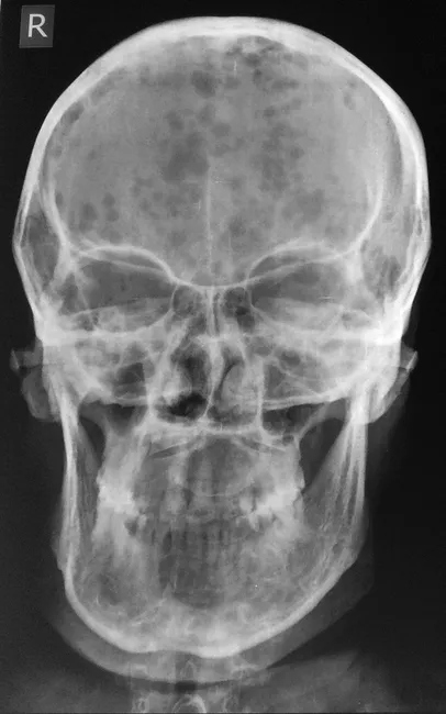
This skull radiograph illustrates findings typical of plasma cell myeloma. Modality: plain skull X-ray (radiography); view: frontal skull projection, with the right side marker visible. The calvarium shows multiple small, uniform, sharply demarcated punched-out lytic lesions throughout the cranial bones, without a sclerotic rim or periosteal new bone formation. In several regions, the lesions extend toward the mandible, reflecting medullary bone destruction with cortical involvement in advanced sites. Generalized osteopenia is evident, resulting in diffuse reduced bone density beyond the focal lucencies. There is no visible acute fracture or reactive sclerosis. The pattern—punched-out lacunae in a background of osteopenia—is highly suggestive of multiple myeloma and is more typical of plasma cell neoplasms than osteoblastic metastases. Clinically, the patient presented with weakness, fatigue, backache, weight loss, and anemia, consistent with CRAB features (hypercalcemia, renal impairment, anemia, bone lesions). The imaging supports differential considerations including metastatic disease and Langerhans cell histiocytosis, but the distribution and appearance favor a myelomatous process. This radiograph provides visual corroboration for staged disease assessment and guides further diagnostics (MRI/CT, skeletal survey, serum protein electrophoresis, bone marrow biopsy). Case courtesy of Radiopaedia, rID 40195. The image exemplifies classic myeloma-related skeletal destruction and underscores the importance of early oncologic workup.

This lateral skull radiograph (X-ray) demonstrates the classic 'punched-out' or 'moth-eaten' appearance characteristic of multiple myeloma. The image reveals numerous well-circumscribed, round-to-oval radiolucent (lytic) lesions of varying sizes distributed across the calvarium, including the frontal, parietal, and occipital bones. These lesions show no evidence of peripheral sclerotic margins, indicating aggressive bone resorption without significant reactive bone formation. The calvarial diploe is notably involved, showing a generalized decrease in bone density and disruption of the normal trabecular pattern. Clinical significance of these findings includes the hallmark skeletal manifestations of plasma cell dyscrasias, where monoclonal plasma cells infiltrate the bone marrow, triggering osteoclast activity. This diagnostic image serves as an educational example of hematologic malignancy-associated osteolysis and is a critical component of a skeletal survey for staging conditions like multiple myeloma or metastatic disease.
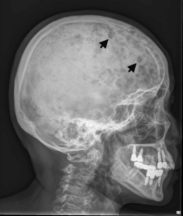
This lateral skull X-ray (radiography) demonstrates a classic 'rain-drop' or 'punched-out' appearance of the calvarium. Numerous small, well-circumscribed, round-to-oval radiolucent (darker) lytic lesions of varying sizes are scattered throughout the skull vault, particularly involving the frontal and parietal bones. These lesions show no reactive sclerotic margins, indicating rapid bone resorption typical of plasma cell dyscrasias like multiple myeloma or osteolytic metastatic disease (such as metastatic breast carcinoma). The image also captures the facial bones, upper cervical spine, and dental hardware. Multiple radiopaque dental implants and restorative work are visible in the maxilla and mandible. The overall diagnostic quality is high, highlighting key radiographic signs of systemic bone destruction for medical education in oncology and radiology.
"multiple myeloma"[MeSH Terms] AND treatment
multiple myeloma overview diagnosis treatment 2025
Multiple Myeloma
- Chromosomal translocations fusing the IgH locus (14q32) to oncogenes such as cyclin D1/D3, dysregulating cell proliferation
- IL-6 (produced by bone marrow stromal fibroblasts/macrophages) drives plasma cell proliferation
- Later cytogenetic hits: RAS mutations, MYC abnormalities, 17p/1p deletions and 1q amplification (associated with adverse prognosis)
- Bone disease: neoplastic plasma cells raise RANKL expression (and lower osteoprotegerin, OPG) on stromal cells/osteoblasts, activating osteoclasts, while cytokines like DKK1, IL-3, and IL-7 suppress osteoblasts. The net effect is pure osteolytic bone destruction, hypercalcemia, and pathologic fractures.
- Renal injury: light chain ("myeloma") cast nephropathy from Bence Jones proteins precipitating in distal tubules, plus hypercalcemia, AL amyloidosis (~10% of patients), light chain deposition disease, and Fanconi syndrome
- Immune defects: functional antibody production is suppressed despite elevated total immunoglobulin, causing high infection risk
- Calcium elevation (hypercalcemia from bone resorption)
- Renal insufficiency (cast nephropathy)
- Anemia (marrow infiltration)
- Bone disease (lytic lesions, osteopenia, fractures)
- Plus radiculopathy/cord compression from vertebral collapse, recurrent bacterial infections from functional hypogammaglobulinemia, and neurologic complications
- Progression to plasma cell leukemia is suggested by hepatic infiltration, pleural effusion, or ascites; multiple extramedullary plasmacytomas also signal aggressive disease
- Transplant-eligible, newly diagnosed: induction triplet (e.g., bortezomib/lenalidomide/dexamethasone) followed by autologous stem cell transplantation, then lenalidomide (± bortezomib in high-risk disease) maintenance.
- Transplant-ineligible: VRd (bortezomib/lenalidomide/dexamethasone) for about 9 months followed by lenalidomide maintenance, or DRd (daratumumab/lenalidomide/dexamethasone) continued until progression.
- Relapsed/refractory disease: almost all patients eventually relapse. Options include immunomodulatory drugs (lenalidomide, pomalidomide, thalidomide), proteasome inhibitors (bortezomib, carfilzomib, ixazomib), and CD38/SLAMF7-targeted monoclonal antibodies (daratumumab, isatuximab, elotuzumab), used in combination regimens tailored to prior therapy and eligibility for further transplant.
- Newer options gaining traction include bispecific antibodies (e.g., teclistamab) and BCMA-targeted CAR T-cell therapy for heavily pretreated relapsed/refractory disease.
- A 2025 meta-analysis on non-transplant-eligible newly diagnosed myeloma (PMID: 41027476) and a 2025 review on treatment outcomes in developing countries (PMID: 41078459) reinforce triplet/quadruplet regimens as standard, with access disparities noted in resource-limited settings.
- A 2024 systematic review of RCT outcomes in myeloma (PMID: 39368634) highlights ongoing emphasis on quality-of-life endpoints alongside survival.
- Real-world data reported by the International Myeloma Foundation show teclistamab achieving 65% response rates and 18-month median overall survival in heavily pretreated relapsed/refractory patients, and early CAR-T (zevorcabtagene autoleucel) data from China showing promise in similarly refractory disease.