
Collagen synthesis biochemistry
Reading File
Collagen synthesis is a multi-step process occurring both intracellularly and extracellularly. Here's a quick overview while I search the textbooks:
- Intracellular: transcription/translation of pro-alpha chains, hydroxylation of proline/lysine (requires vitamin C), glycosylation, triple helix formation into procollagen
- Extracellular: cleavage of propeptides to tropocollagen, cross-linking by lysyl oxidase (requires copper) into fibrils
- Key disorders: scurvy (vitamin C deficiency), Ehlers-Danlos syndrome, osteogenesis imperfecta
Searching sources...
Finding Sources
Finding Sources
I found excellent sources. Let me read the key sections from Lippincott Biochemistry and Harper's, plus grab the diagram image.
Reading File
Reading File
Reading File
Excellent content. Let me now read the full Lippincott biosynthesis section and fetch the collagen synthesis diagram image.
Reading File
Loading Image
Loading Image
Let me read the remaining Lippincott text on cross-linking, and also the Harper's section on diseases.
Reading File
Reading File
I now have all the material needed for a thorough, well-illustrated answer. Here it is:
Collagen Synthesis - Biochemistry
Collagen is the most abundant protein in the human body (~30% of total protein), forming the structural scaffold of connective tissue, bone, skin, tendons, and basement membranes. Its synthesis is one of the most complex known, involving at least 8 enzyme-catalyzed post-translational steps across two compartments - intracellular and extracellular.
Step-by-Step Synthesis Pathway
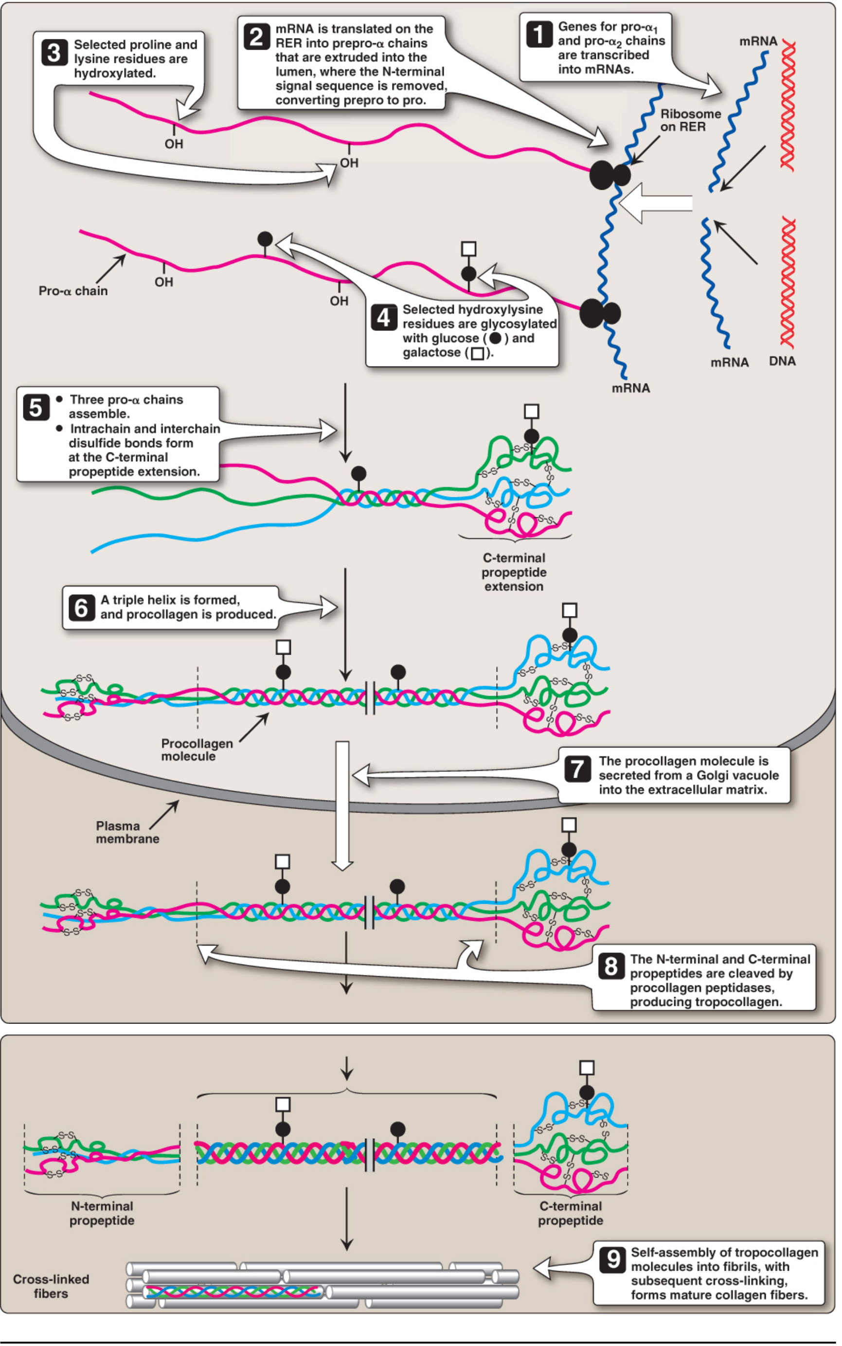
INTRACELLULAR STEPS (in RER and Golgi)
Step 1 - Gene transcription and translation (prepro-α chain formation)
- Over 30 genes encode collagens (prefixed COL, e.g., COL1A1, COL1A2).
- The initial polypeptide is a prepro-α chain with an N-terminal signal sequence that targets it to the rough ER (RER).
- The signal sequence is cleaved in the RER lumen, yielding the pro-α chain.
- Pro-α chains contain N- and C-terminal propeptide extensions flanking the central collagen domain.
- Characteristic repeating sequence: -Gly-X-Y- (X = often proline; Y = often hydroxyproline or hydroxylysine).
- Glycine (every 3rd residue) is essential because it is the only amino acid small enough to fit at the center of the triple helix. Proline "kinks" the chain, pre-forming the helical conformation.
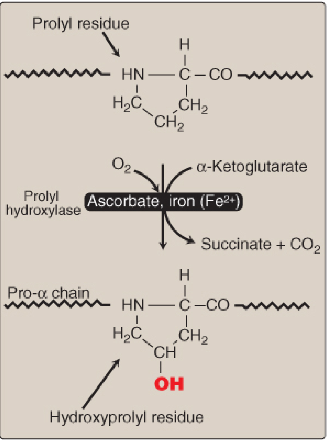
Step 2 - Hydroxylation of proline and lysine
- Proline and lysine in the Y-position undergo hydroxylation to form hydroxyproline and hydroxylysine.
- Enzymes: prolyl hydroxylase and lysyl hydroxylase
- Cofactors required: O₂, Fe²⁺, α-ketoglutarate, and vitamin C (ascorbic acid)
- Vitamin C's role: keeps iron in the reduced Fe²⁺ state so the hydroxylases remain active. Without vitamin C, iron oxidizes to Fe³⁺, the enzymes fail, and hydroxylation stops.
- Hydroxyproline maximizes inter-chain hydrogen bonds stabilizing the triple helix.
- Hydroxylysine is the anchor point for glycosylation and later cross-linking.
Step 3 - Glycosylation
- Hydroxylysine residues are glycosylated with glucose and galactose by glycosyltransferases.
- This must occur before triple helix formation (the helix blocks enzyme access).
Step 4 - Assembly into procollagen (triple helix formation)
- Three pro-α chains self-assemble, initiating at the C-terminal propeptide via interchain disulfide bonds.
- The triple helix then "zips" from C-terminus toward the N-terminus, producing procollagen - a triple-stranded helical molecule with non-helical propeptide extensions at both ends.
- The procollagen is packaged into secretory vesicles in the Golgi and exported.
EXTRACELLULAR STEPS
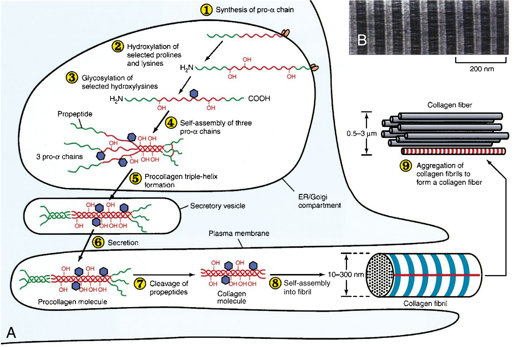
Step 5 - Cleavage of propeptides → tropocollagen
- Extracellular procollagen peptidases (N- and C-procollagen peptidases) cleave off both propeptide extensions.
- The product is tropocollagen (~300 nm long, ~1.5 nm wide), the basic structural unit.
- Clinical note: deficiency of N-procollagen peptidase (ADAMTS2) causes dermatosparaxis EDS - thin irregular fibrils, severely fragile and sagging skin.
Step 6 - Fibril formation
- Tropocollagen molecules spontaneously self-assemble into fibrils in a parallel, staggered arrangement (each molecule overlapping the next by ~3/4 of its length), producing the characteristic 67 nm banding pattern seen on electron microscopy.
- Fibril diameter ranges from 10-300 nm.
Step 7 - Cross-link formation (maturation)
- Lysyl oxidase (a copper-dependent extracellular enzyme) oxidatively deaminates the ε-amino groups of lysine and hydroxylysine residues, producing reactive aldehydes (allysine and hydroxyallysine).
- These aldehydes spontaneously condense with adjacent lysine/hydroxylysine residues to form covalent aldimine and aldol cross-links.
- Cross-linking provides tensile strength; it is maximal in tendons and bone.
- Multiple fibrils aggregate into collagen fibers (0.5-3 µm diameter), visible under light microscopy.
Summary Table of Steps and Cofactors
| Step | Location | Enzyme/Event | Cofactor |
|---|---|---|---|
| Translation of prepro-α chain | RER | Ribosomes | - |
| Signal peptide cleavage | RER lumen | Signal peptidase | - |
| Prolyl hydroxylation | RER lumen | Prolyl hydroxylase | O₂, Fe²⁺, Vitamin C, α-KG |
| Lysyl hydroxylation | RER lumen | Lysyl hydroxylase | O₂, Fe²⁺, Vitamin C, α-KG |
| Glycosylation of hydroxylysine | RER/Golgi | Glycosyltransferases | Glucose, galactose |
| Triple helix assembly | RER/Golgi | Spontaneous (after disulfide bond) | C-propeptide disulfide bonds |
| Secretion | Golgi → ECM | Exocytosis | - |
| Propeptide cleavage | ECM | N/C-procollagen peptidases | - |
| Fibril self-assembly | ECM | Spontaneous | - |
| Cross-link formation | ECM | Lysyl oxidase | Copper (Cu²⁺) |
Key Cofactors - Exam Essentials
| Cofactor | Role | Deficiency Disease |
|---|---|---|
| Vitamin C | Keeps Fe²⁺ for prolyl/lysyl hydroxylase | Scurvy (unstable triple helix, fragile vessels, gum disease, poor wound healing, perifollicular hemorrhages) |
| Copper (Cu²⁺) | Cofactor for lysyl oxidase (cross-linking) | Menkes disease (kinky hair disease) - defective Cu transport |
| Fe²⁺ | Cofactor for hydroxylases | Deficiency impairs hydroxylation (overlaps with vitamin C deficiency) |
Collagen Types and Tissue Distribution
| Type | Chain Composition | Location | Disease If Mutated |
|---|---|---|---|
| I | [α1(I)]₂α2(I) | Bone, skin, tendon, most connective tissue | Osteogenesis imperfecta, aEDS |
| II | [α1(II)]₃ | Cartilage, vitreous humor | Severe chondrodysplasia |
| III | [α1(III)]₃ | Skin, blood vessels, bowel | Vascular EDS (spontaneous artery/bowel rupture) |
| IV | Network-forming | Basement membranes (glomerulus, ear, eye) | Alport syndrome |
| V | Heterotrimeric | Skin, cornea | Classical EDS |
Diseases of Collagen Synthesis
Scurvy (Vitamin C deficiency)
- Hydroxylation of proline/lysine fails → unstable triple helices → defective pro-α chains
- Gradual loss of existing collagen plus inability to synthesize new collagen
- Perifollicular hemorrhages, ecchymoses, gum disease, loose teeth, poor wound healing
Osteogenesis Imperfecta (OI)
- Mutations in COL1A1 or COL1A2 (type I collagen)
- Glycine substitutions in the Gly-X-Y repeat disrupt triple helix packing ("protein suicide")
- Brittle bones, blue sclerae, hearing loss (8 recognized types)
Ehlers-Danlos Syndrome (EDS)
- 13 subtypes per 2017 International Classification
- Classical EDS: type V collagen defect (skin hyperextensibility, joint hypermobility)
- Vascular EDS: type III collagen defect (COL3A1) - fragile vessels, risk of spontaneous rupture
- Kyphoscoliotic EDS: lysyl hydroxylase deficiency - progressive scoliosis, muscle weakness
- Dermatosparaxis EDS: ADAMTS2 (N-procollagen peptidase) deficiency - sagging, fragile skin
Alport Syndrome
- Mutations in COL4A3, COL4A4, COL4A5 (type IV collagen)
- X-linked and autosomal forms
- Hereditary nephritis, sensorineural hearing loss, ocular abnormalities
Lathyrism
- Toxin from Lathyrus odoratus (sweet pea seeds) irreversibly inhibits lysyl oxidase
- Impairs cross-linking → skeletal and vascular problems
Regulators of Collagen Synthesis
| Factor | Effect |
|---|---|
| Vitamin C | Increases (enables hydroxylation) |
| TGF-β | Increases synthesis |
| IGF-1, IGF-2 | Increases synthesis |
| IFN-γ | Decreases type I procollagen mRNA |
| Glucocorticoids | Inhibit procollagen gene transcription → decreased synthesis |
Sources:
- Biochemistry, 8th ed. - Lippincott Illustrated Reviews, pp. 144-152 (collagen structure and biosynthesis)
- Harper's Illustrated Biochemistry, 32nd Ed., pp. 613-614 (genetic diseases of collagen)
- Sabiston Textbook of Surgery, p. 398 (ECM collagen fibril formation and clinical relevance)
This is a shared conversation. Sign in to Orris to start your own chat.