
Gout
gout management hyperuricemia
gout tophi urate crystals joint
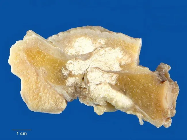
Imaging modality and technique: Gross pathology photograph of a dissected hand/phalangeal segment. The specimen shows chalky white tophi deposits (monosodium urate crystals) within the distal interphalangeal joint region and surrounding soft tissues, with visible superficial chronic inflammatory changes. The material appears friable and is characteristic of urate crystal aggregation within periarticular tissues, tendons, ligaments, and skin. The DIP joint surfaces show cartilage erosion and juxta-articular bone irregularity due to chronic inflammatory destruction. The tophi are large, with fibrous encapsulation and overlying skin thickening; in advanced disease tophi may ulcerate the overlying epidermis, discharging chalky material. The image demonstrates joint involvement commonly seen in chronic gout, though large tophaceous specimens are less common today due to modern therapy. Associated findings include synovial thickening and chronic fibrosis leading to restricted motion. The clinical history notes gouty arthritis of the great toe; the same monosodium urate deposition process may involve other joints, including fingers near the hand. This image serves educational and diagnostic documentation purposes to illustrate features of chronic tophaceous gout, including periarticular soft tissue tophi, articular cartilage erosion, juxta-articular bone destruction, and potential skin involvement. It is useful for pathology teaching, radiology correlation, and differential diagnosis discussions of gout vs CPPD.
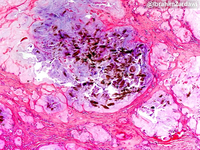
Imaging modality: Light microscopy of a hematoxylin and eosin (H&E) stained paraffin-embedded tissue section. Primary subject: Tissue with monosodium urate (MSU) crystal deposition consistent with gouty tophus formation. Specimen: Soft tissue biopsy with periarticular involvement; center shows brownish crystalline clusters representing surviving MSU crystals, while the surrounding tissue exhibits amorphous pink to amphophilic deposits after processing. Visual features: Focal granulomatous inflammatory milieu surrounds needle-shaped crystals that are dark brown in this preparation; crystals may appear refractile and needle-like within the central cluster. In paraffin-embedded material, MSU crystals are water-soluble and commonly dissolve during aqueous processing, leaving behind eosinophilic amorphous residues; however, at very high tissue concentrations, crystals may persist, as observed in the image center. Diagnostic significance: Histologic demonstration of urate deposition confirms local gout involvement and supports differentiation from other crystal arthropathies; persistence of crystals after processing indicates high tissue urate burden and can reflect delayed or inefficient processing. Clinical correlation: Correlate with patient history of hyperuricemia or gout, joint pain, tophi formation, and responsiveness to urate-lowering therapy. Potential use cases: educational illustration of gout pathology, research on crystal-induced inflammation, and aiding differential diagnosis in soft-tissue lesions.
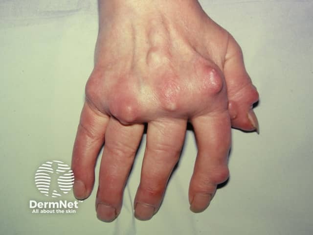
This clinical photograph depicts a dorsal view of the hand with visible gouty tophi over the metacarpophalangeal joints of several digits. The image documents soft tissue nodules characteristic of chronic tophaceous gout. The tophi are subcutaneous, firm masses projecting over the dorsal MCP region, with overlying skin that may appear erythematous or thinned. In advanced disease, chalky white crystalline material may be present within the nodules, reflecting monosodium urate crystal deposition. The distribution is periarticular to the finger joints and may be bilateral and progressive. Pathophysiologically, persistent hyperuricemia promotes MSU crystal deposition with a granulomatous inflammatory response, leading to nodular masses that can cause joint erosion and functional impairment. Clinically, these findings confirm chronic gout and signal high urate burden, risk of joint destruction, and possible renal involvement. This image is educational for rheumatology and dermatology, illustrating cutaneous tophi, aiding differential diagnosis from rheumatoid nodules or xanthomas, and underscoring the role of urate-lowering therapy, diet modification, and renal evaluation. Suitable for medical education, case documentation, and teaching deposition patterns of urate crystals.
Gout
Definition & Epidemiology
Pathogenesis
Uric Acid Metabolism
- De novo pathway - synthesis from non-purine precursors
- Salvage pathway - recycling of free purine bases from diet or DNA/RNA degradation
- Reduced excretion (most common, ~90% of primary gout) - uric acid is normally filtered by the glomerulus, nearly completely reabsorbed in the proximal tubule, and a small fraction secreted distally
- Overproduction - less common; can result from enzymatic defects or rapid cell turnover
Primary vs. Secondary Gout
| Type | Cause |
|---|---|
| Primary (90%) | Idiopathic reduced renal excretion |
| Secondary | Drugs (thiazides, cyclosporin), chronic renal disease, tumor lysis syndrome, Lesch-Nyhan syndrome (HGPRT deficiency) |
Mechanism of Crystal-Induced Inflammation
- MSU crystals precipitate in supersaturated synovial fluid
- Resident macrophages phagocytose crystals → activate the NLRP3 inflammasome (cytosolic sensor)
- Inflammasome activates caspase-1 → generates active IL-1β
- IL-1β recruits neutrophils into the joint
- Neutrophils release cytokines, free radicals, and proteases
- Crystals damage phagolysosomal membranes → release of lysosomal enzymes
- Result: acute arthritis, typically self-limited over days to weeks
Clinical Stages
1. Asymptomatic Hyperuricemia
2. Acute Intermittent Gout
- Onset: Sudden, severe pain - often waking the patient from sleep
- Classic site: First metatarsophalangeal (MTP) joint = podagra (50% of initial attacks)
- Other sites: Midfoot, ankle, knee, wrist, hand
- Features: Intense erythema, swelling, warmth, tenderness (even to light touch)
- Systemic: Fever may be present - raises concern for septic arthritis
- Up to 20% of patients have polyarticular involvement or associated bursitis/tenosynovitis
- Self-limited: Peak symptoms in 1-2 days, resolution within a week
- Patient is asymptomatic between attacks
3. Chronic Tophaceous Gout
- Develops when joints are no longer pain-free between attacks
- Tophi form: aggregates of MSU crystals + granulomatous inflammatory tissue in synovium, periarticular tissues, subcutaneous tissues, bursae
- Tophi appear as gritty, chalk-like nodules (generally painless)
- Cartilage erosion, bony destruction, and joint dysfunction develop
- Skin overlying tophi may ulcerate, discharging chalky material
Clinical Images
Diagnosis
Definitive: Arthrocentesis (Joint Aspiration)
- Recommended for all first episodes; may be omitted in established gout with low septic arthritis risk
- Synovial fluid shows negatively birefringent, needle-shaped MSU crystals under polarizing microscopy (appear yellow when parallel to the slow vibration axis, blue when perpendicular)
- WBC often markedly elevated (>20,000/μL in acute attack)
Serum Uric Acid
- Largely unhelpful acutely - gouty attacks can occur with normal serum uric acid (may fall during acute phase)
- Elevated uric acid alone is nonspecific (19% of people have asymptomatic hyperuricemia)
- Still useful for monitoring ULT
Renal Function
- Important because gout is associated with renal insufficiency and many treatments are nephrotoxic
Imaging
| Modality | Finding |
|---|---|
| Plain X-ray | Acute: soft-tissue swelling only. Chronic: asymmetric, "punched-out" sclerotic erosions outside the joint capsule (overhanging edge sign) |
| Ultrasound | Double contour sign: hyperechoic line along cartilage surface (crystals) + hyperechoic bony surface. Tophi appear as "wet clumps of sugar" - heterogeneous center with hypoechoic rim |
| DECT (dual-energy CT) | Can color-code urate deposits; useful in difficult cases |
| MRI | Tophi appear as intermediate-to-low signal masses on T1; can demonstrate bone erosion |
- Grainger & Allison's Diagnostic Radiology, p. 1125
Management
Acute Attack
| Drug | Dose/Notes |
|---|---|
| NSAIDs (first-line) | Indomethacin, naproxen, or ibuprofen. Start promptly; continue 24h after symptom resolution. Avoid in peptic ulcer disease, GI bleeding, renal insufficiency |
| Colchicine | Inhibits microtubule formation → inhibits crystal-induced inflammation. Low-dose preferred (1.2 mg then 0.6 mg 1h later). Contraindicated in renal/hepatic insufficiency; narrow therapeutic window; GI side effects common |
| Corticosteroids | Prednisone 40 mg/day x 5-7 days (oral), or intra-articular injection (most effective; avoid if septic arthritis cannot be excluded). Intra-articular preferred for monoarticular disease |
| Combination therapy | Intra-articular steroid + colchicine or NSAID for severe/debilitating cases |
- Do NOT start ULT during an acute attack; do continue pre-existing ULT
- Do NOT combine oral corticosteroids with NSAIDs (GI risk)
Long-term Urate-Lowering Therapy (ULT)
| Drug | Class | Notes |
|---|---|---|
| Allopurinol | Xanthine oxidase inhibitor (purine analog) | First-line. Start low (100-300 mg/day), uptitrate. Reduce dose in renal impairment. Risk of DRESS/SJS (HLA-B*5801 screening in high-risk populations). Caution with azathioprine |
| Febuxostat | Xanthine oxidase inhibitor (non-purine) | For allopurinol-intolerant patients. 40-80 mg/day; no dose adjustment for renal impairment. Higher CV/all-cause mortality than allopurinol in high-CV-risk patients (CARES trial) - use with caution |
| Probenecid | Uricosuric | Increases renal urate excretion. Avoid in uric acid stones or low eGFR |
| Pegloticase | Recombinant pegylated uricase | Converts uric acid to allantoin (more soluble). Reserved for refractory tophaceous gout. FDA-approved 2010. Risk of antibody formation → infusion reactions and loss of efficacy |
Prophylaxis During ULT Initiation
- Starting ULT can precipitate acute flares (mobilization of urate stores)
- Give low-dose colchicine (0.5-0.6 mg/day) or low-dose NSAID for 3-6 months when initiating ULT
Lifestyle & Dietary Measures
- Limit purine-rich foods: red meat, organ meats, shellfish, anchovies
- Reduce alcohol (especially beer)
- Avoid fructose-rich beverages
- Stay well hydrated
- Achieve healthy weight (bariatric surgery reduces urate in obese patients per recent systematic review, PMID 39520613)
Complications
- Urate nephropathy / nephrolithiasis (uric acid kidney stones)
- Chronic arthritis with joint destruction
- Tophi with skin ulceration
- Cardiovascular disease association (independent risk factor)
- Hearing loss - gout and hyperuricemia have been associated with ear function impairment (systematic review, PMID 38736350)
Key Differentials
| Condition | Key Distinguishing Features |
|---|---|
| Septic arthritis | Gram stain/culture positive; fever more prominent; no crystals |
| Pseudogout (CPPD) | Positively birefringent, rhomboid/rod-shaped calcium pyrophosphate crystals; typically knee/wrist; chondrocalcinosis on X-ray |
| Cellulitis | No joint fluid crystals; skin flora; responds to antibiotics |
| Reactive arthritis | Preceding infection; asymmetric oligoarthritis; HLA-B27 association |