

answer
cervical rib X-ray radiograph
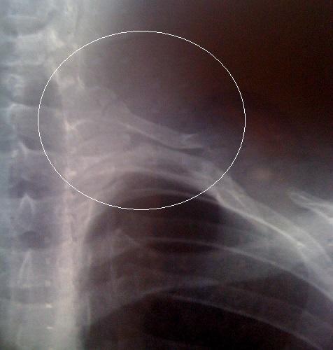
This diagnostic image is an anteroposterior (AP) chest X-ray radiography focusing on the superior thoracic aperture and cervical-thoracic junction. The radiograph reveals the presence of a cervical rib (supernumerary rib), highlighted within a white circular annotation. The cervical rib originates from the seventh cervical vertebra (C7) and appears as a curved, radiopaque bony structure extending laterally and inferiorly toward the first rib. The surrounding anatomy includes the cervical and upper thoracic spine, multiple standard ribs, and the clavicle. The visual evidence illustrates a congenital anatomical variation that is clinically significant as a potential cause of thoracic outlet syndrome (TOS) due to compression of neurovascular structures. The image is intended for educational use in radiology and musculoskeletal anatomy to help learners identify skeletal anomalies and understand their relationship to adjacent neurovascular pathways.
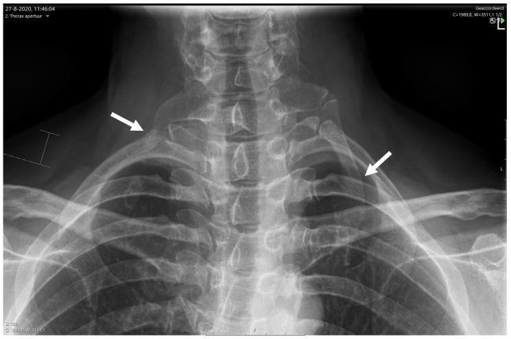
Anteroposterior (AP) X-ray of the lower cervical spine and thoracic inlet, demonstrating bilateral cervical ribs. A white arrow on the right side indicates a short, incomplete cervical rib arising from the C7 vertebra. A corresponding white arrow on the left side highlights a larger, complete cervical rib. Both accessory ribs show distinct articulation with the superior aspect of the first rib. The radiograph clearly visualizes the anatomical relationship between the cervical vertebrae, the thoracic aperture, and the clavicles. This finding is of clinical significance in the evaluation of Neurogenic Thoracic Outlet Syndrome (NTOS) and Gilliatt-Sumner hand, as these congenital anomalies can lead to compression of the lower trunk of the brachial plexus or subclavian vessels within the scalene triangle. The image provides an essential diagnostic baseline for multidisciplinary care pathways in orthopedic and neurological assessment.
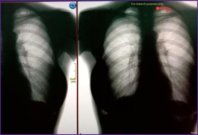
This diagnostic image displays a side-by-side comparison of a posterior-anterior (PA) chest X-ray, illustrating the detection of a congenital cervical rib. The left panel shows the original radiograph, while the right panel highlights the pathology using red artificial intelligence (AI) annotation. Anatomical landmarks include the thoracic spine, rib cage, bilateral clavicles, and radiolucent lung fields. In the right-side image, a small, accessory bony structure (cervical rib) is visible originating from the transverse process of the seventh cervical vertebra (C7), superior to the first thoracic rib. This finding is clinically significant as it may be associated with thoracic outlet syndrome due to potential compression of the brachial plexus or subclavian vessels. The visual demonstrates the application of deep learning algorithms in identifying musculoskeletal anomalies and incidental pulmonary findings during routine fluorographic or radiographic screenings.
X-ray Diagnosis: Cervical Rib
1. Clinical Condition Shown (1 mark)
2. Two Occupations Commonly Associated (1 mark)
- Tailors / seamstresses (prolonged arm elevation and repetitive upper limb use)
- Porters / labourers (carrying heavy loads on the shoulder compresses neurovascular structures at the thoracic outlet)
3. Classical Characteristic Clinical Sign (1 mark)
- The patient takes a deep breath, extends the neck, and rotates the chin toward the affected side.
- The radial pulse becomes feeble or disappears on the affected side.
- This is positive due to compression of the subclavian artery over the cervical rib.