
About the white matter of cerebrum
I'll read the medical library skill first, then search for cerebrum white matter content.
Reading File
Now let me search the library for white matter of cerebrum content in parallel:
Finding Sources
Finding Sources
Good. Now let me search specifically for anatomy textbook content on cerebral white matter fiber types:
Finding Sources
Finding Sources
Good finds. Let me read the most relevant sources - the Kaplan & Sadock "White Matter Tracts" section and the Neuroanatomy book, plus search for anatomy-specific content:
Reading File
Reading File
Finding Sources
Excellent content found. Let me read more from the Kaplan & Sadock white matter tracts section and also get the Kaplan white matter + cerebral connectivity section:
Reading File
Reading File
Excellent - very rich content. Let me now fetch the key diagrams and also get the internal capsule image:
Loading Image
Loading Image
Loading Image
Excellent - great images acquired. Let me also fetch the association fibers diagram and get remaining text:
Reading File
Loading Image
I now have comprehensive content from multiple authoritative textbooks and excellent diagrams. Let me compile the full answer.
White Matter of the Cerebrum
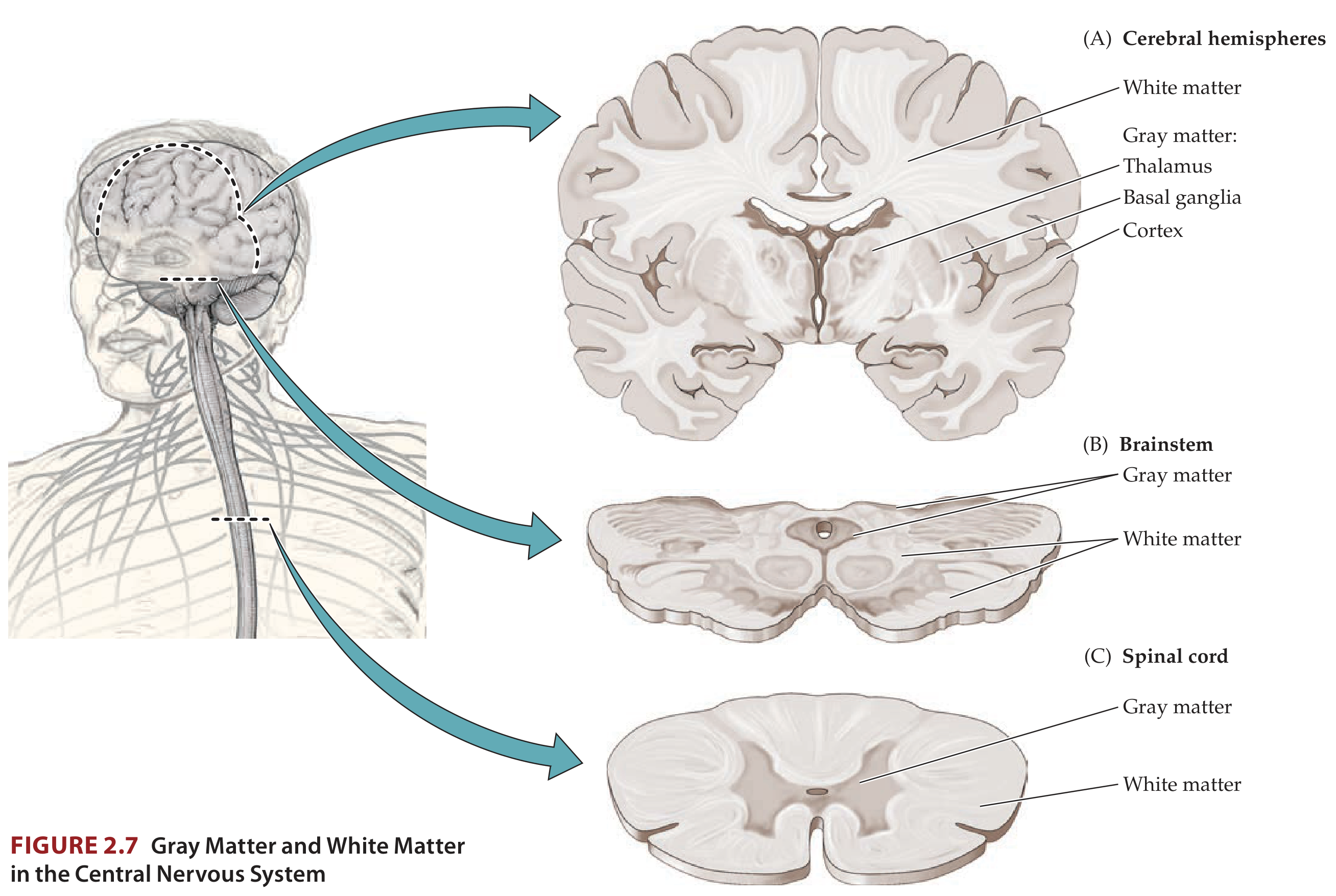
The white matter of the cerebrum lies deep to the cortical gray matter and consists almost entirely of myelinated axons - hence its characteristic white color. It occupies approximately 42% of the volume of the cerebral hemispheres in humans. The great majority of these fibers serve cortico-cortical connectivity rather than projections between cortex and subcortical sites (for example, thalamic input accounts for only about 5% of the total input into primary sensory cortex, the rest coming from other cortical areas).
Distribution in the CNS: In the cerebral hemispheres, gray matter cortex is on the outside while white matter is on the inside - the opposite arrangement to the spinal cord, where white matter is peripheral and gray matter is central.
Types of Fibers
Cerebral white matter fibers are grouped into three major categories:
1. Projection Fibers
These connect the cerebral cortex to subcortical structures and vice versa:
- Corticofugal fibers originate in the cortex and project downward to subcortical targets (e.g., corticothalamic, corticopontine, corticospinal, corticobulbar tracts).
- Corticopetal fibers originate outside the telencephalon and project up to the cortex (e.g., thalamocortical fibers).
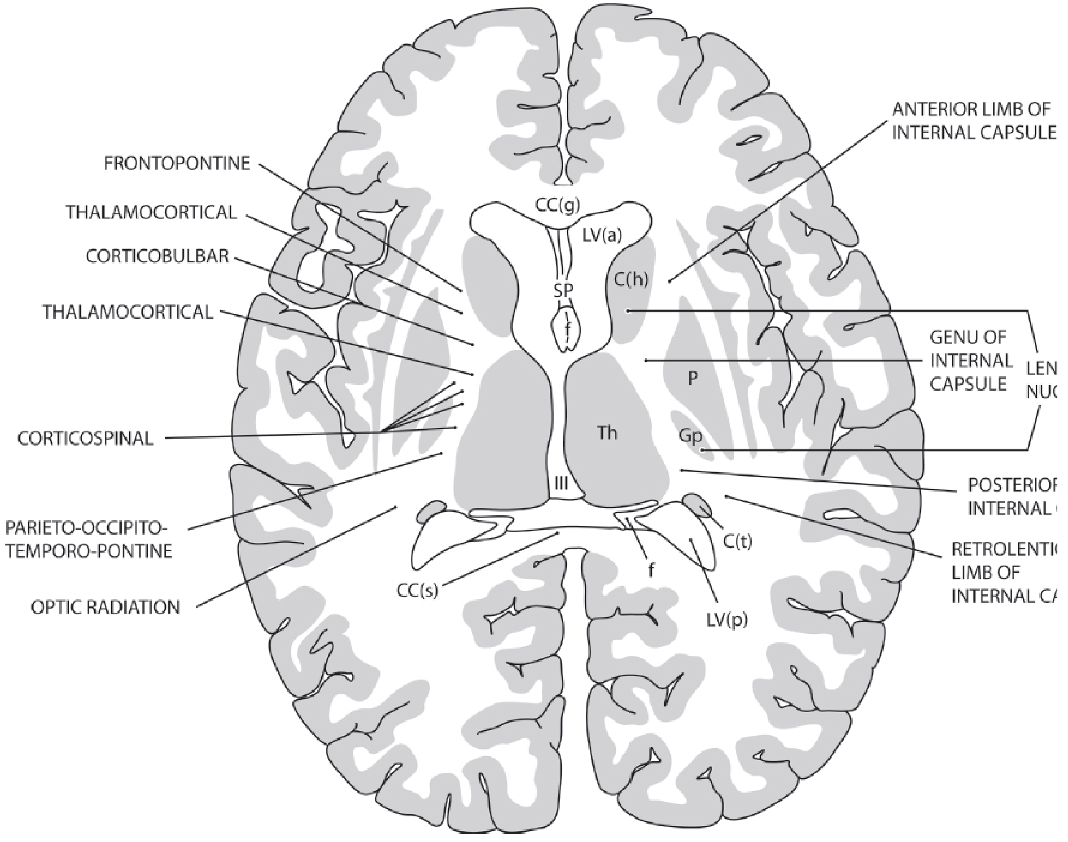
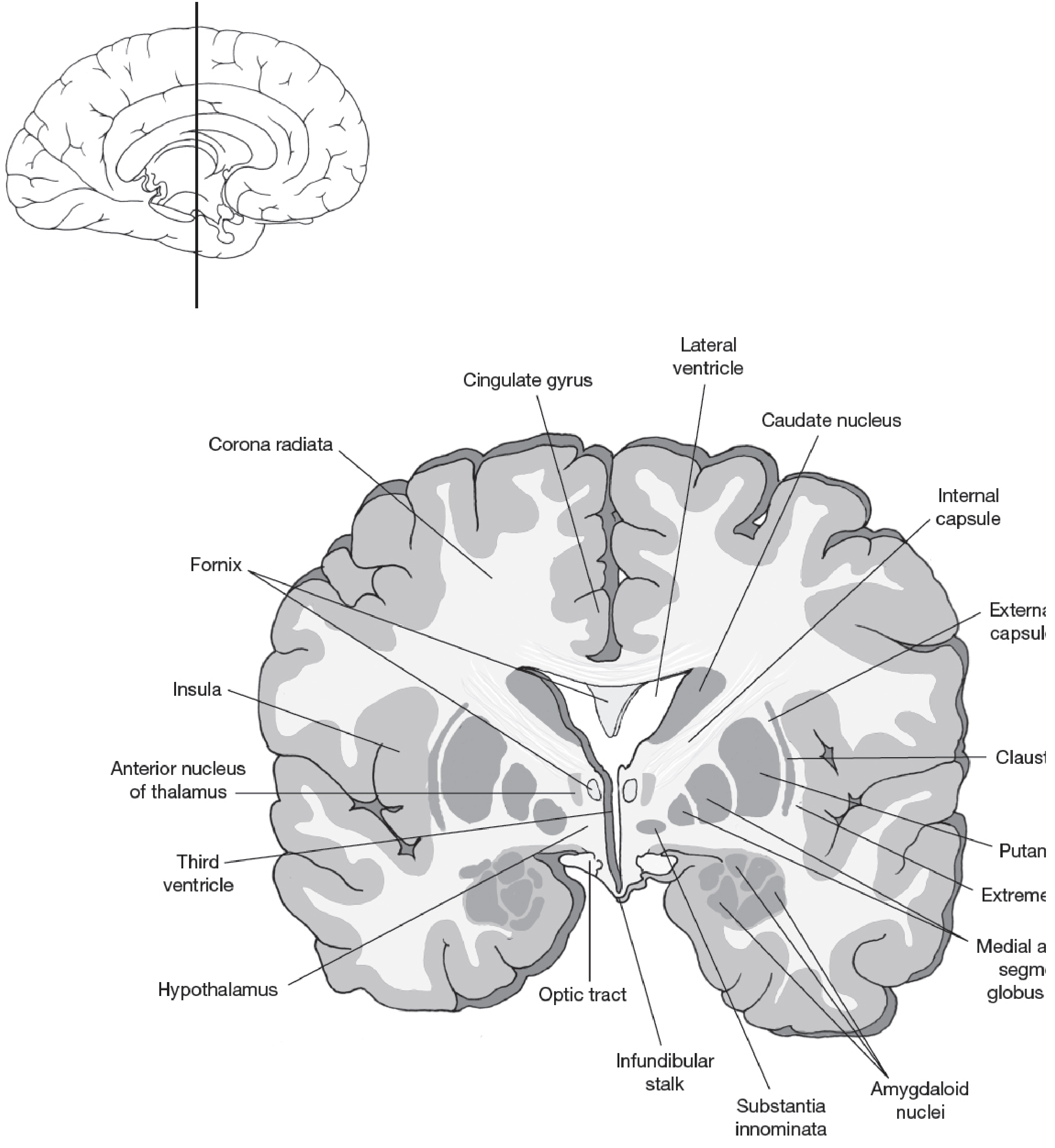
All projection fibers traveling between cortex and subcortical structures converge into the internal capsule and then fan out above it as the corona radiata.
Internal Capsule (5 parts)
| Part | Location | Contents |
|---|---|---|
| Anterior limb | Between lenticular nucleus and caudate head | Frontopontine fibers; thalamofrontal fibers |
| Genu | Intersection of anterior + posterior limbs | Corticobulbar fibers |
| Posterior limb | Between lenticular nucleus and thalamus | Corticospinal fibers |
| Retrolenticular limb | Behind the lenticular nucleus | Optic radiation (lateral geniculate → primary visual cortex) |
| Sublenticular limb | Below the lenticular nucleus | Auditory radiation (medial geniculate → primary auditory cortex) |
2. Commissural Fibers
These interconnect corresponding areas in the two cerebral hemispheres.
Corpus Callosum
The largest fiber bundle in the brain, containing roughly 300 million axons. It connects homotopic (same-position) cortical regions across hemispheres, though heterotopic connections also exist. Almost all cortical regions connect via the corpus callosum, with exceptions: the hand area of motor/somatosensory cortex and the primary visual cortex.
It has four named parts:
- Rostrum - most anterior, tapers inferiorly
- Genu - curves anteriorly and dorsally; carries higher-order cognitive/sensory information from prefrontal, temporal, and parietal cortices
- Body - largest part; carries visual, auditory, and somatosensory information
- Splenium - enlarged rounded posterior end; also carries prefrontal/parietal higher-order information
Anterior Commissure
A compact bundle caudal to the corpus callosum, crossing the midline in front of the fornix. It interconnects the two temporal lobes and fibers from the anterior olfactory nucleus.
Smaller Commissures
- Posterior commissure - connects caudal portions of the diencephalon
- Hippocampal commissure - interconnects the two hippocampal formations
3. Association Fibers
These connect cortical areas within the same hemisphere and are subdivided by length:
Short Association Fibers (U-fibers)
Connect adjacent gyri by looping under the bottom of a sulcus, forming a characteristic "U" shape. These are notably spared in diseases like Binswanger disease and CADASIL.
Long Association Fiber Tracts
| Tract | Connections | Notes |
|---|---|---|
| Superior longitudinal fasciculus | Frontal, parietal, and occipital cortices (lateral hemisphere, above insula) | Main dorsal intrahemispheric highway |
| Arcuate fasciculus | Superior/middle frontal gyri ↔ temporal lobe (and parietal/occipital via its superior portion) | Key for language connectivity |
| Uncinate fasciculus | Orbitofrontal cortex ↔ anterior temporal cortex | Curves around the lateral sulcus |
| Inferior occipitofrontal fasciculus | Occipital lobe ↔ frontal lobe | Courses ventrally and laterally |
| Inferior longitudinal fasciculus | Temporal lobe ↔ occipital lobe | |
| Cingulum | Frontal and parietal lobes ↔ parahippocampal gyrus | Lies in white matter under cingulate gyrus |
Other association bundles include the external capsule (between claustrum and putamen) and the extreme capsule (between claustrum and insular cortex).
Summary Classification (Five-Type Framework)
A useful clinical framework groups white matter fibers into five types:
- Long intrahemispheric tracts - arcuate, uncinate, cingulum, etc. (described above)
- Long projection systems - corona radiata, internal capsule, medial forebrain bundle, thalamic peduncle, corticospinal/corticopontine tracts
- U-fibers - short juxtacortical fibers (spared in many leukoencephalopathies)
- Specific projection pathways - mammillothalamic tract, fornix (interruption → amnesia)
- Commissural pathways - corpus callosum, anterior/posterior commissures, massa intermedia
Clinical Relevance
- Stroke/internal capsule lesions: A small infarct in the posterior limb of the internal capsule can produce a dense contralateral hemiplegia because of the compact concentration of corticospinal fibers there.
- Binswanger disease / CADASIL: Hypertensive small vessel disease destroys deep white matter and long corticocortical fibers while typically sparing U-fibers, producing subcortical dementia with slow processing and impaired executive function.
- Multiple sclerosis: Demyelination of white matter tracts produces varied deficits depending on which tracts are involved.
- Schizophrenia: DTI studies show abnormalities in virtually every major white matter tract - corpus callosum, internal capsule, cingulum, uncinate fasciculus, and fornix.
- Alzheimer disease / Autism: Reduced cross-sectional area of the corpus callosum is reported in both conditions.
- Corpus callosotomy: Surgical severing of the corpus callosum to treat refractory epilepsy produces the "split-brain" syndrome, demonstrating the role of commissural fibers in hemispheric integration.
Sources: Kaplan & Sadock's Comprehensive Textbook of Psychiatry (White Matter Tracts, p. 197-203; White Matter and Cerebral Connectivity, p. 1436-1438); Neuroanatomy through Clinical Cases, 3rd Ed. (p. 46-47)
This is a shared conversation. Sign in to Orris to start your own chat.