
ventricular tachycardia ECG wide complex tachycardia
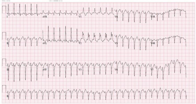
A 12-lead electrocardiogram (ECG) demonstrating a regular wide complex tachycardia (WCT). The rhythm is characterized by a rapid ventricular rate and broad QRS complexes, indicating delayed ventricular depolarization. Key morphological findings include a Right Bundle Branch Block (RBBB) pattern, notably visible as an RSR' or notched morphology in the right precordial leads (V1, V2). There is significant left axis deviation (LAD), with predominantly negative QRS complexes in the inferior leads (II, III, and aVF) and positive complexes in leads I and aVL. This combination of RBBB morphology and left axis deviation in the context of wide complex tachycardia is a classic presentation of idiopathic left ventricular tachycardia, also known as Belhassen tachycardia or verapamil-sensitive fascicular ventricular tachycardia. The rhythm appears monomorphic, and the baseline shows no clearly identifiable P waves, suggesting atrioventricular dissociation or retrograde conduction common in ventricular arrhythmias.
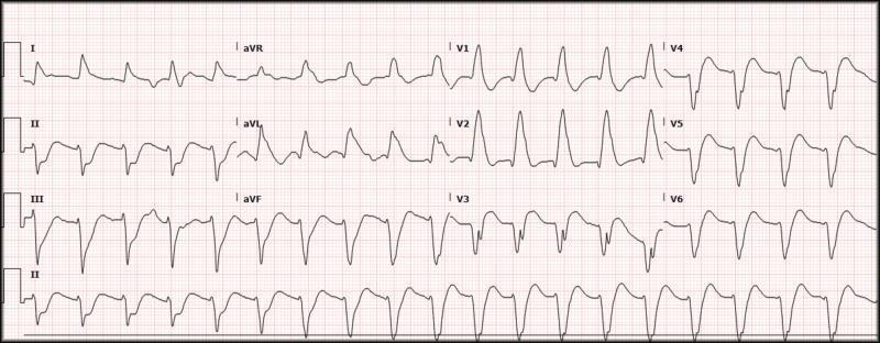
This diagnostic image is a 12-lead electrocardiogram (ECG) demonstrating a wide-complex tachycardia with a ventricular rate of approximately 116 beats per minute and a QRS duration of approximately 200 ms. The ECG shows a regular rhythm with significantly widened QRS complexes and bizarre morphology, suggestive of a ventricular origin or severe intraventricular conduction delay. In the precordial leads, V1 and V2 exhibit tall, peaked R-waves with slurred upstrokes, while V4 through V6 show deep S-waves and discordant T-waves. The limb leads (I, II, III, aVR, aVL, aVF) demonstrate varied morphology with notching and slurring. No clear P-waves are discernible preceding the QRS complexes, which is characteristic of ventricular tachycardia or a highly aberrant supraventricular rhythm. The clinical focus of this visual is to illustrate the features of wide-complex tachycardia in an emergency clinical setting, emphasizing the importance of recognizing prolonged QRS intervals and atypical wave morphology for rapid triage and Advanced Cardiovascular Life Support (ACLS) intervention.
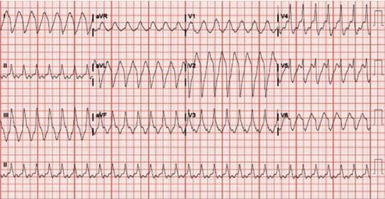
A 12-lead electrocardiogram (ECG) demonstrating a regular, wide-complex tachycardia. The ventricular rate is approximately 170-174 beats per minute with a significantly prolonged QRS duration of approximately 270 ms. The rhythm is monomorphic, showing consistent QRS morphology across all leads. Distinct P waves are not clearly visible, appearing to be obscured by the wide QRS complexes or the subsequent T waves. The tracing shows a classic 'sine-wave' or slurred appearance in several leads, particularly the precordial leads V2-V4, which is characteristic of severe QRS widening. The clinical context suggests a differential diagnosis including ventricular tachycardia, supraventricular tachycardia with aberrancy (bundle branch block), or toxicological effects causing slowed intraventricular conduction, such as flecainide toxicity. The image serves as a key educational example for identifying wide-complex tachycardias and assessing QRS morphology and intervals in an acute cardiology or emergency medicine setting.
ECG Interpretation
- Machine: GE MAC 600-1.02
- Date: 25-Jul-2026, 22:36:45
- Paper speed: 25.0 mm/s | Gain: 10.0 mm/mV | Filter: 0.16-20 Hz / 50 Hz
- Heart rate: 118 bpm (printed on ECG)
- Patient: ~41 years old
Systematic Analysis
| Parameter | Finding |
|---|---|
| Rate | 118 bpm (tachycardic) |
| Rhythm | Regular |
| P waves | Not clearly identifiable / dissociated from QRS complexes |
| QRS width | Wide (>120 ms) - broad, bizarre morphology |
| QRS morphology | Large amplitude, markedly widened complexes; dominant negative QRS in V1 (LBBB-like pattern); tall broad monophasic R in V5-V6 |
| Axis | Likely left-axis deviation based on lead morphology |
| ST segments | Discordant - opposite to QRS direction (as expected in wide-complex tachycardia) |
| T waves | Discordant (opposite to QRS) |
| AV dissociation | Possible - P waves not preceding QRS |
Primary Interpretation
- Rate 118 bpm - within the VT range (typically 120-300 bpm; amiodarone-slowed VT can present at 110-130 bpm per [Tintinalli's Emergency Medicine, p. 156])
- Wide, bizarre QRS complexes (>120 ms) with consistent beat-to-beat morphology - hallmark of monomorphic VT
- No clearly identifiable P waves preceding QRS complexes - AV dissociation is the most reliable criterion for VT
- LBBB-like morphology in precordial leads (dominant negative in V1, broad R in V5-V6) - suggests right ventricular origin or septal VT
- Discordant ST-T changes - T waves point opposite to the main QRS deflection
Differential Diagnosis
| Diagnosis | Features favoring | Features against |
|---|---|---|
| Monomorphic VT (most likely) | Wide QRS, regular rhythm, no clear P waves, rate ~118 | Rate slightly slower than classic VT |
| SVT with aberrancy (BBB) | Regular rhythm | No preceding P waves identifiable |
| Antidromic AVRT (WPW) | Wide QRS, regular | Pre-excitation delta waves not classic here |
| Accelerated idioventricular rhythm | Rate ~118 | Rate 118 is borderline - AIVR usually 60-100 bpm |
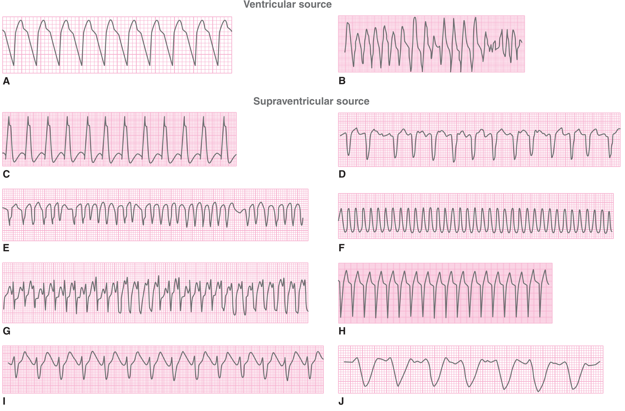
Reference ECG Comparison
Clinical Urgency & Next Steps
- Synchronized DC cardioversion immediately (ACLS protocol)
- 12-lead ECG confirmed
- IV access, continuous monitoring
- Amiodarone 150 mg IV over 10 min, then 1 mg/min infusion (first-line for hemodynamically stable VT)
- Lidocaine 1-1.5 mg/kg IV as alternative
- Do NOT give verapamil or adenosine empirically in wide-complex tachycardia of unknown origin (may precipitate hemodynamic collapse in VT)
- Urgent cardiology consultation
- Identify and treat reversible causes: electrolyte abnormalities (K+, Mg2+), ischemia, drug toxicity, hypoxia
Disclaimer: This interpretation is based on visual analysis of the uploaded ECG image. Clinical correlation, formal reporting by a cardiologist, and assessment of the patient's hemodynamic status are essential before any treatment decisions are made.
Patient has biliary sepsis
Revised ECG Interpretation in the Context of Biliary Sepsis
What Biliary Sepsis Does to the ECG
| Sepsis Complication | ECG Effect |
|---|---|
| Tachycardia (fever, high-output state) | Sinus rate 100-130+ bpm |
| Hyperkalemia (tissue breakdown, acidosis, AKI) | Peaked T waves → wide QRS → P-wave loss → sine wave |
| Metabolic acidosis | Worsens QRS widening; simulates BBB |
| Sepsis-induced cardiomyopathy | LV dysfunction, LBBB-like pattern |
| Electrolyte depletion (Mg2+, K+) | Arrhythmia risk |
Revised Primary Interpretation
- Rate of exactly 118 bpm (a sinus-range tachycardia, not the typical VT rate of 140-180 bpm)
- Biliary sepsis as the clinical background
- Wide, bizarre QRS complexes
- Potential loss or burial of P waves within the preceding T waves (common when HR >100 and PR/QT intervals shift)
Revised Differential Diagnosis (Ranked by Probability in This Context)
| Rank | Diagnosis | Why |
|---|---|---|
| 1 | Sinus tachycardia + metabolic wide QRS (hyperkalemia / acidosis) | Sepsis mechanism; rate 118 fits; wide QRS explained metabolically |
| 2 | Sinus tachycardia + new LBBB (sepsis cardiomyopathy) | LBBB can develop acutely in severe sepsis |
| 3 | Monomorphic VT | Still possible - sepsis is a trigger for VT |
| 4 | SVT with aberrancy | Less likely given no prior BBB history implied |
Immediate Critical Investigations
- Serum electrolytes urgently - K+, Na+, Mg2+, Ca2+
- Hyperkalemia K+ >6.5 mmol/L → immediate calcium gluconate/chloride
- Arterial blood gas - pH, HCO3-, lactate
- Blood cultures x2 (if not already done)
- Renal function (AKI common in biliary sepsis - worsens K+ handling)
- LFTs, bilirubin (confirm biliary obstruction severity)
- Troponin (sepsis-induced myocardial injury)
- Bedside echocardiography - assess LV function, pericardial effusion
Treatment Priorities
For the Sepsis (Source Control is Paramount)
- IV broad-spectrum antibiotics immediately (e.g. piperacillin-tazobactam + metronidazole, or carbapenem if severe)
- Aggressive IV fluid resuscitation (cautiously - monitor for pulmonary edema)
- ERCP with biliary drainage within 24 hours (urgent if Reynolds' pentad present: Charcot's triad + shock + confusion)
- ICU-level monitoring
For the ECG Abnormality
- Do NOT give antiarrhythmics empirically until electrolytes are known
- If hyperkalemia confirmed: Calcium gluconate 10% (10 mL IV) immediately → insulin/dextrose → sodium bicarbonate (if acidotic) → consider dialysis if severe AKI
- If K+ and acid-base are normal and the rhythm persists → reconsider primary VT → amiodarone
- Repeat ECG after electrolyte correction - if wide QRS normalizes, the diagnosis is confirmed metabolic
Critical Caution
If this is hyperkalemic wide-QRS tachycardia, giving amiodarone, lidocaine, or other antiarrhythmics is dangerous and will not fix the problem. The treatment is treating the metabolic cause - calcium, bicarbonate, insulin/dextrose. Only after ruling out metabolic causes should antiarrhythmics be considered.