
Carpel Tunnel and it's applied
carpal tunnel syndrome anatomy median nerve
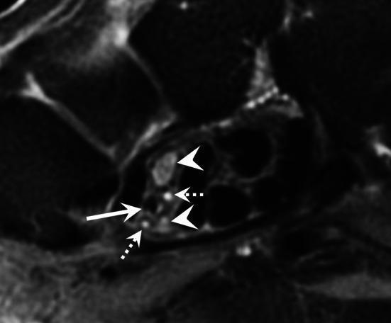
This diagnostic axial T2-weighted MRI scan depicts the cross-sectional anatomy of the carpal tunnel, focusing on anatomical variations and vascular pathology. The image highlights a bifid median nerve, represented by two distinct nerve trunks (indicated by arrowheads) instead of a single nerve bundle. Positioned between or adjacent to these nerve trunks is a persistent median artery (indicated by a straight solid arrow) which is occluded. The occlusion is evidenced by the absence of a normal vascular flow void and altered intraluminal signal intensity on this T2-weighted sequence. Additionally, small satellite veins of the median artery are visible as tiny, hyperintense circular structures (indicated by dotted arrows). This clinical finding is significant in the context of carpal tunnel syndrome, as both the bifid nerve variation and the presence of a persistent median artery (especially when thrombosed or aneurysmal) can increase the volume of contents within the carpal tunnel, potentially leading to median nerve compression.
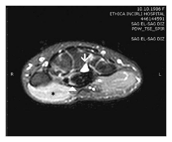
This diagnostic image is a fat-suppressed proton density (PD) axial MRI of the wrist, highlighting the carpal tunnel anatomy and associated pathology. The image demonstrates a high-signal intensity space-occupying lesion (marked by a white arrow) located within the carpal tunnel, consistent with a cystic mass such as a ganglion. The median nerve (indicated by a white arrowhead) shows increased signal intensity and morphological flattening, suggestive of compressive neuropathy or carpal tunnel syndrome. Additionally, there is a prominent area of high signal intensity within the thenar muscles (marked by an asterisk), which is visually distinct from the lower signal of adjacent musculature. This feature is a classic radiological sign of acute muscle denervation resulting from median nerve compromise. The image serves as an educational example of secondary carpal tunnel syndrome, illustrating the relationship between an anatomical obstruction, nerve inflammation, and the subsequent neuromuscular impact on the thenar eminence.
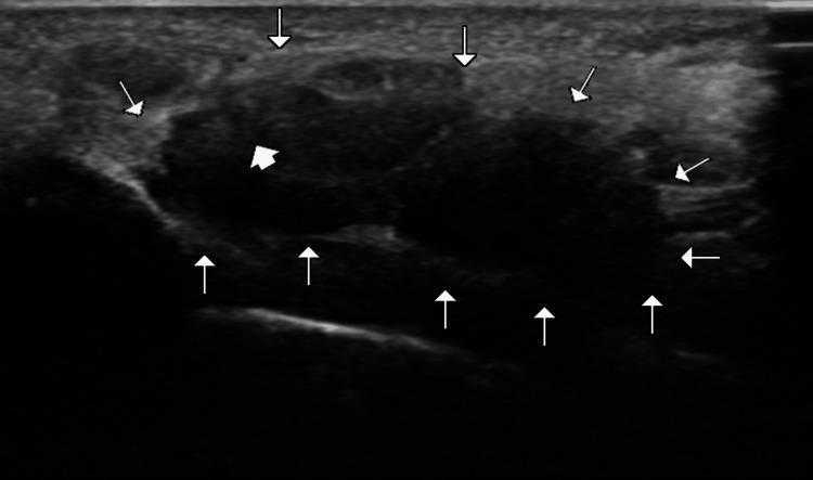
This diagnostic image is a transverse ultrasound of the wrist, focusing on the carpal tunnel. The image demonstrates the cross-sectional anatomy of the carpal tunnel, with its boundaries delineated by multiple white arrowheads. These arrowheads highlight the arch-shaped space formed by the deep carpal bones and the superficial, echogenic flexor retinaculum. Within the tunnel, a thick white arrow points to the median nerve, which presents as a relatively hypoechoic, oval structure with a fascicular internal texture. Surrounding the nerve are the flexor tendons, visible as more heterogeneous and slightly more echogenic structures within the tunnel space. This imaging modality and view are used in clinical practice for diagnosing carpal tunnel syndrome, assessing median nerve cross-sectional area, and identifying space-occupying lesions. The focus is on neuromuscular and musculoskeletal diagnostic features relevant to radiology and physical medicine.
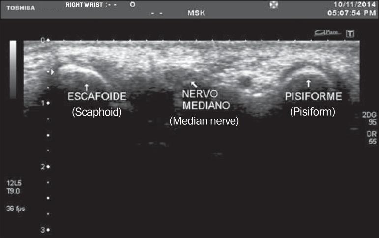
This diagnostic image is a transverse musculoskeletal (MSK) ultrasound of the right wrist at the level of the carpal tunnel inlet. The image captures the median nerve (labeled 'Nervo Mediano') positioned centrally between two bony landmarks: the scaphoid (labeled 'Escafoide') on the radial side and the pisiform (labeled 'Pisiforme') on the ulnar side. The bones appear as hyperechoic, curved surfaces with posterior acoustic shadowing. The median nerve is visualized as an oval, relatively hypoechoic structure with a speckled internal texture, characteristic of a honeycomb appearance formed by nerve fascicles. This imaging modality and view are clinically standard for evaluating the median nerve cross-sectional area (MNA) to diagnose Carpal Tunnel Syndrome (CTS). The anatomical relationship between the nerve and the bony floor of the carpal tunnel is clearly demonstrated for medical educational purposes regarding wrist anatomy and diagnostic sonography.
"carpal tunnel syndrome"[MeSH Terms] AND treatment
Carpal Tunnel Syndrome (CTS) - Complete Clinical Review
1. Anatomy of the Carpal Tunnel
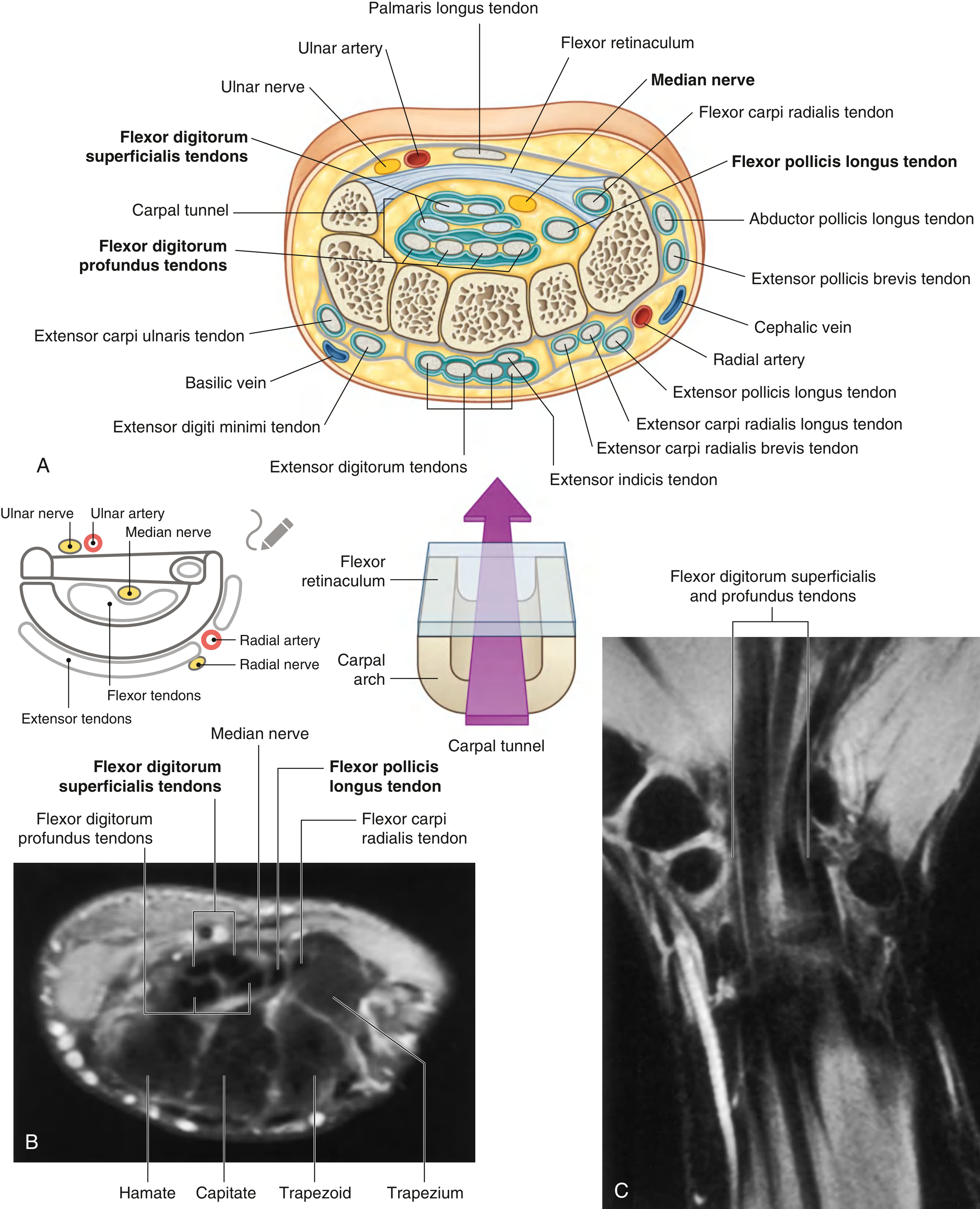
- Floor and walls: The concave arch of the 8 carpal bones (scaphoid and trapezium on the radial side; pisiform and hook of hamate on the ulnar side)
- Roof: The flexor retinaculum (transverse carpal ligament)
| Structure | Count |
|---|---|
| Median nerve | 1 |
| Flexor digitorum superficialis tendons | 4 |
| Flexor digitorum profundus tendons | 4 |
| Flexor pollicis longus tendon | 1 |
Note: The ulnar nerve and ulnar artery do NOT pass through the carpal tunnel - they travel through Guyon's canal medially. The flexor carpi radialis has its own separate compartment.
2. Definition and Epidemiology
- Sex: Female predominance (~3:1)
- Age: ~50% of patients are 40-60 years
- Prevalence: Most common compression neuropathy overall
- Occasionally diagnosed in children (rare)
3. Etiology and Risk Factors
Primary (Idiopathic)
Secondary Causes - Conditions that increase tunnel pressure or nerve vulnerability:
| Category | Conditions |
|---|---|
| Systemic/Metabolic | Diabetes mellitus, hypothyroidism, acromegaly, gout, multiple myeloma, amyloidosis |
| Inflammatory | Rheumatoid arthritis, other inflammatory arthritis |
| Structural | Osteoarthritis, synovial cysts, ganglion cysts, carpal bone fractures/malunion |
| Physiological | Pregnancy (usually resolves after delivery), obesity |
| Occupational | Repetitive wrist motion (e.g., typing, assembly work) |
| Infective/Other | Tuberculosis, congestive heart failure |
4. Clinical Features
Symptoms
- Nocturnal paresthesia - classic and often the presenting complaint; patients are woken from sleep
- Paresthesia/numbness in thumb, index, middle fingers, and radial half of ring finger (median nerve distribution)
- Symptoms relieved by "flick sign" - shaking or dangling the hand
- Forearm and elbow pain - poorly localized, aching, aggravated by activity
- Occasional shoulder or proximal arm pain (brachialgia)
- Weakness and clumsiness of the hand (late)
Signs
| Sign | Description | Notes |
|---|---|---|
| Tinel's sign | Percussion over volar wrist → radiating paresthesia in median distribution | Indicates nerve irritation |
| Phalen's test | Wrist held in complete flexion for 1-2 min → reproduction of symptoms | Sensitivity ~74%, false-positive ~25% |
| Carpal compression test | Direct pressure over carpal tunnel → symptoms | More specific than Tinel's |
| Thenar atrophy | Wasting of abductor pollicis brevis, opponens pollicis | Late sign - advanced disease |
| Decreased sensation | Objective loss in distal thumb, index, middle fingers | Late sign |
5. Investigations
1. Nerve Conduction Studies (NCS) - Gold Standard
- Prolonged distal motor latency across the carpal canal
- Prolonged distal sensory latency - often the earliest finding
- Bilateral studies are recommended, especially for occupational claims or atypical presentations
- In patients with classic clinical findings, diagnosis can be made on clinical grounds alone without NCS
2. EMG
- Polyphasic reinnervation potentials in the abductor pollicis brevis
- Used to assess severity and rule out concurrent cervical radiculopathy or thoracic outlet syndrome
3. Imaging
- Ultrasound: Demonstrates increased cross-sectional area of the median nerve at the carpal tunnel inlet, nerve flattening, and identifies space-occupying lesions
- MRI: Shows nerve signal change, thenar denervation edema (high signal on T2), and structural causes (ganglion, bifid nerve with persistent median artery)
4. Screening Blood Tests
- Fasting blood glucose / HbA1c (diabetes)
- TSH (hypothyroidism)
- Inflammatory markers, RF (rheumatoid arthritis)
- Calcium, protein electrophoresis (myeloma, amyloid)
6. Differential Diagnosis
- Cervical radiculopathy (C6, C7) - check if pain/numbness extends to arm/neck, check reflexes
- Thoracic outlet syndrome - positional symptoms, vascular signs
- Pronator teres syndrome - median nerve compression in the forearm (involves palmar cutaneous branch, so thenar skin sensation is affected)
- Cubital tunnel syndrome - ulnar distribution (ring and little fingers)
- Peripheral polyneuropathy - bilateral, distal stocking-glove pattern
7. Treatment
Conservative (First-line)
- Neutral position (not >10° extension), worn especially at night
- Prevents provocative wrist positioning during sleep
- Use sparingly during the day to prevent secondary muscle weakness
- Effective for mild compression; limited long-term efficacy
- For pain control (short-term)
- 100-200 mg/day - some benefit reported; efficacy not confirmed in RCTs
- Landmark technique: needle inserted at 45° at distal palmar crease, medial to palmaris longus tendon, aimed toward the index finger
- Palmaris longus is absent in ~14% of population
- Ultrasound guidance preferred: transverse view of median nerve, needle from the ulnar side, inject close to nerve under the flexor retinaculum
- Preparation: triamcinolone acetonide, methylprednisolone 5-10 mg with a 25-gauge needle
- Short-term efficacy: good; long-term: mixed results
- Risk of permanent median nerve injury if injected into the nerve
- Best indicated when: condition is temporary (pregnancy), surgery must be deferred
- Recent evidence (PMID 38753671) confirms platelet-rich plasma (PRP) injection shows promise; corticosteroid remains standard
Surgical Treatment
- Failed conservative management
- Objective sensory loss or thenar atrophy (early surgery recommended)
- Confirmed diagnosis on NCS
- Incision ≤3 cm, paralleling the skin creases of the palm
- Division of the flexor retinaculum (transverse carpal ligament)
- Generally preferred where available (Goldman-Cecil Medicine)
- Smaller incision, faster return to activity
- Equivalent outcomes to open approach
- Re-exploration, neurolysis, tendon synovectomy
8. Applied Anatomy Summary
| Applied Point | Clinical Relevance |
|---|---|
| Palmaris longus as landmark | Used for injection guidance; absent in 14% - look for it before using as landmark |
| Palmar cutaneous branch exits proximal to tunnel | Thenar skin sensation PRESERVED in CTS (unlike pronator syndrome) |
| Tunnel volume is fixed | Any space-occupying lesion or swelling → compression |
| Flexor retinaculum is the roof | Surgical/endoscopic division = decompression |
| Bifid median nerve + persistent median artery | Anatomical variant that predisposes to CTS (seen on MRI/US) |
| Recurrent (motor) branch of median nerve | Exits just distal to the tunnel - at risk in surgical release |
9. Special Situations
- Pregnancy: CTS is common; treat conservatively (splinting, short-term injection); resolves post-partum in most cases
- Bilateral CTS: Strongly suggestive of systemic cause (diabetes, hypothyroidism, amyloid) - screen thoroughly
- CTS in elderly: Unexplained painful asymmetric neuropathy in an elderly patient should prompt exclusion of malignancy (small cell lung cancer in >80% of paraneoplastic cases)