
Pathology, Clinical Features, Investigations and Management of Carcinoma of Breast.
breast carcinoma histology pathology
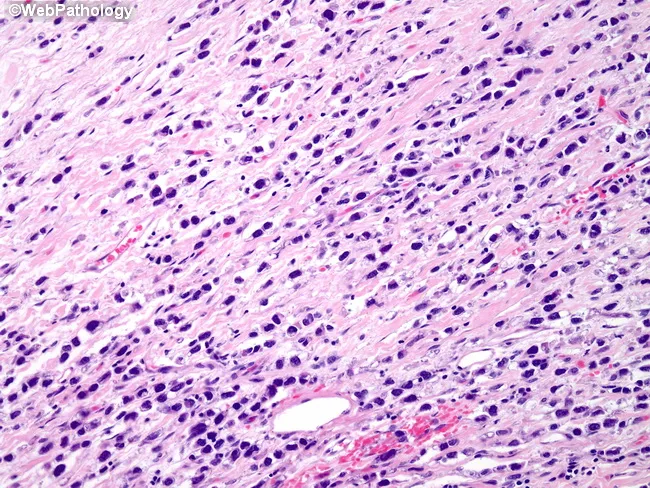
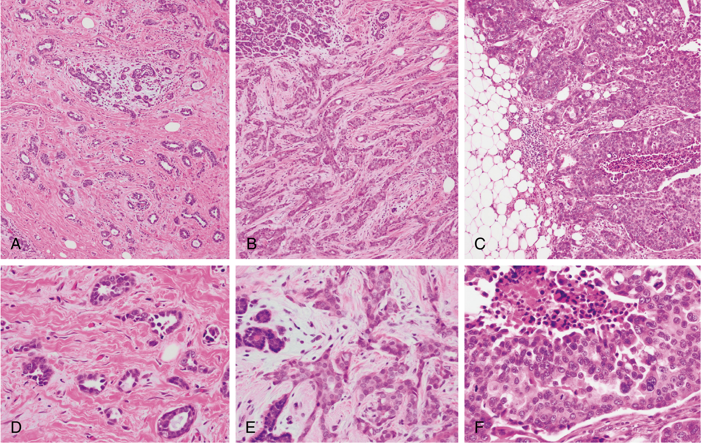
Breast histopathology image analyzed by bright-field light microscopy of formalin-fixed, paraffin-embedded tissue stained with Hematoxylin and Eosin (H&E). The lesion demonstrates invasive lobular carcinoma with a pleomorphic nuclear phenotype. Tumor cells are arranged in the classic lobular pattern with infiltrative, discohesive cells traversing a desmoplastic stroma, but display marked nuclear pleomorphism. Nuclei vary considerably in size and shape, show irregular contours, and are notably hyperchromatic. Cytoplasm is typically scant, and mitotic activity may be low to moderate depending on the field. The architectural pattern may include Indian-file strands, targetoid arrangements, and single-cell infiltration, consistent with lobular histology, but the prominent pleomorphism distinguishes it from conventional ILC. Background breast parenchyma often shows fibrous stroma and mild chronic inflammatory cells. No evident necrosis is observed in this field, though focal areas of cytoplasmic detail may be limited by processing. Clinically, pleomorphic lobular carcinoma is considered a high-grade variant with potential for more aggressive behavior than classic ILC, with implications for prognosis and treatment, including adjuvant endocrine therapy given the common hormone receptor positivity. This image is useful for pathology education, differential diagnosis training (distinguishing lobular from ductal carcinomas), and tumor grading in breast pathology.
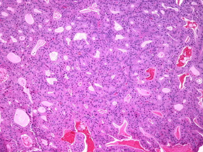
Breast tissue histology prepared as a formalin-fixed paraffin-embedded section stained with hematoxylin and eosin and examined by light microscopy. The specimen represents mammary parenchyma with ducts exhibiting papillary architecture consistent with papillary carcinoma; within several ducts there is apocrine metaplasia characterized by tall columnar cells with abundant eosinophilic granular cytoplasm and prominent apical snouts. The papillary fronds display fibrovascular cores, and the epithelial lining shows mild cytologic atypia without conspicuous mitotic activity in the field. The stroma is fibrous with scattered periductal lymphocytes; luminal spaces contain variable secretions. The differential diagnosis includes benign intraductal papilloma versus papillary carcinoma with apocrine differentiation; recognition of apocrine features is important to avoid misinterpretation as benign changes and to guide further workup. Clinically, papillary lesions of the breast require correlation with imaging and receptor studies; apocrine differentiation may be seen in older patients and can influence immunohistochemical profiling, including androgen receptor expression. This image is pertinent for education on breast tumor histopathology and serves as a reference for pathology residents, fellows, and radiology–pathology correlation cases. Potential applications include diagnostic training, differential diagnosis exercises, and quality assurance reviews in breast pathology. Correlation with gross findings and imaging enhances diagnostic confidence.
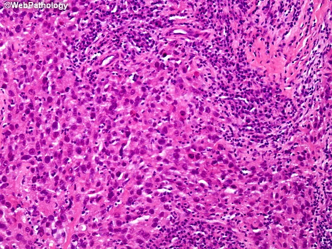
Comprehensive Description: Histopathology image of breast tissue showing medullary carcinoma. A bright-field light microscopy slide stained with hematoxylin and eosin reveals sheets of large, pleomorphic, anaplastic tumor cells arranged in solid, syncytial clusters with indistinct cell borders. The neoplastic cells often possess prominent nucleoli and abundant cytoplasm. Surrounded by a dense lymphoplasmacytic inflammatory infiltrate, the tumor demonstrates a characteristic high-grade appearance with frequent mitotic figures. Occasional tumor giant cells and focal spindle cell-like transformation may be evident. Necrosis may be present in focal areas, while calcifications are typically absent. The tumor margins may appear well circumscribed within dense fibrous stroma, with variable desmoplasia. Immunophenotype of medullary carcinoma often shows triple-negative status (ER-, PR-, HER2-) and a basal-like molecular pattern, though prognosis can be relatively favorable compared to other triple-negative breast cancers. Clinically, this histology is important to distinguish from invasive ductal carcinoma of no special type (IDC-NST) and from other medullary-like variants. The image is educational for surgical pathology, radiology-pathology correlation, and oncologic decision-making, illustrating histologic architecture (syncytial growth), lymphoid-rich stroma, necrosis, and lack of calcified deposits. This description supports differential diagnosis, prognosis estimation, and therapeutic planning in breast cancer cohorts and pathology teaching sets.
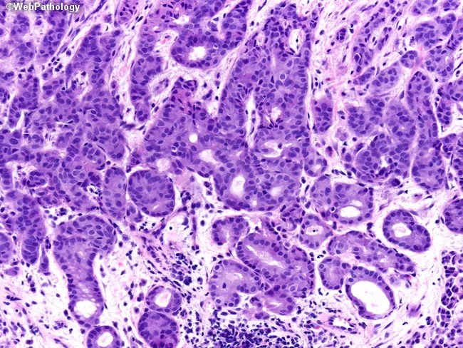
Comprehensive description: Histopathology micrograph of breast lesion showing apocrine carcinoma with glandular differentiation. The specimen is a formalin-fixed, paraffin-embedded tissue section stained with Hematoxylin and Eosin and viewed under bright-field illumination at high magnification (approximately 400× total). Tumor cells are arranged in cohesive nests and small ducts with occasional cribriform and acinar patterns, reflecting glandular differentiation. The epithelial cells exhibit abundant eosinophilic, granular cytoplasm and large, round to ovoid nuclei with conspicuous nucleoli. Apical decapitation secretions are evident on the top-middle aspect of the field, a characteristic feature of apocrine differentiation. Mitoses are variable and nuclear atypia ranges from mild to marked, with occasional prominent nucleoli and intraluminal secretions. The surrounding stroma shows a desmoplastic reaction in places, with occasional inflammatory infiltrate. No definite necrosis or lymphovascular invasion is clearly visible in this field, though assessment requires full sections. Immunohistochemical profiling is typically pursued to confirm apocrine differentiation and receptor status (ER/PR negative, AR positive; HER2 varied). This image is relevant for diagnostic education, differential diagnosis (apocrine carcinoma vs. ductal carcinoma with apocrine features), and radiology-pathology correlation in breast pathology, surgical pathology teaching, and histology training. This description supports searchability for education, research, and clinical workflow optimization in pathology practice.
breast cancer staging TNM mammography
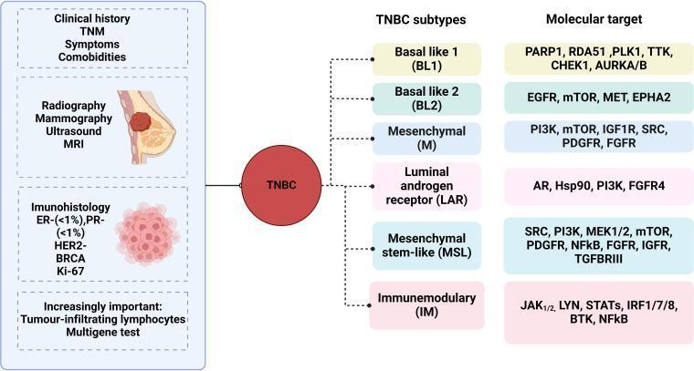
This medical infographic illustrates the diagnostic workflow and molecular classification of Triple-Negative Breast Cancer (TNBC). The diagram is divided into three horizontal sections. On the left, the diagnostic sequence includes: 1) Clinical history (TNM staging, symptoms, comorbidities); 2) Radiography (mammography, ultrasound, MRI); 3) Immunohistochemistry (ER/PR <1%, HER2 negative, BRCA, Ki-67); and 4) Emerging markers (Tumor-infiltrating lymphocytes and multigene testing). The central node, labeled 'TNBC', branches into six distinct molecular subtypes: Basal-like 1 (BL1), Basal-like 2 (BL2), Mesenchymal (M), Luminal Androgen Receptor (LAR), Mesenchymal Stem-like (MSL), and Immunomodulatory (IM). The right column lists corresponding molecular targets for each subtype, such as PARP1 and PLK1 for BL1; EGFR and mTOR for BL2; and AR and Hsp90 for the LAR subtype. This educational diagram demonstrates the heterogeneity of TNBC and the shift toward precision medicine by identifying specific signaling pathways and therapeutic targets like PI3K, SRC, and JAK1/2 across different genomic signatures.
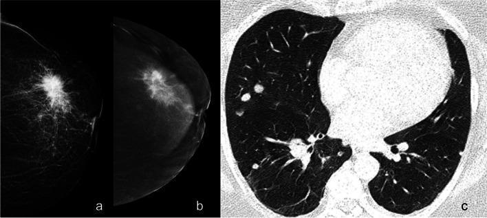
This composite diagnostic image illustrates breast cancer staging using various imaging modalities. Panel (a) is a mammogram showing a large, high-density, irregularly shaped mass in the left breast characterized by prominent radiating spicules, a classic sign of malignancy. Panel (b) shows contrast-enhanced mammography (CEM), which reveals intense enhancement of the primary lesion and provides clearer visualization of disease extent, including involvement of the retroareolar ducts. Panel (c) is a thoracic axial contrast-enhanced CT (CE-CT) scan in a lung window, demonstrating multiple bilateral, well-circumscribed pulmonary nodules. These findings collectively illustrate primary breast adenocarcinoma with secondary lung metastases. The educational focus is on the multi-modal assessment of cancer, demonstrating how unenhanced imaging (a), contrast-enhanced breast imaging (b), and systemic staging scans (c) contribute to defining the clinical stage and disease burden in oncology patients.
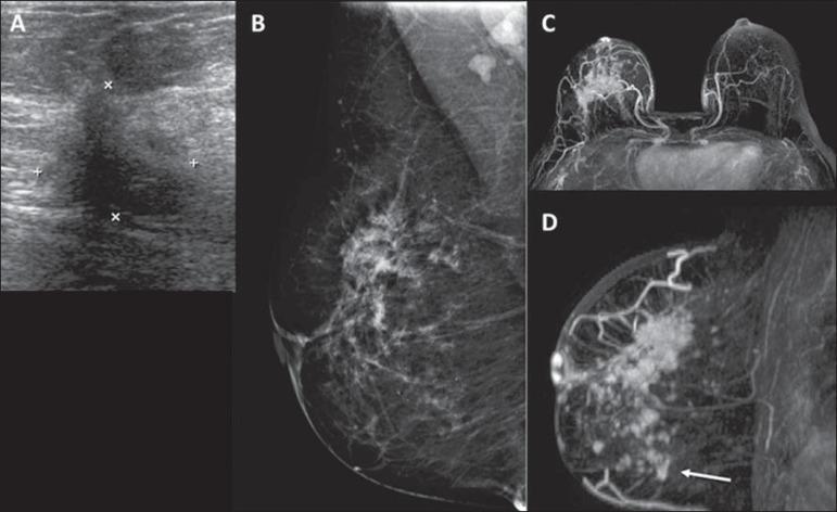
This composite diagnostic image illustrates breast cancer staging using multiple modalities including ultrasound (A), mammography (B), and contrast-enhanced MRI (C, D). Image A (ultrasound) displays an irregular, hypoechoic solid mass in the upper outer quadrant, characterized by ill-defined borders and acoustic shadowing, marked by calipers. Image B (mammogram) shows a corresponding focal asymmetry and architectural distortion in the right breast. Images C and D are Maximum Intensity Projection (MIP) MRI scans demonstrating extensive contrast enhancement. While ultrasound and mammography localized the primary tumor, the MRI reveals a significantly larger area of malignant involvement, including satellite lesions extending into the lower quadrants (indicated by an arrow in D). This comparison highlights the clinical significance of MRI in accurately assessing tumor extent and identifying multicentric or multifocal disease that may be occult on conventional imaging. Target educational concepts include breast imaging BIRADS features, diagnostic modality correlation, and preoperative tumor mapping in surgical oncology.
"breast cancer" AND management
Carcinoma of the Breast
1. EPIDEMIOLOGY
- Luminal A - ER+/HER2-, low grade, best prognosis
- Luminal B - ER+/HER2+ or high-grade ER+/HER2-
- HER2-enriched - HER2 amplified, ER-
- Basal-like/Triple Negative (TNBC) - ER-/PR-/HER2-, most aggressive
- Normal-like and Claudin-low subtypes
2. RISK FACTORS
Modifiable
| Risk Factor | Relative Risk |
|---|---|
| Obesity (BMI >30) | RR = 1.29 (postmenopausal) |
| HRT use >10 years | RR = 1.2 |
| Nulliparity / first pregnancy >35 yrs | Increased |
| No breastfeeding | Increased |
| Alcohol (heavy >4 drinks/day) | RR = 1.46 |
| Radiation exposure | RR = 6 |
| Smoking (>25 cigarettes/day) | RR = 1.14 |
Non-Modifiable
- Increasing age
- Female sex
- BRCA1/2 mutations (lifetime risk 40-90% for BRCA1)
- Family history (first-degree relative)
- Early menarche, late menopause
- Atypical ductal/lobular hyperplasia
- Previous breast cancer
- Dense breast tissue
Key Hereditary Genes (Robbins, Cotran & Kumar)
| Gene | % of Hereditary Cancers | Cancer Risk (by age 70) | Notes |
|---|---|---|---|
| BRCA1 | ~55% | 40-90% | Majority are TNBC |
| BRCA2 | ~35% | 30-60% | Majority ER-positive |
| PALB2 | <1% | 30-60% | Can be ER+ or TNBC |
| TP53 (Li-Fraumeni) | <1% | 50-60% | Often ER+ and HER2+ |
| PTEN (Cowden) | <1% | 20-80% | Apocrine differentiation |
| CDH1 | <1% | ~50% | Lobular type |
| ATM | ~5% | 15-30% | Moderate penetrance |
3. PATHOLOGY
A. Precursor / In-Situ Lesions
- Malignant epithelial cells confined within the basement membrane of the duct
- Subtypes: comedocarcinoma (with central necrosis - the hallmark), cribriform, micropapillary, solid
- Classified by nuclear grade: low, intermediate, high
- High-grade DCIS (comedo type) is associated with central necrosis and calcifications visible on mammography
- Proliferation of neoplastic cells filling and distending lobular acini
- Often multicentric and bilateral
- More a marker of increased risk (~10-fold) than an obligate precursor
- Cells are small, uniform, non-cohesive (loss of E-cadherin expression)
B. Invasive (Infiltrating) Carcinoma - Classification (Schwartz's Principles of Surgery, Foote and Stewart)
| Type | Frequency | Key Features |
|---|---|---|
| Invasive Ductal Carcinoma - No Special Type (NST/IDC) | ~80% | Hardirregular mass, desmoplastic stroma, heterogeneous |
| Invasive Lobular Carcinoma (ILC) | ~10% | Single-file ("Indian file") pattern, discohesive cells, lacks E-cadherin |
| Medullary Carcinoma | ~4% | Syncytial growth, lymphoplasmacytic infiltrate, well-circumscribed, often TNBC |
| Mucinous (Colloid) Carcinoma | ~2% | Clusters of tumor cells floating in mucin pools, better prognosis |
| Tubular Carcinoma | ~2% | Well-formed tubules, low grade, excellent prognosis |
| Papillary Carcinoma | ~2% | Papillary fronds, often intracystic |
| Metaplastic Carcinoma | Rare | Squamous/spindle cell differentiation, often TNBC |
C. Special Types
- Eczematous, chronic ulcerating lesion of the nipple-areola complex
- Pathognomonic: large pale vacuolated Paget cells in the rete pegs
- Almost always associated with underlying DCIS; may have invasive component
- Differentiated from melanoma by CEA staining (positive in Paget's) vs. S-100 (positive in melanoma)
- Clinical diagnosis: erythema, skin warmth, peau d'orange involving >1/3 of the breast surface
- Caused by dermal lymphatic invasion by tumor emboli (NOT an inflammatory process)
- Corresponds to T4d in TNM staging
- Very aggressive, requires neoadjuvant chemotherapy first; mastectomy is contraindicated upfront
D. Histological Grading - Nottingham Score (Robbins, Cotran & Kumar)
| Parameter | Score 1 | Score 2 | Score 3 |
|---|---|---|---|
| Tubule formation | >75% | 10-75% | <10% |
| Nuclear pleomorphism | Small, uniform | Moderate variation | Marked variation |
| Mitotic count | Low | Moderate | High |
- Grade 1 (Score 3-5): Well differentiated - tubular pattern, uniform nuclei
- Grade 2 (Score 6-7): Moderately differentiated - mixed tubular + solid nests
- Grade 3 (Score 8-9): Poorly differentiated - solid sheets, necrosis, high mitoses
E. Molecular Pathogenesis
- Most arise from luminal epithelial cells
- Key driver: ER signaling → PIK3CA mutations most common
- Low chromosomal instability, CDH1 loss in lobular type
- HER2 amplification on chromosome 17q12
- HER2 protein overexpression → constitutive tyrosine kinase activation
- High grade, high proliferative index
- BRCA1 mutation strongly associated
- TP53 mutations, high chromosomal instability, high grade
- CD44+ breast cancer stem cell markers
F. Spread
- Primarily to axillary lymph nodes (levels I, II, III)
- Inner quadrant tumors may spread to internal mammary nodes
- Contralateral axillary involvement = metastatic disease
- Neoangiogenesis occurs at tumor size ~1-2 mm (10^5 cells)
- Sites (in order): lumbar vertebrae > neck of femur > thoracic vertebrae > ribs > skull
- Also: liver, lungs, brain, adrenal glands, ovaries
- Bone metastases are predominantly osteolytic (occasionally osteosclerotic)
- Extensive marrow replacement → leukoerythroblastic anaemia
4. CLINICAL FEATURES
Symptoms and Signs (Bailey and Love, 28th Edition)
- Peau d'orange - lymphoedema of skin due to dermal lymphatic blockage
- Skin dimpling or tethering (Cooper's ligament involvement)
- Ulceration (advanced disease)
- Satellite skin nodules
- Erythema/warmth (inflammatory carcinoma)
- Nipple retraction or distortion
- Nipple discharge (blood-stained or serous)
- Paget's disease (eczematous nipple rash)
- Palpable axillary lymph nodes
- Arm edema (advanced nodal disease)
- Bone pain, pathological fractures
- Dyspnoea (lung metastases/pleural effusion)
- Jaundice, hepatomegaly (liver metastases)
- Headache, neurological deficits (brain metastases)
- Hypercalcaemia (bone metastases)
5. INVESTIGATIONS
Triple Assessment (the gold standard approach)
| Component | Modality |
|---|---|
| 1. Clinical examination | History + physical exam |
| 2. Imaging | Mammography ± Ultrasound ± MRI |
| 3. Pathology | Core needle biopsy / FNAC |
Imaging
- First-line investigation for women >35-40 years
- Features of malignancy:
- Spiculated mass with irregular margins
- Asymmetric density
- Clustered microcalcifications (present in ~50% of non-palpable cancers)
- Architectural distortion
- Sensitivity ~90% for palpable lesions; reduces mortality by 20-25% in screened women ≥50 years
- Preferred in younger women (<35) with dense breast tissue
- Distinguishes solid from cystic lesions
- Guides needle biopsy
- Evaluates axilla for nodal disease
- Highest sensitivity for breast cancer detection
- Used for: extent of disease, contralateral breast assessment, post-neoadjuvant response, high-risk screening (BRCA carriers), implant patients
- MRI reveals satellite lesions and multicentric disease occult on mammography
Biopsy / Pathology
- Core needle biopsy (CNB): Preferred - provides histological diagnosis, grade, ER/PR/HER2 status
- FNAC: Cytology only; cannot distinguish in-situ from invasive; used when CNB not feasible
- Stereotactic/vacuum-assisted biopsy: For non-palpable/calcification-only lesions
- Excisional biopsy: Reserved for small unresolved lesions; diagnostic + therapeutic
Biomarker Assessment (mandatory on every biopsy)
- ER and PR (immunohistochemistry): Positive if ≥1% nuclear staining; guides hormonal therapy
- HER2 (IHC + FISH): 3+ IHC or gene amplification by FISH = positive; guides anti-HER2 therapy
- Ki-67: Proliferation index; helps classify luminal A vs. B
- BRCA1/2 testing: For hereditary cases, triple-negative tumors, young patients
Staging Investigations
- Blood tests: FBC, LFTs, ALP, Ca²⁺, LDH, CEA, CA 15-3
- Chest X-ray / CT chest-abdomen-pelvis: Systemic metastases
- Bone scan (isotope): Suspected bone metastases (bone pain, raised ALP)
- PET-CT: Most accurate for staging and response assessment
- Liver ultrasound / CT: Hepatic metastases
6. TNM STAGING (UICC-AJCC 8th Edition)
Primary Tumor (T)
| Stage | Criteria |
|---|---|
| Tis | DCIS or Paget's disease (no invasive component) |
| T1mi | ≤1 mm |
| T1a | >1 mm but ≤5 mm |
| T1b | >5 mm but ≤10 mm |
| T1c | >10 mm but ≤20 mm |
| T2 | >20 mm but ≤50 mm |
| T3 | >50 mm |
| T4a | Extension to chest wall |
| T4b | Skin ulceration/peau d'orange/satellite nodules |
| T4c | T4a + T4b |
| T4d | Inflammatory carcinoma |
Regional Nodes (N)
- N0 = No regional nodal metastases
- N1 = Mobile ipsilateral axillary LN (Level I/II)
- N2 = Fixed/matted axillary LN, or internal mammary without axillary
- N3 = Infraclavicular (Level III), or internal mammary + axillary, or supraclavicular
Distant Metastasis (M)
- M0 = No distant metastasis
- M1 = Distant metastasis present
Stage Grouping
| Stage | TNM | 5-Year Survival |
|---|---|---|
| 0 | Tis N0 M0 | ~99% |
| I | T1 N0 M0 | ~90-100% |
| II | T2-3 N0-1 M0 | ~70-80% |
| III | T4 or N2-3 M0 | ~40-60% |
| IV | Any T, Any N, M1 | ~20-25% |
7. MANAGEMENT
A. Surgery
- Excision of tumor with 1-2 mm clear margins + adjuvant radiotherapy
- Equivalent survival to mastectomy for early-stage disease
- Patient preference, tumor-to-breast ratio, and ability to achieve clear margins determine eligibility
- Contraindications: multicentric disease, previous breast RT, inability to achieve clear margins, inflammatory carcinoma
- Simple (total) mastectomy: Removal of all breast tissue including nipple-areola complex
- Modified radical mastectomy (MRM) - Patey's operation: Total mastectomy + axillary clearance, preserving pectoralis major (standard procedure)
- Radical mastectomy (Halsted): Historical; includes pectoralis major removal - rarely performed now
- Skin/nipple-sparing mastectomy: For prophylactic surgery or when oncologically safe
- Sentinel Lymph Node Biopsy (SLNB): Standard for clinically node-negative patients
- Dual technique: radioisotope (Tc-99m) + blue dye (methylene blue/patent blue) or indocyanine green
- If sentinel node negative → no further axillary surgery required
- If 1-2 positive sentinel nodes + BCS + RT → axillary RT may replace ALND (Z0011 criteria)
- Contraindications: inflammatory BC, T4 disease, previous breast/chest wall surgery
- Axillary Lymph Node Dissection (ALND): For biopsy-proven positive nodes or >3 positive sentinel nodes
- Level I + II dissection standard; Level III only if enlarged Level I/II nodes
- Preserves: axillary vein, thoracodorsal vessels, long thoracic nerve (to serratus anterior), thoracodorsal nerve
- Immediate (at time of mastectomy) or delayed (6-12 months post-adjuvant treatment)
- Options:
- Implant-based (silicone gel implants, tissue expander → implant)
- Autologous tissue flaps: TRAM (transverse rectus abdominis myocutaneous), DIEP (deep inferior epigastric perforator), latissimus dorsi flap
B. Radiotherapy
- Post-BCS: Whole breast irradiation (standard dose: 40 Gy/15 fractions or 50 Gy/25 fractions) reduces local recurrence by ~50-70%
- Post-mastectomy RT (PMRT): Indicated for T3/T4 tumors, ≥4 positive nodes, positive margins
- Boost: Additional dose (10-16 Gy) to tumor bed in high-risk patients
- Chest wall + nodal RT: After mastectomy in node-positive disease
C. Systemic Therapy
Neoadjuvant (Pre-operative) Chemotherapy
- HER2-positive tumors >5 mm (+ targeted therapy)
- Triple-negative breast cancer (TNBC)
- Inflammatory breast cancer
- Premenopausal women with axillary node metastasis
- Downstage to allow BCS or resectability
Adjuvant Chemotherapy
- Indicated for: Node-positive, high-grade, ER-negative, large tumors (>2 cm), HER2+, TNBC
- Regimens: AC x4 → T (paclitaxel) x4; or FEC-D; or TC (docetaxel + cyclophosphamide) for lower-risk
Hormonal (Endocrine) Therapy (ER/PR positive tumors)
- Premenopausal: Tamoxifen (SERM, 20 mg/day) x 5-10 years ± ovarian suppression (GnRH agonist: goserelin)
- Postmenopausal: Aromatase inhibitors (anastrozole, letrozole, exemestane) preferred over tamoxifen; lower recurrence rates
- Neoadjuvant hormonal: Elderly/frail ER+ patients unfit for chemotherapy; response evident in 3-6 months
Anti-HER2 Targeted Therapy
- Trastuzumab (Herceptin): Monoclonal antibody against HER2; given for 1 year adjuvantly; combined with chemotherapy in neoadjuvant setting
- Pertuzumab: Added to trastuzumab + taxane for HER2+ neoadjuvant (dual blockade)
- Lapatinib: Tyrosine kinase inhibitor; for metastatic HER2+ after trastuzumab failure
- T-DM1 (ado-trastuzumab emtansine): Antibody-drug conjugate; residual disease post-neoadjuvant in HER2+
- T-DXd (trastuzumab deruxtecan): More potent ADC; now first-line metastatic HER2+ disease
TNBC / BRCA-Mutated Therapies
- Platinum chemotherapy (carboplatin) added to neoadjuvant regimen for BRCA-mutated TNBC
- Olaparib/Talazoparib (PARP inhibitors): Adjuvant for BRCA1/2 germline mutated HER2-negative high-risk breast cancer
- Pembrolizumab (anti-PD1): Neoadjuvant + adjuvant for high-risk TNBC (KEYNOTE-522 trial)
- Capecitabine: Adjuvant for residual disease after neoadjuvant chemotherapy in TNBC
Metastatic Disease
- Goals: disease control, quality of life, symptom management (not curative)
- Endocrine therapy + CDK4/6 inhibitors (palbociclib, ribociclib, abemaciclib) = standard for ER+/HER2- metastatic
- Bone metastases: Bisphosphonates (zoledronic acid) / denosumab to prevent skeletal-related events
- Radiotherapy for symptomatic bone metastases
D. Management Summary by Stage
| Stage | Primary Treatment |
|---|---|
| Stage 0 (DCIS) | BCS + RT ± tamoxifen; or mastectomy |
| Stage I-II | BCS + RT or mastectomy ± SLNB; adjuvant systemic therapy based on molecular profile |
| Stage III (operable) | Neoadjuvant chemotherapy → surgery → adjuvant therapy |
| Stage III (inflammatory) | Neoadjuvant chemotherapy → MRM → adjuvant RT + systemic therapy |
| Stage IV | Systemic therapy (chemotherapy/hormonal/targeted); surgery/RT for palliation |
8. PROGNOSIS - KEY PROGNOSTIC FACTORS
- Axillary lymph node status - single most important prognostic factor
- Tumor size (T stage)
- Histological grade (Nottingham)
- ER/PR/HER2 status
- Lymphovascular invasion
- Molecular subtype (Luminal A best, TNBC worst)
- Ki-67 proliferation index
- Genomic scores (Oncotype DX, MammaPrint) - guide chemotherapy decisions in ER+/HER2- early-stage disease
- Robbins, Cotran & Kumar: Pathologic Basis of Disease, pp. 974-990
- Bailey and Love's Short Practice of Surgery, 28th Edition, Chapter 58, pp. 952-970
- Schwartz's Principles of Surgery, 11th Edition, Chapter 17