
Imaging techniques and treatment of cp angle tumor in detail according to standard ENT textbook in simplified manner
cerebellopontine angle tumor MRI acoustic neuroma vestibular schwannoma
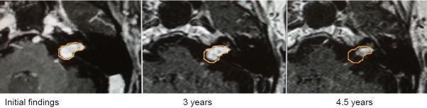
This diagnostic imaging sequence consists of three axial, contrast-enhanced T1-weighted MRI frames showing the longitudinal progression of a vestibular schwannoma (acoustic neuroma) at three stages: initial findings, 3-year follow-up, and 4.5-year follow-up. The tumor is located at the cerebellopontine angle, involving both the internal auditory canal (intrameatal) and the adjacent cistern (extrameatal). In the 'Initial findings,' the lesion shows robust contrast enhancement and a distinct extrameatal mass. At the 3-year mark, there is a subtle reduction in volume. By the 4.5-year follow-up, significant regression is evident; the extrameatal component has nearly completely disappeared, and the overall contrast enhancement intensity is diminished. Orange outlines highlight the tumor's margins across the series to assist in visual comparison. This comparison illustrates spontaneous or treatment-induced regression of a vestibular schwannoma, focusing on the changes in morphology and extrameatal extension over a multi-year timeline.
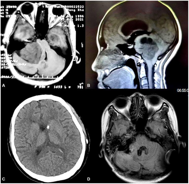
This composite of neuroimaging studies illustrates the progression and surgical management of a vestibular schwannoma. Images A (axial T1-weighted MRI) and B (sagittal T1-weighted MRI) demonstrate a large, well-circumscribed, solid mass measuring approximately 6x5 cm in the right cerebellopontine angle (CPA). The mass exerts a significant mass effect, resulting in severe compression of the brainstem, distortion of the fourth ventricle, and obstructive hydrocephalus. Secondary findings include cerebellar tonsillar herniation. Image C (axial CT scan) shows the placement of a ventriculoperitoneal (V-P) shunt, evidenced by the hyperdense shunt catheter within the enlarged ventricles, which are beginning to show signs of decompression. Image D (postoperative axial T1-weighted MRI) confirms the complete surgical resection of the CPA tumor, restoration of the normal anatomy of the brainstem and posterior fossa, and resolution of the associated hydrocephalus. The case highlights the diagnosis of acoustic neuroma and the staged surgical approach involving intracranial pressure management followed by definitive tumor excision.
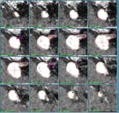
This diagnostic image displays a 4x4 matrix of 16 sequential 1-mm axial MRI slices illustrating a stereotactic radiosurgery treatment plan for an acoustic neuroma (vestibular schwannoma). Each frame shows the anatomical cross-section of the enhancing tumor target, characterized by high-intensity (bright) signal in the cerebellopontine angle. Overlaid on the tumor are two colored contour lines representing different 50% isodose levels (IDL). The outer purple line indicates the planned 50% IDL using standard 4-mm-helmet output factors (OF4/18 = 0.870), while the inner yellow line represents the modified 50% IDL if the output factor were reduced by 20%. The sequence demonstrates the spatial relationship and minimal dosimetric deviation between the two planning scenarios across the target volume. This clinical visual is utilized in radiation oncology and medical physics to assess the sensitivity of dose distributions to variations in collimator output factors, emphasizing treatment precision and quality assurance in intracranial radiosurgery.
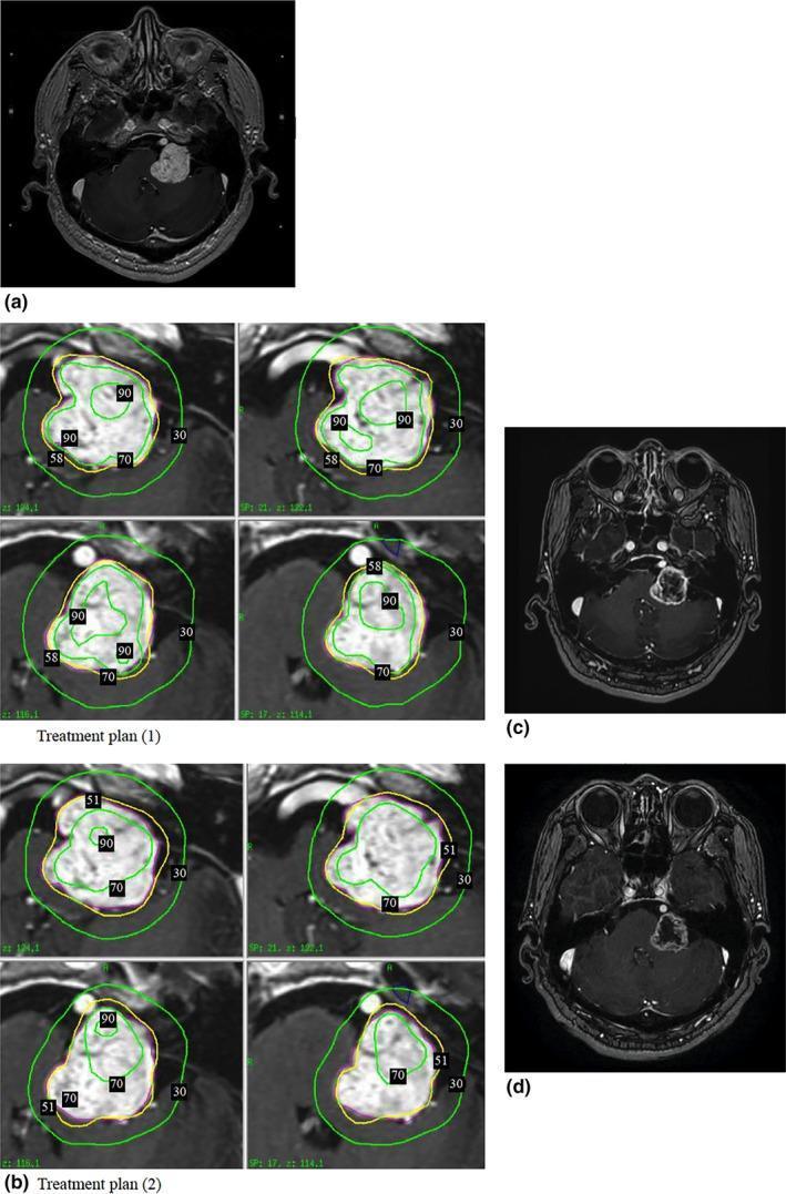
This composite figure illustrates a clinical case of a left-sided acoustic neuroma (vestibular schwannoma) and its response to Gamma Knife Radiosurgery (GKRS). Panel (a) shows a pre-treatment axial T1-weighted contrast-enhanced MRI demonstrating a large, well-defined, intensely enhancing mass in the left cerebellopontine angle with significant compression of the adjacent brainstem and cerebellum. Panel (b) provides a detailed comparison of two radiation treatment plans, displaying isodose curves (e.g., 90%, 70%, 58/51%, and 30% lines) superimposed on the tumor volume to demonstrate dose distribution and conformity. Panels (c) and (d) present longitudinal follow-up axial MRIs at 6 and 12 months post-treatment, respectively. These images demonstrate progressive therapeutic effects, including a marked loss of central contrast enhancement (indicating central necrosis or reduced vascularity) and a significant reduction in tumor volume (shrinking from 11.5 cm³ to 8.3 cm³). The images confirm the absence of adverse radiation-induced effects, such as perifocal edema or mass effect increase, highlighting the efficacy of the treatment planning strategy in tumor control and brain tissue preservation.
Cerebellopontine Angle (CPA) Tumors: Imaging & Treatment
What is the CPA?
Types of CPA Tumors
| Tumor | % of CPA tumors |
|---|---|
| Vestibular Schwannoma (VS) (acoustic neuroma) | ~85% |
| Meningioma | ~10% |
| Epidermoid cyst (cholesteatoma) | ~5% |
| Others (facial nerve schwannoma, trigeminal schwannoma, glomus, lipoma) | Rare |
The term "acoustic neuroma" is technically inaccurate - the more precise term is vestibular schwannoma, as it arises from the vestibular (not cochlear) division of CN VIII.
Clinical Features (How the Patient Presents)
- Unilateral sensorineural hearing loss (most common early symptom - in 70% of patients)
- Tinnitus - usually high-pitched, unilateral
- Vestibular imbalance (not acute vertigo - more a chronic unsteadiness)
- Facial numbness (CN V compression)
- Facial weakness (CN VII)
- Headache, vomiting (raised ICP from hydrocephalus)
- Gait ataxia
- Cerebellar signs
Key point: Vertigo (sudden spinning attacks) is uncommon with CPA tumors. This helps distinguish VS from Meniere's disease. - Localization in Clinical Neurology, 8e
IMAGING
1. MRI with Gadolinium - Gold Standard
- Detects even small intracanalicular tumors (confined to the internal auditory canal/IAC)
- Gadolinium-enhanced T1 shows bright, homogeneous enhancement of VS
- Visualizes the tumor's relationship to brainstem, cranial nerves, and vessels
- T1 with gadolinium: Enhancing mass centered on the IAC, widening the porus acusticus
- T2: Hypointense or heterogeneous; FIESTA/CISS (steady-state free precession) sequences give high-resolution detail of nerve-tumor relationships
- Typical appearance: "Ice cream cone" shape - intracanalicular + extrameatal component
- Large tumors may show rim enhancement or cystic degeneration
-
FIESTA/CISS/SPACE sequences (steady-state free precession): Define anatomical relations between tumor and adjacent cranial nerves with high resolution - essential for surgical planning
-
Thin-slice axial T2: Shows fluid in IAC
-
Adams & Victor's Principles of Neurology, p. 684
2. CT Scan
- Less sensitive than MRI for small/intracanalicular tumors
- CT detects VS >2 cm diameter or projecting >1.5 cm into the CPA cistern
- High-resolution CT (HRCT) of petrous bone: Used for surgical planning to assess:
- Position of the sigmoid sinus and jugular bulb
- Degree of mastoid air cell pneumatization
- Position of the facial nerve canal
- CT also shows bony expansion/erosion of the IAC (characteristic of VS)
- Cannot reliably detect small intracanalicular tumors
3. Audiological Tests
- Audiogram: Downward-sloping high-frequency SNHL on the tumor side
- Brainstem Auditory Evoked Response (BAER/ABR): Most sensitive audiologic test for VS - shows prolonged/absent wave V and increased I-V interpeak latency
- Speech discrimination: Poor speech discrimination out of proportion to pure-tone loss is characteristic
- Caloric testing: Reduced or absent caloric response on the affected side ("dead labyrinth")
4. Plain X-rays (Historic)
- Towne's view/Stenver's view: May show enlarged IAC
- Obsolete in modern practice; replaced by MRI
Tumor Size Classification
| Category | Extrameatal diameter |
|---|---|
| Intrameatal (Stage 1) | 0 mm (confined to IAC) |
| Small (Stage 2) | 1-15 mm |
| Medium (Stage 3) | 16-30 mm |
| Large (Stage 4) | >30 mm |
| Giant | >40 mm |
TREATMENT
1. Watch-and-Scan (Conservative Management)
- Small (< 15-20 mm extrameatal), non-cystic VS
- Elderly or medically unfit patients
- Incidentally discovered VS with no or minimal symptoms
- Bilateral VS in NF2 (to preserve hearing as long as possible)
- Annual MRI for 5 years
- Then MRI every 2 years for 4 more years
- Then one final MRI at 5 years (total ~14 years of follow-up)
- If no growth occurs, observation can be ended
- 60-70% of VS remain stable (never grow)
- Intrameatal tumors: 83% remain intrameatal; 17% extend extrameally
- Growth, if it occurs, mostly happens within the first 5 years of diagnosis
- Quality of life is better with observation than with active treatment for small, non-growing VS
- Growth of ≥3 mm in largest extrameatal diameter
- Cystic change
- Symptoms worsen
- Patient preference
2. Surgery
- Large tumors (>30 mm maximal intracranial diameter)
- Growing tumors
- Cystic VS (not suitable for radiotherapy due to risk of sudden rapid growth)
- Young patients with small tumors who want definitive treatment
- Hearing preservation attempted in selected cases
- Continuous facial nerve EMG monitoring
- Brainstem auditory evoked responses (BSER)
- Neuromuscular blocking agents must NOT be used after intubation
A. Translabyrinthine Approach
- Best for: Medium to large VS, when hearing is already non-serviceable
- Access: Through the mastoid and inner ear (labyrinth is sacrificed)
- Advantage: Direct access to the IAC and facial nerve, widest exposure, lower retraction on cerebellum, safest for facial nerve
- Disadvantage: Complete and permanent hearing loss
- Steps (Scott-Brown's):
- Retroauricular skin incision
- Cortical mastoidectomy with wide exposure of middle fossa dura and sigmoid sinus
- Labyrinthectomy (removal of semicircular canals and vestibule)
- Skeletonization of jugular bulb and vertical facial nerve
- U-shaped drilling around the IAC (270-degree removal of bone around meatus)
- Posterior fossa dura opened
- Tumor removed under microsurgical technique
- Wound closed with abdominal fat graft to fill the defect
B. Suboccipital (Retrosigmoid) Approach
- Best for: All sizes; particularly when hearing preservation is attempted
- Access: Through a posterior craniotomy behind the sigmoid sinus
- Advantage: Provides widest exposure of the CPA cistern; allows hearing preservation in tumors <2.5 cm
- Disadvantage: Requires cerebellar retraction; higher risk of postoperative headache; facial nerve identification can be more difficult
- Note: This was the approach favored by Harvey Cushing and Walter Dandy historically; modern variant uses retrosigmoid craniotomy with endoscopic assistance
C. Middle Cranial Fossa Approach
- Best for: Small, purely intracanalicular VS when hearing preservation is desired
- Access: Through the temporal bone superior surface via a temporal craniotomy
- Advantage: Best approach for hearing preservation in intracanalicular/small tumors
- Disadvantage: Limited exposure for larger tumors; risk of temporal lobe retraction
- Possible in ~1/3 of patients with tumors <2.5 cm (Adams & Victor)
- Middle fossa or retrosigmoid approaches used
- Intraoperative BAER monitoring is essential
- Facial nerve function can usually be preserved with intraoperative monitoring
- Facial motor and sensory functions preserved in ~75% of cases
3. Stereotactic Radiosurgery (SRS)
Gamma Knife Radiosurgery (GKRS) - Most Common
- Small to medium VS (< 30 mm) - the role is considered minimal for tumors >30 mm
- Elderly or medically unfit patients
- Patients refusing surgery
- Recurrent tumors after surgery
- Preferred in older patients with few symptoms
- Highly focused radiation delivered in a single high dose (Gamma Knife) or multiple fractions
- Causes DNA damage and vascular thrombosis, arresting tumor growth
- Does NOT "cure" the tumor; goal is tumor control (growth arrest)
- Controls growth in most small-medium VS
- Facial motor and sensory function preserved in ~75% of patients
- After 28 months of observation, no new neurologic deficits in most patients
- Hearing loss and facial weakness rates similar to or lower than surgery in short follow-up
- Cystic VS: NOT suitable (can cause sudden dramatic expansion)
- Long-term hearing outcomes may still deteriorate
- Occasional radiation-induced complications (edema, new neurological deficits)
- Some tumors continue to grow despite treatment
- Preferred over surgery for recurrent tumors (Adams & Victor)
- Radiation delivered in multiple fractions
- Used for tumors near sensitive structures
- Covered elsewhere in the literature (beyond this scope)
- In NF2 patients with bilateral VS, the anti-angiogenic agent bevacizumab has shown tumor size reduction in preliminary reports
- Not a first-line treatment
Summary Decision-Making Table
| Tumor Size | Hearing Status | Recommended Treatment |
|---|---|---|
| Intrameatal, small (<15 mm), non-growing | Good hearing | Watch and scan |
| Small (<15 mm), growing | Good hearing | SRS or middle fossa surgery |
| Small-medium (15-30 mm) | Poor hearing | Translabyrinthine surgery or SRS |
| Small-medium (15-30 mm) | Good hearing | Retrosigmoid surgery (hearing preservation) |
| Large (>30 mm) | Any | Surgery (translabyrinthine or retrosigmoid) |
| Cystic VS | Any | Surgery (SRS contraindicated) |
| NF2 associated | Any | Individual assessment |
Complications of Treatment
Surgical Complications
- Facial nerve palsy (most feared) - rates depend on tumor size
- Hearing loss (inevitable in translabyrinthine; possible in retrosigmoid/MCF)
- CSF leak (through wound or as rhinorrhea via eustachian tube)
- Meningitis
- Bleeding/hematoma
- Hydrocephalus (obstructive, due to fourth ventricle compromise in large tumors)
Post-Radiosurgery Complications
- Radiation-induced facial numbness/weakness
- Trigeminal neuropathy
- Tumor swelling (transient)
- Small risk of malignant transformation (extremely rare)
Other CPA Tumors - Brief Notes
- Meningioma: Does NOT arise from the IAC; does not cause early hearing loss; off-center on MRI; flat dural tail sign; treated surgically
- Epidermoid cyst (cholesteatoma): Irregular pearly white lesion; does NOT enhance; "cauliflower" appearance on MRI; treated surgically (complete removal is curative)
- Trigeminal schwannoma: Initial symptom is facial pain/numbness rather than hearing loss
- Glomus jugulare: Pulsatile tinnitus, lower cranial nerve palsies; highly vascular; "salt and pepper" appearance on MRI
- Scott-Brown's Otorhinolaryngology Head & Neck Surgery, Vol. 2 - Chapters 101 & 102 (Vestibular Schwannoma; Surgical Approaches to the CPA)
- Adams & Victor's Principles of Neurology, 12th Ed., Chapter 30 (p. 683-685)
- Localization in Clinical Neurology, 8e