
Define base excess How do the kidney compensate for acid base imbalance
Base excess (BE) is the amount of strong acid or base needed to titrate 1 liter of blood to a pH of 7.40 at 37°C with a PaCO2 of 40 mmHg. A normal BE is -2 to +2 mEq/L. A positive BE (>+2) indicates metabolic alkalosis; a negative BE (<-2, also called base deficit) indicates metabolic acidosis. It reflects the non-respiratory component of acid-base status.
Renal compensation works by adjusting bicarbonate (HCO3-) reabsorption and H+ excretion - primarily in the proximal tubule, thick ascending limb, and collecting duct. This is slower than respiratory compensation (takes 3-5 days for full effect).
Searching sources...
Reading File
Now I'll search the medical library for both topics in parallel:
Finding Sources
Finding Sources
Finding Sources
Reading File
Finding Sources
Reading File
Reading File
Reading File
Reading File
Reading File
Now let me get the Ganong's Physiology section on renal acid-base handling, which is likely the most detailed:
Reading File
Reading File
I now have comprehensive content from Ganong's Physiology. Let me also fetch the Siggaard-Andersen nomogram image, which is directly relevant to base excess:
Loading Image
Reading File
I now have all the information needed for a thorough, well-sourced answer. Let me compile it.
Base Excess - Definition
Base excess (BE) is defined as the amount of acid or base (in mEq/L) that must be added to 1 liter of blood to return the pH to 7.40, at a PaCO2 of 40 mmHg, full O2 saturation, and 37°C. Critically, it also adjusts for noncarbonic buffering in the blood.
- Normal range: -2 to +2 mEq/L
- Positive BE (> +2): Metabolic alkalosis - excess base present
- Negative BE (< -2, also called base deficit): Metabolic acidosis - base deficient
BE was introduced by Siggaard-Andersen specifically to isolate the metabolic component of an acid-base disturbance from the respiratory component. It is derived from the Siggaard-Andersen curve nomogram and requires a hemoglobin concentration measurement.
The standard bicarbonate (read from the nomogram at PCO2 = 40 mmHg) is closely related - it represents what the HCO3- would be after eliminating any respiratory component, making it an index of metabolic alkalosis or acidosis. BE goes a step further by indicating exactly how much acid or base would restore 1 L of blood to normal acid-base composition.
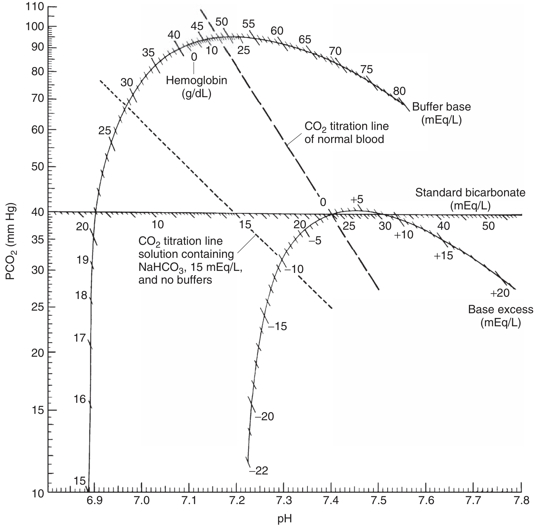
Siggaard-Andersen nomogram showing Base Excess (bottom curved scale), Standard Bicarbonate, Buffer Base, and CO2 titration lines - Ganong's Review of Medical Physiology, 26th Ed.
Note: A base deficiency cannot be corrected simply by calculating the difference between normal standard bicarbonate (24 mEq/L) and the actual value and giving that amount of NaHCO3 - because bicarbonate is distributed throughout a larger volume than plasma alone.
Renal Compensation for Acid-Base Imbalance
The kidneys regulate pH by two interrelated mechanisms:
- Reabsorbing filtered HCO3- (mainly in the proximal tubule)
- Secreting H+ ions into tubular fluid (in exchange for Na+)
This is slower than respiratory compensation - it begins within hours but takes 3-5 days for full effect. Because of this delay, respiratory acid-base disorders are classified as "acute" (before renal compensation) or "chronic" (after renal compensation).
1. Renal Compensation for Respiratory Acidosis (high PaCO2)
Mechanism:
- Elevated PaCO2 raises intracellular CO2 in tubular cells → more H2CO3 forms via carbonic anhydrase → more H+ available for secretion
- Renal tubular H+ secretion increases, removing H+ from the body
- Even though plasma HCO3- is already elevated, HCO3- reabsorption is further increased (since HCO3- is reabsorbed by exchange for H+)
- Net result: plasma HCO3- rises further, partially restoring pH toward 7.40
- Cl- excretion increases, plasma Cl- falls as plasma HCO3- rises
Expected compensation: HCO3- rises ~3.5 mEq/L per 10 mmHg rise in PaCO2 (chronic)
2. Renal Compensation for Respiratory Alkalosis (low PaCO2)
Mechanism:
- Low PaCO2 → less CO2 in tubular cells → H+ secretion is hindered
- HCO3- reabsorption is depressed → HCO3- is lost in urine
- Plasma HCO3- falls further, pulling pH back toward normal
Expected compensation: HCO3- falls ~5 mEq/L per 10 mmHg fall in PaCO2 (chronic)
3. Renal Compensation for Metabolic Acidosis (low HCO3-)
This is the most mechanistically detailed scenario, involving three tubular buffering reactions:
Step 1 - H+ secretion / HCO3- reabsorption:
The anions that replace HCO3- in plasma are filtered alongside Na+ (to maintain electrochemical neutrality). Tubular cells secrete H+ in exchange for Na+; for each H+ secreted, one Na+ and one HCO3- are added back to blood.
Step 2 - Titratable acid (phosphate buffer):
Secreted H+ reacts with HPO4²- in tubular fluid → H2PO4- (excreted as titratable acid). This prevents urinary pH from hitting the limiting minimum of 4.5 too quickly.
Step 3 - Ammonium (NH4+) excretion:
Secreted H+ reacts with NH3 (produced by tubular cells from glutamine) → NH4+ is excreted. This is the major mechanism allowing large H+ loads to be cleared. In chronic acidosis:
- Glutamine synthesis in the liver increases
- Glutamine is delivered to the kidneys as substrate for NH4+ production
- NH3 secretion adapts upward over days
- Metabolism of glutamine also generates α-ketoglutarate → decarboxylated → new HCO3- enters the bloodstream, directly buffering the acid load
The overall renal reaction (e.g., for H2SO4 added to blood):
2NaHCO3 + H2SO4 → Na2SO4 + 2H2CO3
The kidney reverses this:
Na2SO4 + 2H2CO3 → 2NaHCO3 + 2H+ + SO4²- (H+ excreted as titratable acidity + NH4+)
4. Renal Compensation for Metabolic Alkalosis (high HCO3-)
- Elevated plasma HCO3- → increased filtered load of HCO3-
- More renal H+ secretion is needed just to reabsorb this increased load
- When plasma HCO3- exceeds 26-28 mEq/L, HCO3- appears in urine (renal threshold exceeded)
- The rise in PCO2 (from respiratory compensation of metabolic alkalosis) actually inhibits renal compensation slightly by facilitating acid secretion
- Net result: HCO3- is excreted, plasma HCO3- falls toward normal
Summary Table
| Disorder | Primary Change | Renal Response | Effect |
|---|---|---|---|
| Respiratory acidosis | ↑ PaCO2 | ↑ H+ secretion, ↑ HCO3- reabsorption | ↑ plasma HCO3- |
| Respiratory alkalosis | ↓ PaCO2 | ↓ H+ secretion, ↓ HCO3- reabsorption | ↓ plasma HCO3- (HCO3- lost in urine) |
| Metabolic acidosis | ↓ HCO3- | ↑ H+ excretion (titratable acid + NH4+), new HCO3- generated | Restores plasma HCO3- |
| Metabolic alkalosis | ↑ HCO3- | HCO3- excreted in urine | ↓ plasma HCO3- |
Sources: Ganong's Review of Medical Physiology, 26th Ed. | Morgan & Mikhail's Clinical Anesthesiology, 7th Ed. | Sabiston Textbook of Surgery, 21st Ed.
Describe the counter current mechanism in the kidney.Discuss the renal protection strategies during perioperative period
The countercurrent mechanism involves two components: the countercurrent multiplier (loop of Henle) which creates a medullary osmotic gradient, and the countercurrent exchanger (vasa recta) which maintains it. Together they allow concentration of urine up to 1200 mOsm/L. Perioperative renal protection focuses on optimizing hemodynamics, avoiding nephrotoxins, and using pharmacological adjuncts.
Searching sources...
Reading File
Finding Sources
Finding Sources
Finding Sources
Finding Sources
Finding Sources
Finding Sources
Good hits. Now I'll read the key sections in parallel:
Reading File
Reading File
Reading File
Reading File
Reading File
Reading File
Reading File
Loading Image
Let me get the full countercurrent diagram URL and the remaining perioperative renal protection content:
Reading File
Reading File
Loading Image
Reading File
Reading File
Loading Image
Now let me get the key perioperative renal protection strategies section and the vasa recta exchanger content:
Reading File
Reading File
I now have comprehensive content from both Guyton & Hall and Morgan & Mikhail. I have everything I need for a complete answer. Let me compile it now.
Part 1: The Countercurrent Mechanism in the Kidney
The countercurrent mechanism has two distinct but complementary components: the countercurrent multiplier (loop of Henle) and the countercurrent exchanger (vasa recta). Together they establish and maintain a hyperosmotic medullary interstitium that allows urine concentration up to 1200-1400 mOsm/L.
A. Countercurrent Multiplier - Loop of Henle
The goal is to build a progressive osmotic gradient from cortex (~300 mOsm/L) to papilla (~1200 mOsm/L).
Key structural properties (from tubule characteristics table):
| Segment | Active NaCl Transport | Water Permeability | NaCl Permeability | Urea Permeability |
|---|---|---|---|---|
| Thin descending limb | None | High (AQP-1) | Low | Low |
| Thin ascending limb | None | None | Moderate (passive) | Moderate |
| Thick ascending limb | High (Na-K-2Cl pump) | None | None | None |
| Inner medullary collecting duct | Moderate | +ADH | None | +ADH |
Stepwise generation of the medullary gradient:
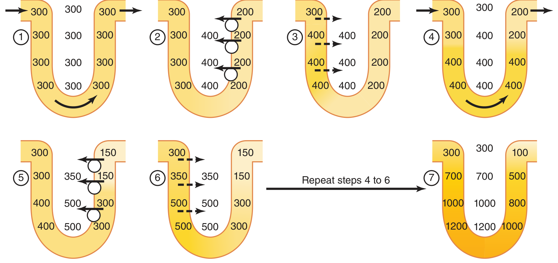
Figure: Countercurrent multiplier in the loop of Henle. Steps 1-7 show progressive concentration from 300 mOsm/L to 1200 mOsm/L - Guyton and Hall Textbook of Medical Physiology, 14e
Step-by-step mechanism:
- The loop fills with isosmotic fluid at 300 mOsm/L (equal to plasma)
- The thick ascending limb actively pumps Na+/K+/2Cl- out into the interstitium (via the NKCC2 carrier), creating a 200 mOsm/L gradient between tubular lumen and interstitium. The thick ascending limb is impermeable to water, so solute leaves without water following - this is the "single effect"
- Water osmotically leaves the descending limb (which is highly water-permeable via AQP-1 aquaporins) to equilibrate with the now-hypertonic interstitium. Tubular fluid in the descending limb becomes concentrated
- This hyperosmotic fluid from the descending limb flows around the bend into the ascending limb
- The thick ascending limb pumps ions out again, raising interstitial osmolarity further
- Descending limb fluid equilibrates again with the higher interstitial osmolarity
- Steps 4-6 repeat - this "multiplication" of the single 200 mOsm effect progressively concentrates the medullary interstitium to 1200-1400 mOsm/L at the papilla
The process is called the countercurrent multiplier because the tubular flows are in opposite directions (counter), and the repeated cycling multiplies the initial small solute gradient into a large cortico-papillary gradient.
Role of Urea
Urea contributes 40-50% of medullary osmolarity (~500-600 mOsm/L) during maximal urine concentration.
- In the presence of ADH, the inner medullary collecting duct becomes permeable to urea (via UT-A1 and UT-A3 transporters), allowing urea to diffuse into the medullary interstitium
- This urea then enters the thin descending limb (via UT-A2), travels up through the ascending limb and distal tubule, back down the collecting duct, and recirculates - progressively concentrating
- Urea recirculation is an essential component of the concentrating mechanism
B. ADH and the Collecting Duct - Final Urine Concentration
The medullary gradient alone is useless without ADH:
- Without ADH: Collecting duct is impermeable to water. Hypotonic fluid (100-140 mOsm/L) exits the loop and dilute urine is excreted
- With high ADH: ADH inserts aquaporin-2 (AQP-2) channels into the apical membrane of the collecting duct. Water is reabsorbed down the osmotic gradient. Final urine osmolarity approaches 1200 mOsm/L, equal to the medullary interstitium
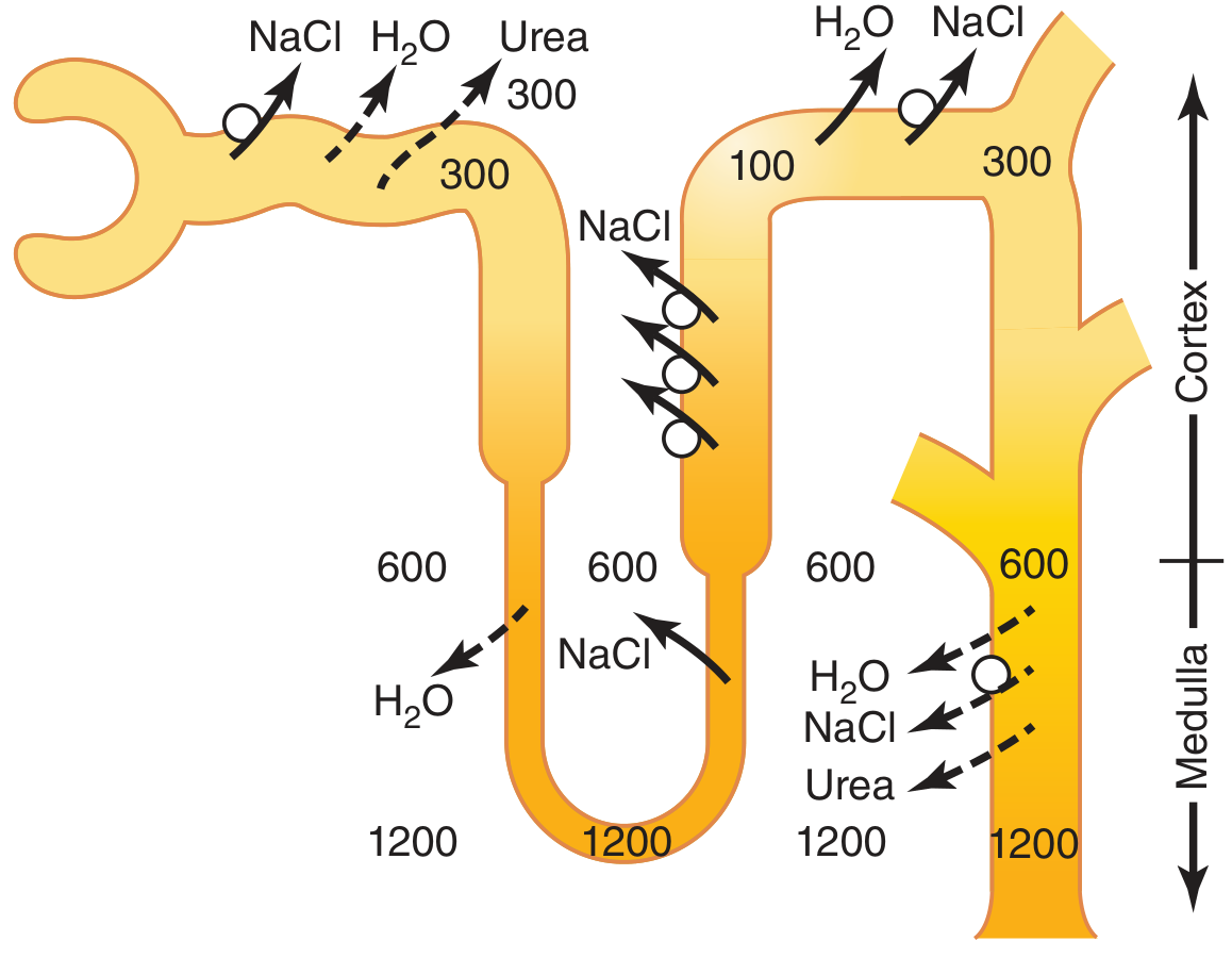
Formation of concentrated urine when ADH levels are high. The gradient goes from 300 (cortex) to 1200 mOsm/L (papilla) - Guyton and Hall, 14e
C. Countercurrent Exchanger - Vasa Recta
The vasa recta are hairpin-shaped capillaries running parallel to the loops of Henle. They preserve the medullary gradient rather than creating it.
Why they are needed: If ordinary capillaries perfused the medulla, blood flowing in would dilute the interstitium and wash out the gradient.
Mechanism:
- Medullary blood flow is deliberately low - less than 5% of total renal blood flow - minimizing solute washout
- As blood descends into the medulla, solutes (NaCl, urea) passively diffuse in and water diffuses out - blood becomes hyperosmotic to match the interstitium
- As blood ascends back toward the cortex, solutes diffuse back out into the interstitium, and water re-enters the blood
- The U-shaped hairpin architecture ensures that solutes picked up on the way down are returned on the way up - there is little net solute removal from the interstitium
"Although large amounts of fluid and solute are exchanged across the vasa recta, there is little net dilution of the concentration of the interstitial fluid at each level of the renal medulla because of the U shape of the vasa recta capillaries." - Guyton and Hall, 14e
Important: If vasodilators or high arterial pressure markedly increase medullary blood flow, solutes are "washed out" and urine-concentrating ability falls - even with maximal ADH levels.
Part 2: Perioperative Renal Protection Strategies
Why perioperative AKI matters
Acute kidney injury (AKI) affects 1-5% of general surgical patients and up to 30% of cardiac/vascular surgery patients. Mortality from postoperative AKI can exceed 50-70%. The KDIGO definition: rise in creatinine ≥0.3 mg/dL within 48 hours, or ≥1.5x baseline within 7 days, or urine output <0.5 mL/kg/hr for 6 hours.
High-risk patients: Preexisting CKD, diabetes, hypertension, liver disease, age >55 years, cardiac/aortic/vascular surgery, obstructive jaundice, sepsis, hypovolemia.
How anesthesia and surgery threaten renal function
Indirect (hemodynamic) effects:
- Most anesthetics cause dose-dependent hypotension via cardiac depression and vasodilation
- When MAP falls below the autoregulatory threshold, renal blood flow (RBF) and GFR fall proportionally
- Spinal/epidural anesthesia reduces sympathetic tone → venous pooling → decreased cardiac output
Stress response/endocrine effects:
- Pain, surgical stimulation, anxiety → catecholamine surge → renal arterial constriction → reduced RBF and GFR
- Release of ADH (non-osmotic), aldosterone, angiotensin II → sodium and water retention (perioperative oliguria is partly physiological)
Direct nephrotoxins:
- NSAIDs (ketorolac): block vasodilatory prostaglandins, reducing RBF - especially dangerous in low-flow states
- Aminoglycosides, radiocontrast agents, ACE inhibitors, hydroxyethyl starch
Renal Protection Strategies
1. Optimize Fluid Management
- Maintain normovolemia - hypovolemia is the most preventable cause of perioperative AKI
- Balanced crystalloids (Plasma-Lyte, lactated Ringer's) are preferred over 0.9% normal saline, since hyperchloremia from large-volume saline causes renal vasoconstriction and metabolic acidosis
- Exception: use saline in patients with alkalosis/hypochloremia; avoid LR in hyperkalemia
- Avoid hydroxyethyl starch in critically ill patients or those with preexisting renal impairment - associated with increased AKI risk and death
- Goal-directed fluid therapy using stroke volume/cardiac output monitoring intraoperatively
2. Maintain Renal Perfusion Pressure
- Keep MAP within the autoregulatory range (65-80 mmHg; higher targets in hypertensive patients)
- Use vasopressors (norepinephrine) to treat anesthesia-induced hypotension when volume is adequate
- Avoid excessive PEEP in ventilated patients (reduces venous return and cardiac output)
3. Avoid and Remove Nephrotoxins
- Stop NSAIDs preoperatively; substitute alternative analgesics
- Use contrast-sparing imaging where possible; pre-hydrate before contrast administration
- Avoid/dose-adjust aminoglycosides; monitor levels
- N-acetylcysteine has not shown benefit in the perioperative setting (except potentially for radiocontrast nephropathy)
4. Drugs - What Doesn't Work and What May Help
The following have no proven benefit for renal protection despite continued use:
- Mannitol: No evidence of protection or conversion from oliguric to nonoliguric failure. High doses are actually nephrotoxic. May mask hypovolemia by increasing urine output
- Low-dose dopamine ("renal dose"): No protection against AKI despite causing natriuresis and diuresis
- Fenoldopam (dopamine-1 agonist): Does not reduce AKI or need for dialysis
- Loop diuretics (furosemide): No protection - loop diuretics impair the countercurrent gradient by blocking the NKCC2 pump in the thick ascending limb
- Sodium bicarbonate infusion: Not proven effective
Possibly beneficial/under investigation:
- Dexmedetomidine: Some evidence of renoprotection via anti-inflammatory, anti-sympathetic mechanisms
- Remote ischemic preconditioning: Brief cycles of limb ischemia/reperfusion before aortic or cardiac surgery
- Cold renal perfusion (4°C): Used in vascular surgery with juxtarenal/suprarenal cross-clamping; metabolic suppression reduces ischemic injury
5. Anesthetic Agent Considerations
- Volatile agents: Isoflurane, desflurane, sevoflurane generally decrease renal vascular resistance. Sevoflurane compound A accumulates at low fresh gas flows in rats but no clinical injury in humans - use ≥2 L/min FGF as a precaution
- Ketamine: May relatively preserve RBF during hemorrhagic hypovolemia
- Propofol/opioids: Minimal direct renal effects
6. Surgical Considerations
- Aortic cross-clamping: Suprarenal/juxtarenal clamping causes direct renal ischemia; minimize clamp time; consider sequential/rapid clamping, renal cooling, or perfusion
- Avoid prolonged supine lithotomy position which can increase intra-abdominal pressure
- Treat intraoperative sepsis aggressively
7. Monitoring
- Hourly urine output (target >0.5 mL/kg/hr) - an early warning sign but sensitive to non-renal factors
- Serum creatinine equilibrates 48-72 hours after GFR changes - a late marker
- Novel biomarkers (NGAL, KIM-1, cystatin C) detect tubular injury earlier but not yet routine
- BUN:creatinine ratio >15:1 suggests prerenal azotemia (tubular reabsorption of urea increases)
Summary
| Strategy | Goal |
|---|---|
| Balanced crystalloid volume optimization | Prevent hypovolemia and hyperchloremic acidosis |
| Vasopressors to maintain MAP | Keep above autoregulatory threshold |
| Avoid NSAIDs, contrast, aminoglycosides | Remove nephrotoxic insults |
| No mannitol/low-dose dopamine/furosemide for prophylaxis | Avoid false security and harm |
| Balanced anesthetic technique | Minimize hemodynamic instability |
| Renal cooling in aortic surgery | Metabolic suppression during ischemia |
| Goal-directed therapy (SV/CO monitoring) | Individualize fluid management |
Sources: Guyton and Hall Textbook of Medical Physiology, 14e | Morgan & Mikhail's Clinical Anesthesiology, 7e | Mulholland and Greenfield's Surgery, 7e
This is a shared conversation. Sign in to Orris to start your own chat.